RPE65 Antibody (401.8B11.3D9) - BSA Free
Novus Biologicals | Catalog # NB100-355
Key Product Details
Validated by
Knockout/Knockdown
Species Reactivity
Validated:
Human, Mouse, Rat, Porcine, Bovine, Canine, Chicken, Primate, Xenopus
Cited:
Human, Mouse, Rat, Porcine, Avian - Chicken, Canine, Primate
Applications
Validated:
Knockout Validated, Immunohistochemistry, Immunohistochemistry-Paraffin, Immunohistochemistry-Frozen, Western Blot, Flow Cytometry, Immunofluorescence, Immunocytochemistry/ Immunofluorescence, Simple Western, Immunoprecipitation, In vivo assay, CyTOF-ready
Cited:
Immunohistochemistry-Paraffin, Immunohistochemistry-Frozen, Western Blot, Flow Cytometry, Immunocytochemistry/ Immunofluorescence, Simple Western, IF/IHC
Label
Unconjugated
Antibody Source
Monoclonal Mouse IgG1 kappa Clone # 401.8B11.3D9
Format
BSA Free
Loading...
Product Specifications
Immunogen
This RPE65 Antibody (401.8B11.3D9) was developed against bovine RPE65 microsomal membrane proteins. [UniProt# Q28175]
Reactivity Notes
Primate reactivity reported in scientific literature (PMID: 31660416).
Localization
Cytoplasmic, Microsomeal and Cell Membrane
Marker
Retinal Pigment Epithelium Marker
Clonality
Monoclonal
Host
Mouse
Isotype
IgG1 kappa
Theoretical MW
65 kDa.
Disclaimer note: The observed molecular weight of the protein may vary from the listed predicted molecular weight due to post translational modifications, post translation cleavages, relative charges, and other experimental factors.
Disclaimer note: The observed molecular weight of the protein may vary from the listed predicted molecular weight due to post translational modifications, post translation cleavages, relative charges, and other experimental factors.
Scientific Data Images for RPE65 Antibody (401.8B11.3D9) - BSA Free
![Western Blot: RPE65 Antibody (401.8B11.3D9)BSA Free [NB100-355]](https://resources.rndsystems.com/images/products/RPE65-Antibody-401-8B11-3D9-BSA-Free-Western-Blot-NB100-355-img0013.jpg "Western Blot: RPE65 Antibody (401.8B11.3D9)BSA Free [NB100-355]")
Western Blot: RPE65 Antibody (401.8B11.3D9)BSA Free [NB100-355]
RPE65-Antibody-401-8B11-3D9-BSA-Free-Western-Blot-NB100-355-img0013.jpg![Immunocytochemistry/ Immunofluorescence: RPE65 Antibody (401.8B11.3D9) - BSA Free [NB100-355]](https://resources.rndsystems.com/images/products/RPE65-Antibody-401-8B11-3D9-BSA-Free-Immunocytochemistry-Immunofluorescence-NB100-355-img0014.jpg "Immunocytochemistry/ Immunofluorescence: RPE65 Antibody (401.8B11.3D9) - BSA Free [NB100-355]")
Immunocytochemistry/ Immunofluorescence: RPE65 Antibody (401.8B11.3D9) - BSA Free [NB100-355]
Immunocytochemistry/Immunofluorescence: RPE65 Antibody (401.8B11.3D9) - BSA Free [NB100-355] - Expression of eye-specific markers in the induced eye-like structures induced from lignin-added ES cells. (a) Higher-magnification image of the RPE like structure induced from ESCs after 12-day culture. (b) Immunostaining of eye- like structures. Eye- like structures induced from ESCs after 12-day culture were stained with antibodies against RPE65 (red) and nuclei were stained with DAPI solution (blue). Scale bar: a = 50 um, b,d = 200 um, c,e = 100 um. PLoS One. 2013 Jun 21;8(6):e66376. doi: 10.1371/journal.pone.0066376.![Immunohistochemistry-Paraffin: RPE65 Antibody (401.8B11.3D9) - BSA Free [NB100-355]](https://resources.rndsystems.com/images/products/RPE65-Antibody-401-8B11-3D9-BSA-Free-Immunohistochemistry-Paraffin-NB100-355-img0012.jpg "Immunohistochemistry-Paraffin: RPE65 Antibody (401.8B11.3D9) - BSA Free [NB100-355]")
Immunohistochemistry-Paraffin: RPE65 Antibody (401.8B11.3D9) - BSA Free [NB100-355]
Immunohistochemistry-Paraffin: RPE65 Antibody (401.8B11.3D9) - BSA Free [NB100-355] - Analysis of FFPE human glioblastoma tissue section using 1:500 dilution of RPE65 [401.8B11.3D9] antibody on a Bond Rx autostainer (Leica Biosystems). The assay involved 20 minutes of heat induced antigen retrieval (HIER) with 10mM sodium citrate buffer (pH 6.0) and endogenous peroxidase quenching using peroxide block. The sections were incubated with primary antibody for 30 minutes. Bond Polymer Refine Detection (Leica Biosystems) and DAB were used for signal detection which followed counterstaining with hematoxylin. Whole slide scanning and capturing of representative images (20X) were performed using Aperio AT2 (Leica Biosystems).![Western Blot: RPE65 Antibody (401.8B11.3D9)BSA Free [NB100-355]](https://resources.rndsystems.com/images/products/RPE65-Antibody-401-8B11-3D9-BSA-Free-Western-Blot-NB100-355-img0008.jpg "Western Blot: RPE65 Antibody (401.8B11.3D9)BSA Free [NB100-355]")
Western Blot: RPE65 Antibody (401.8B11.3D9)BSA Free [NB100-355]
Western Blot: RPE65 Antibody (401.8B11.3D9) - BSA Free [NB100-355] - WB analysis of RPE65 in 20ug lysate of COS7 cells expressing recombinant Human RPE65 (Lane 1) and 5ug of Bovine retinal pigment epithelium membrane fraction (Lane 2). Blot processed for detection with alkaline phosphatase conjugated goat-anti Mouse IgG secondary antibody and NBT/BCIP substrate.![Immunocytochemistry/ Immunofluorescence: RPE65 Antibody (401.8B11.3D9) - BSA Free [NB100-355]](https://resources.rndsystems.com/images/products/RPE65-Antibody-401-8B11-3D9-BSA-Free-Immunocytochemistry-Immunofluorescence-NB100-355-img0006.jpg "Immunocytochemistry/ Immunofluorescence: RPE65 Antibody (401.8B11.3D9) - BSA Free [NB100-355]")
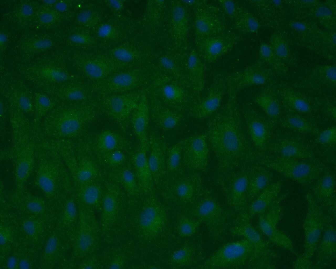
Immunocytochemistry/ Immunofluorescence: RPE65 Antibody (401.8B11.3D9) - BSA Free [NB100-355]
Immunocytochemistry/Immunofluorescence: RPE65 Antibody (401.8B11.3D9) - BSA Free [NB100-355] - ICC-IF analysis of cultured ARPE19 cells, a spontaneously arising human retinal pigment epithelia cell line - 10 minutes fixation in 4% PFA, 10 minutes permeabilization in PBS containing 0.2% Triton X-100 (PBS-T), 1 hour blocking in 10% normal goat serum containing 1% BSA in PBS-T, 1:100 primary antibody dilution in PBS, ON 4C incubation.![Immunohistochemistry-Frozen: RPE65 Antibody (401.8B11.3D9) - BSA Free [NB100-355]](https://resources.rndsystems.com/images/products/RPE65-Antibody-401-8B11-3D9-BSA-Free-Immunohistochemistry-Frozen-NB100-355-img0007.jpg "Immunohistochemistry-Frozen: RPE65 Antibody (401.8B11.3D9) - BSA Free [NB100-355]")
Immunohistochemistry-Frozen: RPE65 Antibody (401.8B11.3D9) - BSA Free [NB100-355]
Immunohistochemistry-Frozen: RPE65 Antibody (401.8B11.3D9) - BSA Free [NB100-355] - Staining of RPE65 in a cryosection of mouse retina tissue using RPE65 antibody (clone 401.8B11.3D9).![Immunohistochemistry: RPE65 Antibody (401.8B11.3D9) - BSA Free [NB100-355]](https://resources.rndsystems.com/images/products/RPE65-Antibody-401-8B11-3D9-BSA-Free-Immunohistochemistry-NB100-355-img0011.jpg "Immunohistochemistry: RPE65 Antibody (401.8B11.3D9) - BSA Free [NB100-355]")
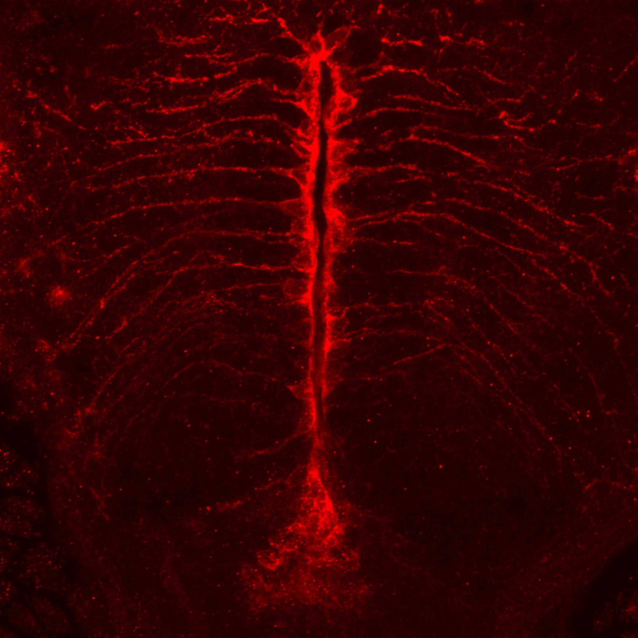
Immunohistochemistry: RPE65 Antibody (401.8B11.3D9) - BSA Free [NB100-355]
Immunohistochemistry: RPE65 Antibody (401.8B11.3D9) - BSA Free [NB100-355] - Zebrafish brain ventricular area labeled with mouse anti-RPE65 (1:100). This image was submitted through a verified customer review.![Simple Western: RPE65 Antibody (401.8B11.3D9)BSA Free [NB100-355]](https://resources.rndsystems.com/images/products/RPE65-Antibody-401-8B11-3D9-BSA-Free-Simple-Western-NB100-355-img0016.jpg "Simple Western: RPE65 Antibody (401.8B11.3D9)BSA Free [NB100-355]")
Simple Western: RPE65 Antibody (401.8B11.3D9)BSA Free [NB100-355]
RPE65-Antibody-401-8B11-3D9-BSA-Free-Simple-Western-NB100-355-img0016.jpg![Knockout Validated: RPE65 Antibody (401.8B11.3D9) - BSA Free [NB100-355]](https://resources.rndsystems.com/images/products/RPE65-Antibody-401-8B11-3D9-BSA-Free-Knockout-Validated-NB100-355-img0015.jpg "Western Blot: RPE65 Antibody (401.8B11.3D9) - BSA Free [NB100-355]")
 - BSA Free [NB100-355] -")
Western Blot: RPE65 Antibody (401.8B11.3D9) - BSA Free [NB100-355] -
Western Blot: RPE65 Antibody (401.8B11.3D9) - BSA Free [NB100-355] - Expression of RPE-specific marker proteins in hPSC-RPE & iPSC-RPE cells.(a–b) Immunoblotting showing the expression of RPE-specific proteins BEST1, RPE65, CRALBP, & the loading control beta -Actin in hPSC-RPE (a) & iPSC-RPE (b) cells. Two gels/blots in the same panel were prepared from the same cell lysate of each PSC-RPE to detect BEST1 + beta -Actin, & RPE65 + CRALBP, respectively. Image collected & cropped by CiteAb from the following publication (https://pubmed.ncbi.nlm.nih.gov/34061021), licensed under a CC-BY license. Not internally tested by Novus Biologicals. - BSA Free [NB100-355] -")
Immunocytochemistry/ Immunofluorescence: RPE65 Antibody (401.8B11.3D9) - BSA Free [NB100-355] -
Immunocytochemistry/ Immunofluorescence: RPE65 Antibody (401.8B11.3D9) - BSA Free [NB100-355] - BMP5 induces ectopic RPE development in the central NR at optic cup stages. (A,B) Peripheral, untreated chick eye at E8 showing pigmentation & MITF expression being restricted to the outer layer of the eye. The arrowheads indicate the peripheral NR. (C-F) Higher magnification of the central chick eye at E8 showing the pigmented RPE, where RPE65 protein is detected (arrows). Faint Wnt2b & strong Vsx2 expression is detected in the central NR. The arrows indicate the RPE. (G,H) Following BMP5 application at E3.5/4, the NR detached (arrowhead) & MITF protein is now detected in the central NR (cNR). Note that a part of the cNR has RPE-like morphology (eRPE). (I-L) Higher magnification images of the eRPE. Ectopic pigment granulae, RPE65 protein & strong Wnt2b expression are detected in this region, whereas Vsx2 expression is downregulated (arrowheads). (M-O) Untreated chick eye at E8 showing pigmentation & MITF (arrow) & VSX2 protein distribution, restricted to the RPE & NR, respectively. (P-R) Following BMP5 application at E3.5/4, ectopic pigmentation & nuclear MITF protein are detected in the inner optic cup (arrowheads), whereas VSX2 protein is only weakly or not detected. The arrows indicate the RPE. (S) Graphical representation of the average thickness of the inner layer of the untreated (control) & BMP5-treated eye at E8. All expression pattern studies included n≥5. Image collected & cropped by CiteAb from the following publication (https://pubmed.ncbi.nlm.nih.gov/28546339), licensed under a CC-BY license. Not internally tested by Novus Biologicals. - BSA Free [NB100-355] -")
Immunohistochemistry: RPE65 Antibody (401.8B11.3D9) - BSA Free [NB100-355] -
Altered RPE structure and cell morphology in JR5558 mice. Immunofluorescence imaging of JR5558 retinal sections with antibodies against GFAP (green) and RPE65 (red) revealed subretinal Müller cell gliosis and altered RPE structure around the lesion area (A), corresponding to the altered outer retina and RPE structure noted in OCT image (B). Arrow and arrowhead highlighting subretinal gliosis and altered RPE, respectively. Phalloidin staining of JR5558 retinal flatmounts revealed the normal morphology of RPE cells at 4 (C) and 8 weeks (D) is changed from 12 weeks onwards (E–F). Dashed box represents area imaged in panel A. Scale bars: 50 μm. Image collected and cropped by CiteAb from the following open publication (https://www.nature.com/articles/s41598-024-66068-z), licensed under a CC-BY license. Not internally tested by Novus Biologicals. - BSA Free [NB100-355] -")
Immunohistochemistry: RPE65 Antibody (401.8B11.3D9) - BSA Free [NB100-355] -
Altered RPE structure and cell morphology in JR5558 mice. Immunofluorescence imaging of JR5558 retinal sections with antibodies against GFAP (green) and RPE65 (red) revealed subretinal Müller cell gliosis and altered RPE structure around the lesion area (A), corresponding to the altered outer retina and RPE structure noted in OCT image (B). Arrow and arrowhead highlighting subretinal gliosis and altered RPE, respectively. Phalloidin staining of JR5558 retinal flatmounts revealed the normal morphology of RPE cells at 4 (C) and 8 weeks (D) is changed from 12 weeks onwards (E–F). Dashed box represents area imaged in panel A. Scale bars: 50 μm. Image collected and cropped by CiteAb from the following open publication (https://www.nature.com/articles/s41598-024-66068-z), licensed under a CC-BY license. Not internally tested by Novus Biologicals.Applications for RPE65 Antibody (401.8B11.3D9) - BSA Free
Application
Recommended Usage
Immunocytochemistry/ Immunofluorescence
1:50 - 1:200
Immunofluorescence
1:50 - 1:200
Immunohistochemistry
1:250
Immunohistochemistry-Frozen
1:250
Immunohistochemistry-Paraffin
1:250-1:500
Immunoprecipitation
reported by customer review
In vivo assay
reported in scientific literature (PMID 32173468)
Western Blot
1 - 2 ug/ml
Application Notes
For Western blot, this antibody has been validated in lysates of bovine RPE membrane and COS7 cells transfected with human RPE65, and in both samples, the antibody recognized a band at ~65 kDa, representing RPE65 protein. Note:- Hamel et al. have reported that in cultured bovine RPE cells, the levels of RPE65 are undetectable in WB after day 14, however, the mRNA levels are detectable by Northern for at least 7 weeks (PMID: 8340400). The observed molecular weight of the protein may vary from the listed predicted molecular weight due to post translational modifications, post translation cleavages, relative charges, and other experimental factors. This antibody is CyTOF ready. See Simple Western Antibody Database for Simple Western validation: tested in iPSC-RPE; separated by size
Reviewed Applications
Read 7 reviews rated 3.4 using NB100-355 in the following applications:
Flow Cytometry Panel Builder
Bio-Techne Knows Flow Cytometry
Save time and reduce costly mistakes by quickly finding compatible reagents using the Panel Builder Tool.
Advanced Features
- Spectra Viewer - Custom analysis of spectra from multiple fluorochromes
- Spillover Popups - Visualize the spectra of individual fluorochromes
- Antigen Density Selector - Match fluorochrome brightness with antigen density

Formulation, Preparation, and Storage
Purification
Protein G purified
Formulation
PBS
Format
BSA Free
Preservative
0.02% Sodium Azide
Concentration
1.0 mg/ml
Shipping
The product is shipped with polar packs. Upon receipt, store it immediately at the temperature recommended below.
Stability & Storage
Aliquot and store at -20C or -80C. Avoid freeze-thaw cycles.
Background: RPE65
Given its essential role in the vision cycle, it is understandable that mutations in RPE65 are associated with a variety of inherited retinal dystrophies (1, 3-6). Leber Congenital Amaurosis (LCA) and retinitis pigmentosa (RP) are two of the most common retinal dystrophies associated with bi-allelic RPE65 gene mutations (5,6). In 2017 the FDA approved an in vivo gene therapy for treatment of RPE65-associated diseases (5,6). The drug Voretigene Neparvovec, also called Luxturna, is delivered sub-retinally and transduces RPE cells with cDNA encoding for normal RPE65 to help restore vision (5,6). There are several promising completed and ongoing clinical trials for treating RPE65-associated diseases using gene replacement therapy (5).
References
1. Kiser, P. D., & Palczewski, K. (2010). Membrane-binding and enzymatic properties of RPE65. Progress in retinal and eye research. https://doi.org/10.1016/j.preteyeres.2010.03.002
2. Uppal, S., Poliakov, E., Gentleman, S., & Redmond, T. M. (2019). RPE65 Palmitoylation: A Tale of Lipid Posttranslational Modification. Advances in experimental medicine and biology. https://doi.org/10.1007/978-3-030-27378-1_88
3. Redmond T. M. (2009). Focus on Molecules: RPE65, the visual cycle retinol isomerase. Experimental eye research. https://doi.org/10.1016/j.exer.2008.07.015
4. Saari J. C. (2016). Vitamin A and Vision. Sub-cellular biochemistry. https://doi.org/10.1007/978-94-024-0945-1_9
5. Miraldi Utz, V., Coussa, R. G., Antaki, F., & Traboulsi, E. I. (2018). Gene therapy for RPE65-related retinal disease. Ophthalmic genetics. https://doi.org/10.1080/13816810.2018.1533027
6. Apte R. S. (2018). Gene Therapy for Retinal Degeneration. Cell. https://doi.org/10.1016/j.cell.2018.03.021
Alternate Names
All-trans-retinyl-palmitate hydrolase, BCO family, member 3, BCO3, EC 3.1.1.64, EC:3.1.1.64, EC:5.3.3.22, LCA2, lutein isomerase, meso-zeaxanthin isomerase, mRPE65, p63, RBP-binding membrane protein, rd12, retinal pigment epithelium specific protein 65, Retinal pigment epithelium-specific 65 kDa protein, retinal pigment epithelium-specific protein 65kDa, retinitis pigmentosa 20 (autosomal recessive), retinoid isomerohydrolase, Retinol isomerase, RP20, RPE65, RPE65, retinoid isomerohydrolase, sRPE65
Gene Symbol
RPE65
Additional RPE65 Products
Product Documents for RPE65 Antibody (401.8B11.3D9) - BSA Free
Certificate of Analysis
To download a Certificate of Analysis, please enter a lot or batch number in the search box below.
Product Specific Notices for RPE65 Antibody (401.8B11.3D9) - BSA Free
This product is for research use only and is not approved for use in humans or in clinical diagnosis. Primary Antibodies are guaranteed for 1 year from date of receipt.
Citations for RPE65 Antibody (401.8B11.3D9) - BSA Free
 Powered by Bioz
Customer Reviews for RPE65 Antibody (401.8B11.3D9) - BSA Free (7)
3.4 out of 5
7 Customer Ratings
Have you used RPE65 Antibody (401.8B11.3D9) - BSA Free?
Submit a review and receive an Amazon gift card!
$25/€18/£15/$25CAN/¥2500 Yen for a review with an image
$10/€7/£6/$10CAN/¥1110 Yen for a review without an image
Submit a review
Customer Images

-(01-ml)_NB100-355_7231.jpg)
Showing
1
-
5 of
7 reviews
Showing All
Filter By:
-
Application: ImmunoprecipitationSample Tested: Bovine RPE cell extracts and 293T cell lysate (overexpressing RPE65)Species: Human and BovineVerified Customer | Posted 05/06/2019Bovine RPE cell extracts: Lane 1 - IP: RPE65 (5 uL antibody, 700 ug total protein), lane 2 - lysate WB: RPE65 (1:1000)We applied this antibody to endogenous RPE65 from bovine RPE cell extracts and human RPE65 overexpressed in 293T cells. In both cases it produced robust immunoprecipitation and good western blot signal. Four stars because it does recognize a relatively strong nonspecific band around 40 kD in 293T cell lysates. Cells lysed in 1% IGEPAL IP: 2.5 uL RPE65 antibody per approximately 350 ug protein using A/G agarose beads WB dilution: 1:1000

-
Application: ImmunocytochemistrySample Tested: ARPE-19 cellsSpecies: HumanVerified Customer | Posted 08/29/2017ARPE-19 monolayer on glass substrateFixative Methanol Blocking Goat Serum Dilution 1:250 Incubation 1h RT
-
Application: ImmunofluorescenceSample Tested: pfa fixed 11day old zebrafish cryosectionsSpecies: OtherVerified Customer | Posted 03/23/201611d zebrafish brain ventricular area labeled with mouse anti-RPE65 (1:100)
-
Application: Immunohistochemistry-FrozenSample Tested: See PMID 24143172Species: OtherVerified Customer | Posted 12/12/2014
-
Application: ImmunofluorescenceSample Tested:Species: OtherVerified Customer | Posted 10/06/2014
-
Application: ImmunofluorescenceSample Tested:Species: HumanVerified Customer | Posted 04/30/2014
-
Application: Western BlotSample Tested: RPE whole cell lysate, Sample Amount: 20-25microgramSpecies: HumanVerified Customer | Posted 12/06/2010
There are no reviews that match your criteria.
Protocols
View specific protocols for RPE65 Antibody (401.8B11.3D9) - BSA Free (NB100-355):
Western Blot Protocol
1. Perform SDS-PAGE on samples to be analyzed, loading 10-25 ug of total protein per lane.
2. Transfer proteins to PVDF membrane according to the instructions provided by the manufacturer of the membrane and transfer apparatus.
3. Stain the membrane with Ponceau S (or similar product) to assess transfer success, and mark molecular weight standards where appropriate.
4. Rinse the blot TBS -0.05% Tween 20 (TBST).
5. Block the membrane in 5% Non-fat milk in TBST (blocking buffer) for at least 1 hour.
6. Wash the membrane in TBST three times for 10 minutes each.
7. Dilute primary antibody in blocking buffer and incubate overnight at 4C with gentle rocking.
8. Wash the membrane in TBST three times for 10 minutes each.
9. Incubate the membrane in diluted HRP conjugated secondary antibody in blocking buffer (as per manufacturer's instructions) for 1 hour at room temperature.
10. Wash the blot in TBST three times for 10 minutes each (this step can be repeated as required to reduce background).
11. Apply the detection reagent of choice in accordance with the manufacturers instructions.
Find general support by application which include: protocols, troubleshooting, illustrated assays, videos and webinars.
- 7-Amino Actinomycin D (7-AAD) Cell Viability Flow Cytometry Protocol
- Antigen Retrieval Protocol (PIER)
- Antigen Retrieval for Frozen Sections Protocol
- Appropriate Fixation of IHC/ICC Samples
- Cellular Response to Hypoxia Protocols
- Chromogenic IHC Staining of Formalin-Fixed Paraffin-Embedded (FFPE) Tissue Protocol
- Chromogenic Immunohistochemistry Staining of Frozen Tissue
- ClariTSA™ Fluorophore Kits
- Detection & Visualization of Antibody Binding
- Extracellular Membrane Flow Cytometry Protocol
- Flow Cytometry Protocol for Cell Surface Markers
- Flow Cytometry Protocol for Staining Membrane Associated Proteins
- Flow Cytometry Staining Protocols
- Flow Cytometry Troubleshooting Guide
- Fluorescent IHC Staining of Frozen Tissue Protocol
- Graphic Protocol for Heat-induced Epitope Retrieval
- Graphic Protocol for the Preparation and Fluorescent IHC Staining of Frozen Tissue Sections
- Graphic Protocol for the Preparation and Fluorescent IHC Staining of Paraffin-embedded Tissue Sections
- Graphic Protocol for the Preparation of Gelatin-coated Slides for Histological Tissue Sections
- ICC Cell Smear Protocol for Suspension Cells
- ICC Immunocytochemistry Protocol Videos
- ICC for Adherent Cells
- IHC Sample Preparation (Frozen sections vs Paraffin)
- Immunocytochemistry (ICC) Protocol
- Immunocytochemistry Troubleshooting
- Immunofluorescence of Organoids Embedded in Cultrex Basement Membrane Extract
- Immunofluorescent IHC Staining of Formalin-Fixed Paraffin-Embedded (FFPE) Tissue Protocol
- Immunohistochemistry (IHC) and Immunocytochemistry (ICC) Protocols
- Immunohistochemistry Frozen Troubleshooting
- Immunohistochemistry Paraffin Troubleshooting
- Immunoprecipitation Protocol
- Intracellular Flow Cytometry Protocol Using Alcohol (Methanol)
- Intracellular Flow Cytometry Protocol Using Detergents
- Intracellular Nuclear Staining Flow Cytometry Protocol Using Detergents
- Intracellular Staining Flow Cytometry Protocol Using Alcohol Permeabilization
- Intracellular Staining Flow Cytometry Protocol Using Detergents to Permeabilize Cells
- Preparing Samples for IHC/ICC Experiments
- Preventing Non-Specific Staining (Non-Specific Binding)
- Primary Antibody Selection & Optimization
- Propidium Iodide Cell Viability Flow Cytometry Protocol
- Protocol for Heat-Induced Epitope Retrieval (HIER)
- Protocol for Liperfluo
- Protocol for Making a 4% Formaldehyde Solution in PBS
- Protocol for VisUCyte™ HRP Polymer Detection Reagent
- Protocol for the Characterization of Human Th22 Cells
- Protocol for the Characterization of Human Th9 Cells
- Protocol for the Fluorescent ICC Staining of Cell Smears - Graphic
- Protocol for the Fluorescent ICC Staining of Cultured Cells on Coverslips - Graphic
- Protocol for the Preparation & Fixation of Cells on Coverslips
- Protocol for the Preparation and Chromogenic IHC Staining of Frozen Tissue Sections
- Protocol for the Preparation and Chromogenic IHC Staining of Frozen Tissue Sections - Graphic
- Protocol for the Preparation and Chromogenic IHC Staining of Paraffin-embedded Tissue Sections
- Protocol for the Preparation and Chromogenic IHC Staining of Paraffin-embedded Tissue Sections - Graphic
- Protocol for the Preparation and Fluorescent ICC Staining of Cells on Coverslips
- Protocol for the Preparation and Fluorescent ICC Staining of Non-adherent Cells
- Protocol for the Preparation and Fluorescent ICC Staining of Stem Cells on Coverslips
- Protocol for the Preparation and Fluorescent IHC Staining of Frozen Tissue Sections
- Protocol for the Preparation and Fluorescent IHC Staining of Paraffin-embedded Tissue Sections
- Protocol for the Preparation of Gelatin-coated Slides for Histological Tissue Sections
- Protocol for the Preparation of a Cell Smear for Non-adherent Cell ICC - Graphic
- Protocol: Annexin V and PI Staining by Flow Cytometry
- Protocol: Annexin V and PI Staining for Apoptosis by Flow Cytometry
- R&D Systems Quality Control Western Blot Protocol
- TUNEL and Active Caspase-3 Detection by IHC/ICC Protocol
- The Importance of IHC/ICC Controls
- Troubleshooting Guide: Fluorokine Flow Cytometry Kits
- Troubleshooting Guide: Immunohistochemistry
- Troubleshooting Guide: Western Blot Figures
- Western Blot Conditions
- Western Blot Protocol
- Western Blot Protocol for Cell Lysates
- Western Blot Troubleshooting
- Western Blot Troubleshooting Guide
- View all Protocols, Troubleshooting, Illustrated assays and Webinars
FAQs for RPE65 Antibody (401.8B11.3D9) - BSA Free
Showing
1
-
5 of
5 FAQs
Showing All
-
Q: Do you recommend an antigen retrieval step for this antibody : RPE65 Antibody (401.8B11.3D9)?. My application is IHC-paraffin cross section!
A:
Yes, we do recommend antigen retrieval step for NB100-355 and in our IHC validation for this product, heat induced method was employed. You may use citrate buffer (10mM Citric Acid, 0.05% Tween 20, pH 6.0) for this purpose. Please view more information on antigen retrieval protocols.
-
Q: For clarification, is the 1:250 the recommended dilution for only IHC-F or all applications preceding IHC-F on the data sheet? If not, is there a recommended dilution for IHC-P? Do you have any information on antigen retrieval? Citrate buffer or higher pH (EDTA)? Temperature? Time? Regarding incubation time, is 1 hour at room temperature or overnight at 4C recommended?
A: The dilution of 1:250 should suffice for IHC-P as well as frozen sections. Unfortunately this item was tested by an outside collaborator who did not provide us with the protocol they used. Generally we recommend using antigen retrieval with citrate buffer and incubating for 1 hour at RT. Some antigens may be tricky and require overnight incubation. As all samples are different, this usually has to be user determined.
-
Q: I am thinking about buying this ab (RPE65 Antibody); you have a nice picture showing how it is working in a saggital retinal section. Could I have the protocol that was used in this labeling? Frozen sections? Antigen retrieval?
A:
Thank you for your email! We do not have a specific protocol for this Ab. Please see the Immunohistochemistry-Frozen Protocol our lab follows. The recommended dilution for IHC-Fr is 1:250 and antigen retrieval is not neccessary as the the protein is located in the cytoplasm.
-
Q: I have one question regarding the immunoblot of the RPE65 I always got two bands in positive control. I am not able to understand why. Please clarify my doubt. I am unsing Novus RPE65 antibody.
A:
RPE65 is subject to several different post-translational modifications that influence its apparent band size in an SDS-PAGE gel. Please see UniProt to learn more (https://www.uniprot.org/uniprotkb/Q16518/entry). The doublet you are seeing (which we also observe) most likely reflects an unmodified and a modified version of the protein. I hope this helps, but please let me know if you have additional questions.
-
Q: We have a question regarding species-specificity for this antibody. Does it work on rat? We'd like to use it on rat eye cryosections. But on the data sheet, only Bovine, Human, Mouse and Porcine were listed.
A: We have not tested RPE65 on rat samples. There is 93% similarity between the rat and bovine proteins, which is very promising but I still cannot guarantee that it will work. If you would like to test this antibody in rat samples, you would qualify for the Innovator's Reward Program which rewards our customers for trying our antibodies in previously untested species and applications.
-
Q: Do you recommend an antigen retrieval step for this antibody : RPE65 Antibody (401.8B11.3D9)?. My application is IHC-paraffin cross section!
A:
Yes, we do recommend antigen retrieval step for NB100-355 and in our IHC validation for this product, heat induced method was employed. You may use citrate buffer (10mM Citric Acid, 0.05% Tween 20, pH 6.0) for this purpose. Please view more information on antigen retrieval protocols.
-
Q: For clarification, is the 1:250 the recommended dilution for only IHC-F or all applications preceding IHC-F on the data sheet? If not, is there a recommended dilution for IHC-P? Do you have any information on antigen retrieval? Citrate buffer or higher pH (EDTA)? Temperature? Time? Regarding incubation time, is 1 hour at room temperature or overnight at 4C recommended?
A: The dilution of 1:250 should suffice for IHC-P as well as frozen sections. Unfortunately this item was tested by an outside collaborator who did not provide us with the protocol they used. Generally we recommend using antigen retrieval with citrate buffer and incubating for 1 hour at RT. Some antigens may be tricky and require overnight incubation. As all samples are different, this usually has to be user determined.
-
Q: I am thinking about buying this ab (RPE65 Antibody); you have a nice picture showing how it is working in a saggital retinal section. Could I have the protocol that was used in this labeling? Frozen sections? Antigen retrieval?
A:
Thank you for your email! We do not have a specific protocol for this Ab. Please see the Immunohistochemistry-Frozen Protocol our lab follows. The recommended dilution for IHC-Fr is 1:250 and antigen retrieval is not neccessary as the the protein is located in the cytoplasm.
-
Q: I have one question regarding the immunoblot of the RPE65 I always got two bands in positive control. I am not able to understand why. Please clarify my doubt. I am unsing Novus RPE65 antibody.
A:
RPE65 is subject to several different post-translational modifications that influence its apparent band size in an SDS-PAGE gel. Please see UniProt to learn more (https://www.uniprot.org/uniprotkb/Q16518/entry). The doublet you are seeing (which we also observe) most likely reflects an unmodified and a modified version of the protein. I hope this helps, but please let me know if you have additional questions.
-
Q: We have a question regarding species-specificity for this antibody. Does it work on rat? We'd like to use it on rat eye cryosections. But on the data sheet, only Bovine, Human, Mouse and Porcine were listed.
A: We have not tested RPE65 on rat samples. There is 93% similarity between the rat and bovine proteins, which is very promising but I still cannot guarantee that it will work. If you would like to test this antibody in rat samples, you would qualify for the Innovator's Reward Program which rewards our customers for trying our antibodies in previously untested species and applications.
Loading...