RSK1 [p Ser380] Antibody (1024F) - Azide and BSA Free
Novus Biologicals | Catalog # MAB79672
Recombinant Monoclonal Antibody
Key Product Details
Validated by
Biological Validation
Species Reactivity
Human
Applications
Western Blot, Immunocytochemistry/ Immunofluorescence
Label
Unconjugated
Antibody Source
Monoclonal Rabbit IgG Clone # 1024F
Format
Azide and BSA Free
Loading...
Product Specifications
Immunogen
Phosphopeptide containing the human RSK1 S380 site
Accession # Q15418
Accession # Q15418
Modification
p Ser380
Clonality
Monoclonal
Host
Rabbit
Isotype
IgG
Scientific Data Images for RSK1 [p Ser380] Antibody (1024F) - Azide and BSA Free
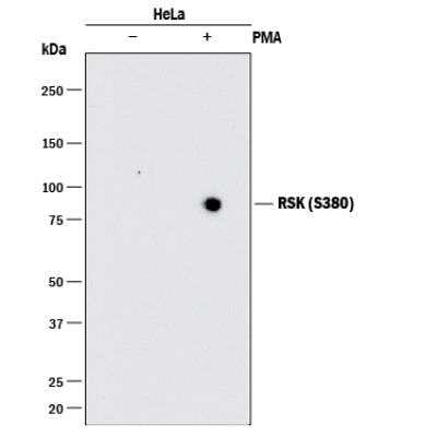
Western Blot: RSK1 [p Ser380] Antibody (1024F) [MAB79672] - Analysis of HeLa -/+ PMA cell lysates with 0.25 ug/ml of hRSK (S380) Monoclonal Antibody followed by HAF008. A specific band was detected for hRSK (S380) at approximately90 kDA (as indicated). This experiment was conducted under reducing conditions and using Immunoblot Buffer Group 1.
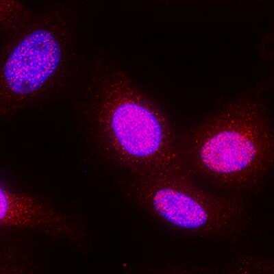
Immunocytochemistry/Immunofluorescence: RSK1 [p Ser380] Antibody (1024F) [MAB79672] - RSK1 [pSer380] was detected in immersion fixed HeLa cells treated with 200 nm of PMA using RSK1 [pSer380] Monoclonal Antibody at 1 ug/ml dilution. Cells were stained using the NorthernLights 557-conjugated Anti-Rabbit IgG Secondary Antibody (red) and counterstained with DAPI (blue). Specific staining was localized to the cytoplasm.
Applications for RSK1 [p Ser380] Antibody (1024F) - Azide and BSA Free
Application
Recommended Usage
Western Blot
0.25 ug/ml
Formulation, Preparation, and Storage
Purification
Protein A or G purified
Formulation
PBS
Format
Azide and BSA Free
Preservative
No Preservative
Concentration
1.0 mg/ml
Shipping
The product is shipped with polar packs. Upon receipt, store it immediately at the temperature recommended below.
Stability & Storage
Store at -20C. Avoid freeze-thaw cycles.
Background: RSK1
Long Name
Ribosomal Protein S6 Kinase I
Alternate Names
HU-1, MAPKAPL1A, RPS6KA1
Gene Symbol
RPS6KA1
Additional RSK1 Products
Product Documents for RSK1 [p Ser380] Antibody (1024F) - Azide and BSA Free
Certificate of Analysis
To download a Certificate of Analysis, please enter a lot or batch number in the search box below.
Product Specific Notices for RSK1 [p Ser380] Antibody (1024F) - Azide and BSA Free
This product is for research use only and is not approved for use in humans or in clinical diagnosis. Primary Antibodies are guaranteed for 1 year from date of receipt.
Related Research Areas
Customer Reviews for RSK1 [p Ser380] Antibody (1024F) - Azide and BSA Free
There are currently no reviews for this product. Be the first to review RSK1 [p Ser380] Antibody (1024F) - Azide and BSA Free and earn rewards!
Have you used RSK1 [p Ser380] Antibody (1024F) - Azide and BSA Free?
Submit a review and receive an Amazon gift card!
$25/€18/£15/$25CAN/¥2500 Yen for a review with an image
$10/€7/£6/$10CAN/¥1110 Yen for a review without an image
Submit a review
Protocols
Find general support by application which include: protocols, troubleshooting, illustrated assays, videos and webinars.
- Appropriate Fixation of IHC/ICC Samples
- Cellular Response to Hypoxia Protocols
- ClariTSA™ Fluorophore Kits
- Detection & Visualization of Antibody Binding
- ICC Cell Smear Protocol for Suspension Cells
- ICC Immunocytochemistry Protocol Videos
- ICC for Adherent Cells
- Immunocytochemistry (ICC) Protocol
- Immunocytochemistry Troubleshooting
- Immunofluorescence of Organoids Embedded in Cultrex Basement Membrane Extract
- Immunohistochemistry (IHC) and Immunocytochemistry (ICC) Protocols
- Preparing Samples for IHC/ICC Experiments
- Preventing Non-Specific Staining (Non-Specific Binding)
- Primary Antibody Selection & Optimization
- Protocol for VisUCyte™ HRP Polymer Detection Reagent
- Protocol for the Fluorescent ICC Staining of Cell Smears - Graphic
- Protocol for the Fluorescent ICC Staining of Cultured Cells on Coverslips - Graphic
- Protocol for the Preparation and Fluorescent ICC Staining of Cells on Coverslips
- Protocol for the Preparation and Fluorescent ICC Staining of Non-adherent Cells
- Protocol for the Preparation and Fluorescent ICC Staining of Stem Cells on Coverslips
- Protocol for the Preparation of a Cell Smear for Non-adherent Cell ICC - Graphic
- R&D Systems Quality Control Western Blot Protocol
- TUNEL and Active Caspase-3 Detection by IHC/ICC Protocol
- The Importance of IHC/ICC Controls
- Troubleshooting Guide: Western Blot Figures
- Western Blot Conditions
- Western Blot Protocol
- Western Blot Protocol for Cell Lysates
- Western Blot Troubleshooting
- Western Blot Troubleshooting Guide
- View all Protocols, Troubleshooting, Illustrated assays and Webinars
Loading...