RUVBL2 Antibody - BSA Free
Novus Biologicals | Catalog # NBP1-40354
![Western Blot: RUVBL2 Antibody [NBP1-40354]](https://resources.rndsystems.com/images/products/RUVBL2-Antibody-Western-Blot-NBP1-40354-img0025.jpg "Western Blot: RUVBL2 Antibody [NBP1-40354]")
Key Product Details
Species Reactivity
Validated:
Human, Mouse
Cited:
Human
Predicted:
Bovine (100%). Backed by our 100% Guarantee.
Applications
Validated:
Immunohistochemistry, Immunohistochemistry-Paraffin, Western Blot, Immunocytochemistry/ Immunofluorescence, Immunoprecipitation, Knockdown Validated
Cited:
Western Blot, Immunocytochemistry/ Immunofluorescence, Knockdown Validated
Label
Unconjugated
Antibody Source
Polyclonal Rabbit IgG
Format
BSA Free
Loading...
Product Specifications
Immunogen
The immunogen for this product maps to a region between residue 1 and 50 of human RuvB-like 2 using the numbering given in entry NP_006657.1 (GeneID 10856).
Clonality
Polyclonal
Host
Rabbit
Isotype
IgG
Scientific Data Images for RUVBL2 Antibody - BSA Free
Western Blot: RUVBL2 Antibody [NBP1-40354]
Western Blot: RUVBL2 Antibody [NBP1-40354] - Whole cell lysate from HeLa (5, 15 and 50 mcg for WB; 1 mg for IP, 20% of IP loaded), 293T (T; 50 mcg), and mouse NIH3T3 (M; 50 mcg) cells. Antibodies: Affinity purified rabbit anti-RuvBL2 antibody used for WB at 0.1 mcg/ml (A) and 1 mcg/ml (B) and used for IP at 10 mcg/mg lysate. RuvBL2 was also immunoprecipitated by rabbit anti-RuvBL2 antibody NBP1-40355 which recognizes a downstream epitope.![Immunohistochemistry-Paraffin: RUVBL2 Antibody [NBP1-40354]](https://resources.rndsystems.com/images/products/RUVBL2-Antibody-Immunohistochemistry-Paraffin-NBP1-40354-img0026.jpg "Immunohistochemistry-Paraffin: RUVBL2 Antibody [NBP1-40354]")
Immunohistochemistry-Paraffin: RUVBL2 Antibody [NBP1-40354]
Immunohistochemistry-Paraffin: RUVBL2 Antibody [NBP1-40354] - Sample: FFPE section of human breast carcinoma. Antibody: Affinity purified rabbit anti- RuvBL2 used at a dilution of 1:1,000 (1ug/ml). Detection: DAB
Immunocytochemistry/ Immunofluorescence: RUVBL2 Antibody [NBP1-40354] -
Immunocytochemistry/ Immunofluorescence: RUVBL2 Antibody [NBP1-40354] - Endogenous RUVBL1 & RUVBL2 colocalize with HA-NP. HeLa cells were transfected with vector control, HA-NP, or HA-VP35. Twenty-four h later, the cells were fixed & processed for immunofluorescence detection of endogenous RUVBL1 or RUVBL2 in the presence of vector control, HA-NP, or HA-VP35. Representative images of (A) endogenous RUVBL1 localization pattern with control vector (top panels), HA-NP (middle panels), or HA-VP35 (bottom panels) & (B) endogenous RUVBL2 localization pattern with control vector (top panels), HA-NP (middle panels), or HA-VP35 (bottom panels) are shown. HA-NP or HA-VP35 (green), RUVBL1/2 (red), & Hoechst 33342 nuclear stain (blue) were visualized by confocal microscopy. Scale bars = 20 µM. Image collected & cropped by CiteAb from the following publication (https://pubmed.ncbi.nlm.nih.gov/31018511), licensed under a CC-BY license. Not internally tested by Novus Biologicals.
Western Blot: RUVBL2 Antibody [NBP1-40354] -
Western Blot: RUVBL2 Antibody [NBP1-40354] - RUVBL1/2 do not effect EBOV minigenome activity. (A) Schematic diagram of the EBOV minigenome system. The EBOV minigenome system consists of six plasmids: Four support plasmids encode replication complex components NP, L, VP35, & VP30. The EBOV minigenome plasmid encodes a firefly luciferase reporter gene flanked by the leader & trailer sequences of EBOV, & the plasmid that encodes Renilla luciferase is used for normalization. (B) Minigenome activity upon the knockdown of either RUVBL1, RUVBL2, or in combination. Below are protein levels confirmed by immunoblot. HeLa cells were transfected with 80 nM scrambled siRNA, 30 nM siRNA targeting RUVBL1, or 50 nM siRNA targeting RUVBL2. Twenty-four h after siRNA addition, the minigenome components were transfected. Forty-eight h later, minigenome reporter activity was measured. (C) Overexpression of FLAG-RUVBL1 & HA-RUVBL2 in the EBOV minigenome. HeLa cells were left untransfected, or transfected with vector control (VC), or increasing amounts of FLAG-RUVBL1 (125, 250, & 500 ng) or HA-RUVBL2 (125, 250, & 500 ng). Twenty-four h after exogenous transfection, the minigenome components were transfected. Forty-eight h later, minigenome reporter activity was measured. Data represent mean ± SEM from one representative experiment (n = 3) of at least three experiments (* p < 0.05). Image collected & cropped by CiteAb from the following publication (https://pubmed.ncbi.nlm.nih.gov/31018511), licensed under a CC-BY license. Not internally tested by Novus Biologicals.Applications for RUVBL2 Antibody - BSA Free
Application
Recommended Usage
Immunocytochemistry/ Immunofluorescence
Reported in scientific literature (PMID: 31018511).
Immunohistochemistry
1:500 - 1:2000
Immunohistochemistry-Paraffin
1:500-1:2000
Immunoprecipitation
5-15 ug/mg lysate
Knockdown Validated
Reported in scientific literature (PMID: 31018511).
Western Blot
1:2000-1:10000
Application Notes
Epitope retrieval with citrate buffer pH6.0 is recommended for FFPE tissue sections.
Reviewed Applications
Read 1 review rated 5 using NBP1-40354 in the following applications:
Formulation, Preparation, and Storage
Purification
Immunogen affinity purified
Formulation
Tris-Citrate/Phosphate (pH 7.0 - 8.0)
Format
BSA Free
Preservative
0.09% Sodium Azide
Concentration
1.0 mg/ml
Shipping
The product is shipped with polar packs. Upon receipt, store it immediately at the temperature recommended below.
Stability & Storage
Store at 4C. Do not freeze.
Background: RUVBL2
Alternate Names
48 kDa TATA box-binding protein-interacting protein, 51 kDa erythrocyte cytosolic protein, EC 3.6.1, EC 3.6.4.12,48 kDa TBP-interacting protein, ECP51TIP49B, erythrocyte cytosolic protein, 51-KD, INO80 complex subunit J, INO80JTAP54-beta, Repressing pontin 52, REPTIN, Reptin 52, Reptin52, RuvB (E coli homolog)-like 2, ruvB-like 2, RuvB-like 2 (E. coli), RVB2, TBP-interacting protein, 48-KD, TIH2, TIP48ECP-51, TIP49b, TIP60-associated protein 54-beta
Entrez Gene IDs
10856 (Human)
Gene Symbol
RUVBL2
Additional RUVBL2 Products
Product Documents for RUVBL2 Antibody - BSA Free
Certificate of Analysis
To download a Certificate of Analysis, please enter a lot or batch number in the search box below.
Product Specific Notices for RUVBL2 Antibody - BSA Free
This product is for research use only and is not approved for use in humans or in clinical diagnosis. Primary Antibodies are guaranteed for 1 year from date of receipt.
Citations for RUVBL2 Antibody - BSA Free
 Powered by Bioz
Customer Reviews for RUVBL2 Antibody - BSA Free (1)
5 out of 5
1 Customer Rating
Have you used RUVBL2 Antibody - BSA Free?
Submit a review and receive an Amazon gift card!
$25/€18/£15/$25CAN/¥2500 Yen for a review with an image
$10/€7/£6/$10CAN/¥1110 Yen for a review without an image
Submit a review
Customer Images
Showing
1
-
1 of
1 review
Showing All
Filter By:
-
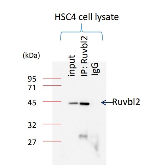
Application: ImmunoprecipitationSample Tested: Oral cancer cell lineSpecies: HumanVerified Customer | Posted 12/07/2017

There are no reviews that match your criteria.
Protocols
Find general support by application which include: protocols, troubleshooting, illustrated assays, videos and webinars.
- Antigen Retrieval Protocol (PIER)
- Antigen Retrieval for Frozen Sections Protocol
- Appropriate Fixation of IHC/ICC Samples
- Cellular Response to Hypoxia Protocols
- Chromogenic IHC Staining of Formalin-Fixed Paraffin-Embedded (FFPE) Tissue Protocol
- Chromogenic Immunohistochemistry Staining of Frozen Tissue
- ClariTSA™ Fluorophore Kits
- Detection & Visualization of Antibody Binding
- Fluorescent IHC Staining of Frozen Tissue Protocol
- Graphic Protocol for Heat-induced Epitope Retrieval
- Graphic Protocol for the Preparation and Fluorescent IHC Staining of Frozen Tissue Sections
- Graphic Protocol for the Preparation and Fluorescent IHC Staining of Paraffin-embedded Tissue Sections
- Graphic Protocol for the Preparation of Gelatin-coated Slides for Histological Tissue Sections
- ICC Cell Smear Protocol for Suspension Cells
- ICC Immunocytochemistry Protocol Videos
- ICC for Adherent Cells
- IHC Sample Preparation (Frozen sections vs Paraffin)
- Immunocytochemistry (ICC) Protocol
- Immunocytochemistry Troubleshooting
- Immunofluorescence of Organoids Embedded in Cultrex Basement Membrane Extract
- Immunofluorescent IHC Staining of Formalin-Fixed Paraffin-Embedded (FFPE) Tissue Protocol
- Immunohistochemistry (IHC) and Immunocytochemistry (ICC) Protocols
- Immunohistochemistry Frozen Troubleshooting
- Immunohistochemistry Paraffin Troubleshooting
- Immunoprecipitation Protocol
- Preparing Samples for IHC/ICC Experiments
- Preventing Non-Specific Staining (Non-Specific Binding)
- Primary Antibody Selection & Optimization
- Protocol for Heat-Induced Epitope Retrieval (HIER)
- Protocol for Making a 4% Formaldehyde Solution in PBS
- Protocol for VisUCyte™ HRP Polymer Detection Reagent
- Protocol for the Fluorescent ICC Staining of Cell Smears - Graphic
- Protocol for the Fluorescent ICC Staining of Cultured Cells on Coverslips - Graphic
- Protocol for the Preparation & Fixation of Cells on Coverslips
- Protocol for the Preparation and Chromogenic IHC Staining of Frozen Tissue Sections
- Protocol for the Preparation and Chromogenic IHC Staining of Frozen Tissue Sections - Graphic
- Protocol for the Preparation and Chromogenic IHC Staining of Paraffin-embedded Tissue Sections
- Protocol for the Preparation and Chromogenic IHC Staining of Paraffin-embedded Tissue Sections - Graphic
- Protocol for the Preparation and Fluorescent ICC Staining of Cells on Coverslips
- Protocol for the Preparation and Fluorescent ICC Staining of Non-adherent Cells
- Protocol for the Preparation and Fluorescent ICC Staining of Stem Cells on Coverslips
- Protocol for the Preparation and Fluorescent IHC Staining of Frozen Tissue Sections
- Protocol for the Preparation and Fluorescent IHC Staining of Paraffin-embedded Tissue Sections
- Protocol for the Preparation of Gelatin-coated Slides for Histological Tissue Sections
- Protocol for the Preparation of a Cell Smear for Non-adherent Cell ICC - Graphic
- R&D Systems Quality Control Western Blot Protocol
- TUNEL and Active Caspase-3 Detection by IHC/ICC Protocol
- The Importance of IHC/ICC Controls
- Troubleshooting Guide: Immunohistochemistry
- Troubleshooting Guide: Western Blot Figures
- Western Blot Conditions
- Western Blot Protocol
- Western Blot Protocol for Cell Lysates
- Western Blot Troubleshooting
- Western Blot Troubleshooting Guide
- View all Protocols, Troubleshooting, Illustrated assays and Webinars
Loading...