SARS-CoV-2 Nucleocapsid Antibody (1C7C7) - Azide and BSA Free
Novus Biologicals | Catalog # NBP3-07043
Recombinant Monoclonal Antibody
Loading...
Key Product Details
Species Reactivity
SARS-CoV-2
Applications
Immunohistochemistry, Western Blot, ELISA
Label
Unconjugated
Antibody Source
Recombinant Monoclonal Mouse IgG2A Clone # 1C7C7 expressed in HEK293
Format
Azide and BSA Free
Loading...
Product Specifications
Immunogen
Recombinant nucleoprotein of SARS-CoV (severe acute respiratory syndrome).The nucleocapsid protein is expressed in the internal nucleocapsid of SARS-CoV-2.
Specificity
This antibody specifically targets an epitope on the SARS-CoV-2 and SARS-CoV nucleocapsid protein. This antibody does not cross-react with the nucleocapsid of other common human coronaviruses, OC43 and 229E
Clonality
Monoclonal
Host
Mouse
Isotype
IgG2A
Scientific Data Images for SARS-CoV-2 Nucleocapsid Antibody (1C7C7) - Azide and BSA Free
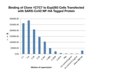
N/A: SARS-CoV-2 Nucleocapsid Antibody (1C7C7) [NBP3-07043] - Exp1293 cells were transfected with expression plasmid coding for SARS-CoV-2 Nucleocapsid and incubated for 48 hours. Cells were permeabilized, incubated with dilutions of supernatant collected from cells transfected with DNA plasmids coding for the heavy and light chains of 1C7 and binding identified by the addition of goat anti-mouse IgG FITC.
 - Azide and BSA Free [NBP3-07043] -")
Western Blot: SARS-CoV-2 Nucleocapsid Antibody (1C7C7) - Azide and BSA Free [NBP3-07043] -
Western Blot: SARS-CoV-2 Nucleocapsid Antibody (1C7C7) - Azide and BSA Free [NBP3-07043] - Lysates were derived from A549 cells (Transduced with ACE2 by adenoviral vector), mock vs. SARS-CoV-2-infected MO12, 24 hours, with 60ug of lysates loaded/lane, Image Courtesy of Jeff Johnson Laboratory, ISMMS.Applications for SARS-CoV-2 Nucleocapsid Antibody (1C7C7) - Azide and BSA Free
Application
Recommended Usage
ELISA
Optimal dilutions of this antibody should be experimentally determined.
Immunohistochemistry
Optimal dilutions of this antibody should be experimentally determined.
Western Blot
Optimal dilutions of this antibody should be experimentally determined.
Formulation, Preparation, and Storage
Purification
Protein A purified
Formulation
0.01M PBS (pH 7.2 - 7.4), 150mM NaCl
Format
Azide and BSA Free
Preservative
No Preservative
Concentration
1 mg/ml
Shipping
The product is shipped with polar packs. Upon receipt, store it immediately at the temperature recommended below.
Stability & Storage
Store at 4C short term. Aliquot and store at -80C long term. Avoid freeze-thaw cycles.
Background: SARS-CoV-2 Nucleocapsid
Following SARS-CoV-2 infection, B cells and T cells display an immune response against nucleocapsid protein and nucleocapsid-specific neutralizing antibodies are produced (5). Interestingly, a study of patients who have recovered from COVID-19 revealed that there were no neutralizing antibodies against nucleocapsid protein present; however, a high presence against neutralizing spike RBD antibodies were detected (5).
References
1. Pillay T. S. (2020). Gene of the month: the 2019-nCoV/SARS-CoV-2 novel coronavirus spike protein. Journal of Clinical Pathology. https://doi.org/10.1136/jclinpath-2020-206658
2. Malik Y. A. (2020). Properties of Coronavirus and SARS-CoV-2. The Malaysian Journal of Pathology.
3. Zhu, G., Zhu, C., Zhu, Y., & Sun, F. (2020). Minireview of progress in the structural study of SARS-CoV-2 proteins. Current Research in Microbial Sciences. https://doi.org/10.1016/j.crmicr.2020.06.003
4. Uniprot (P0DTC9)
5. Shah, V. K., Firmal, P., Alam, A., Ganguly, D., & Chattopadhyay, S. (2020). Overview of Immune Response During SARS-CoV-2 Infection: Lessons From the Past. Frontiers in Immunology. https://doi.org/10.3389/fimmu.2020.01949
Alternate Names
2019-nCoV N Protein, 2019-nCoV Nucleocapsid Protein, COVID-19 N protein, COVID-19 nucleocapsid protein, Human coronavirus Nucleoprotein, nucleocapsid phosphoprotein, Nucleocapsid protein, Nucleoprotein, ORF9; structural protein, SARS-CoV-2, SARS-CoV-2 Nucleocapsid protein, SARS-CoV-2 N protein, SARSCoV2 Nucleoprotein, Severe Acute Respiratory Syndrome Coronavirus 2 Nucleocapsid Protein
Gene Symbol
N
Additional SARS-CoV-2 Nucleocapsid Products
Product Documents for SARS-CoV-2 Nucleocapsid Antibody (1C7C7) - Azide and BSA Free
Certificate of Analysis
To download a Certificate of Analysis, please enter a lot or batch number in the search box below.
Product Specific Notices for SARS-CoV-2 Nucleocapsid Antibody (1C7C7) - Azide and BSA Free
This product is for research use only and is not approved for use in humans or in clinical diagnosis. Primary Antibodies are guaranteed for 1 year from date of receipt.
Customer Reviews for SARS-CoV-2 Nucleocapsid Antibody (1C7C7) - Azide and BSA Free
There are currently no reviews for this product. Be the first to review SARS-CoV-2 Nucleocapsid Antibody (1C7C7) - Azide and BSA Free and earn rewards!
Have you used SARS-CoV-2 Nucleocapsid Antibody (1C7C7) - Azide and BSA Free?
Submit a review and receive an Amazon gift card!
$25/€18/£15/$25CAN/¥2500 Yen for a review with an image
$10/€7/£6/$10CAN/¥1110 Yen for a review without an image
Submit a review
Protocols
Find general support by application which include: protocols, troubleshooting, illustrated assays, videos and webinars.
- Antigen Retrieval Protocol (PIER)
- Antigen Retrieval for Frozen Sections Protocol
- Appropriate Fixation of IHC/ICC Samples
- Cellular Response to Hypoxia Protocols
- Chromogenic IHC Staining of Formalin-Fixed Paraffin-Embedded (FFPE) Tissue Protocol
- Chromogenic Immunohistochemistry Staining of Frozen Tissue
- ClariTSA™ Fluorophore Kits
- Detection & Visualization of Antibody Binding
- ELISA Sample Preparation & Collection Guide
- ELISA Troubleshooting Guide
- Fluorescent IHC Staining of Frozen Tissue Protocol
- Graphic Protocol for Heat-induced Epitope Retrieval
- Graphic Protocol for the Preparation and Fluorescent IHC Staining of Frozen Tissue Sections
- Graphic Protocol for the Preparation and Fluorescent IHC Staining of Paraffin-embedded Tissue Sections
- Graphic Protocol for the Preparation of Gelatin-coated Slides for Histological Tissue Sections
- How to Run an R&D Systems DuoSet ELISA
- How to Run an R&D Systems Quantikine ELISA
- How to Run an R&D Systems Quantikine™ QuicKit™ ELISA
- IHC Sample Preparation (Frozen sections vs Paraffin)
- Immunofluorescent IHC Staining of Formalin-Fixed Paraffin-Embedded (FFPE) Tissue Protocol
- Immunohistochemistry (IHC) and Immunocytochemistry (ICC) Protocols
- Immunohistochemistry Frozen Troubleshooting
- Immunohistochemistry Paraffin Troubleshooting
- Preparing Samples for IHC/ICC Experiments
- Preventing Non-Specific Staining (Non-Specific Binding)
- Primary Antibody Selection & Optimization
- Protocol for Heat-Induced Epitope Retrieval (HIER)
- Protocol for Making a 4% Formaldehyde Solution in PBS
- Protocol for VisUCyte™ HRP Polymer Detection Reagent
- Protocol for the Preparation & Fixation of Cells on Coverslips
- Protocol for the Preparation and Chromogenic IHC Staining of Frozen Tissue Sections
- Protocol for the Preparation and Chromogenic IHC Staining of Frozen Tissue Sections - Graphic
- Protocol for the Preparation and Chromogenic IHC Staining of Paraffin-embedded Tissue Sections
- Protocol for the Preparation and Chromogenic IHC Staining of Paraffin-embedded Tissue Sections - Graphic
- Protocol for the Preparation and Fluorescent IHC Staining of Frozen Tissue Sections
- Protocol for the Preparation and Fluorescent IHC Staining of Paraffin-embedded Tissue Sections
- Protocol for the Preparation of Gelatin-coated Slides for Histological Tissue Sections
- Quantikine HS ELISA Kit Assay Principle, Alkaline Phosphatase
- Quantikine HS ELISA Kit Principle, Streptavidin-HRP Polymer
- R&D Systems Quality Control Western Blot Protocol
- Sandwich ELISA (Colorimetric) – Biotin/Streptavidin Detection Protocol
- Sandwich ELISA (Colorimetric) – Direct Detection Protocol
- TUNEL and Active Caspase-3 Detection by IHC/ICC Protocol
- The Importance of IHC/ICC Controls
- Troubleshooting Guide: ELISA
- Troubleshooting Guide: Immunohistochemistry
- Troubleshooting Guide: Western Blot Figures
- Western Blot Conditions
- Western Blot Protocol
- Western Blot Protocol for Cell Lysates
- Western Blot Troubleshooting
- Western Blot Troubleshooting Guide
- View all Protocols, Troubleshooting, Illustrated assays and Webinars
Loading...