SCP1 Antibody - BSA Free
Novus Biologicals | Catalog # NB300-228
![Immunocytochemistry/ Immunofluorescence: SCP1 Antibody [NB300-228]](https://resources.rndsystems.com/images/products/SCP1-Antibody-Immunocytochemistry-Immunofluorescence-NB300-228-img0003.jpg "Immunocytochemistry/ Immunofluorescence: SCP1 Antibody [NB300-228]")
Key Product Details
Species Reactivity
Validated:
Human, Mouse, Rat, Primate
Cited:
Human, Mouse, Rat, Primate
Applications
Validated:
Immunohistochemistry, Immunohistochemistry-Paraffin, Western Blot, Immunoblotting, Immunocytochemistry/ Immunofluorescence, Simple Western
Cited:
Immunohistochemistry-Paraffin, Western Blot, Immunocytochemistry/ Immunofluorescence
Label
Unconjugated
Antibody Source
Polyclonal Rabbit IgG
Format
BSA Free
Loading...
Product Specifications
Immunogen
A C-terminal synthetic peptide made to the human SCP1 protein sequence. [UniProt# Q15431]
Localization
Nuclear
Clonality
Polyclonal
Host
Rabbit
Isotype
IgG
Scientific Data Images for SCP1 Antibody - BSA Free
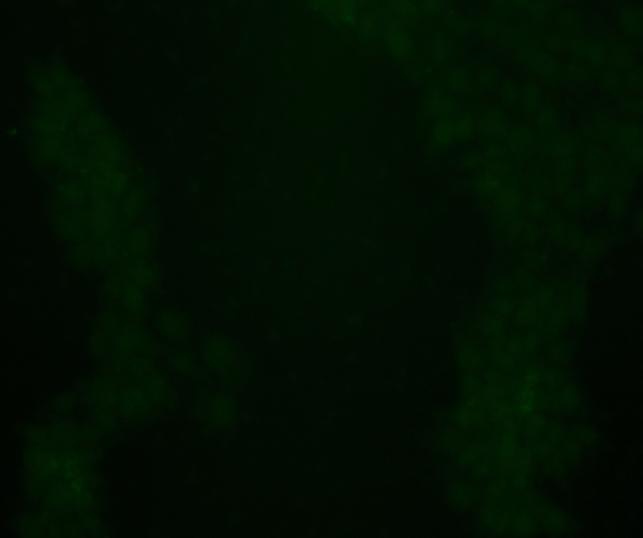
Immunocytochemistry/ Immunofluorescence: SCP1 Antibody [NB300-228]
Immunocytochemistry/Immunofluorescence: SCP1 Antibody [NB300-228] - SCP1 labeled in mouse pachytene preparation (red), using NB 300-228. CDK2 staining, near teleomeres, is also present (green).![Simple Western: SCP1 Antibody [NB300-228]](https://resources.rndsystems.com/images/products/SCP1-Antibody-Simple-Western-NB300-228-img0006.jpg "Simple Western: SCP1 Antibody [NB300-228]")
Simple Western: SCP1 Antibody [NB300-228]
Simple Western: SCP1 Antibody [NB300-228] - Lane view shows a specific band for SCP1 in 0.5 mg/ml of Human Testis (left) and Mouse Testis (right) lysate. This experiment was performed under reducing conditions using the 12-230 kDa separation system. * Non-specific interaction with the 230 kDa Simple Western standard may be seen with this antibodyApplications for SCP1 Antibody - BSA Free
Application
Recommended Usage
Immunoblotting
reported in scientific literature (PMID 27431324)
Immunocytochemistry/ Immunofluorescence
1:300-1:750
Immunohistochemistry-Paraffin
reported in scientific literature (PMID 26858254)
Simple Western
1:200
Western Blot
reported in scientific literature (PMID 23843895)
Application Notes
This SCP1 antibody does not appear to work in Immunohistochemistry on paraffin-embedded sections. *The investigator should determine the optimal working dilution for a specific application.
In Simple Western only 10 - 15 uL of the recommended dilution is used per data point.
See Simple Western Antibody Database for Simple Western validation: Tested in Human Testis and Mouse Testis lysate 0.5 mg/mL, separated by Size, antibody dilution of 1:200. Separated by Size-Wes, Sally Sue/Peggy Sue.
Reviewed Applications
Read 1 review rated 1 using NB300-228 in the following applications:
Formulation, Preparation, and Storage
Purification
Immunogen affinity purified
Formulation
Tris-Citrate/Phosphate (pH 7.0 - 8.0)
Format
BSA Free
Preservative
0.1% Sodium Azide
Concentration
1 mg/ml
Shipping
The product is shipped with polar packs. Upon receipt, store it immediately at the temperature recommended below.
Stability & Storage
Store at 4C. Do not freeze.
Background: SCP1
Alternate Names
Cancer/testis antigen 8, CT8MGC104417, HOM-TES-14, SCP1SCP-1, synaptonemal complex protein 1
Gene Symbol
SYCP1
Additional SCP1 Products
Product Documents for SCP1 Antibody - BSA Free
Certificate of Analysis
To download a Certificate of Analysis, please enter a lot or batch number in the search box below.
Product Specific Notices for SCP1 Antibody - BSA Free
This product is for research use only and is not approved for use in humans or in clinical diagnosis. Primary Antibodies are guaranteed for 1 year from date of receipt.
Citations for SCP1 Antibody - BSA Free
 Powered by Bioz
Customer Reviews for SCP1 Antibody - BSA Free (1)
1 out of 5
1 Customer Rating
Have you used SCP1 Antibody - BSA Free?
Submit a review and receive an Amazon gift card!
$25/€18/£15/$25CAN/¥2500 Yen for a review with an image
$10/€7/£6/$10CAN/¥1110 Yen for a review without an image
Submit a review
Customer Images
Showing
1
-
1 of
1 review
Showing All
Filter By:
-
Application: ImmunocytochemistrySample Tested: Induced pluripotent stem cells overexpressing SYCP1 and MeiocytesSpecies: HumanVerified Customer | Posted 03/26/2024Human iPSCs overexpressing SYCP1, stained at 1:250 dilution of primary antibody.Stained at 1:250 dilution, using donkey anti-rabbit 488 secondary antibody. No signal was observed in human meiotic cells. I also tested this with human iPSCs overexpressing SYCP1 and also observed no signal.
 Bio-Techne ResponseThank you for reviewing our product. We are sorry to hear that this product did not perform as expected. We have been in touch with the customer to resolve this issue according to our Product Guarantee and to the customer’s satisfaction.
There are no reviews that match your criteria.
Protocols
View specific protocols for SCP1 Antibody - BSA Free (NB300-228):
SCP1 Antibody:
Immunofluorescence Procedure
1. Freshly prepared slides are soaked in 1X ADB for 75 minutes.
2. Primary antibodies are added concurrently (SCP1 and CDK2).
3. The primary antibodies are incubated overnight in a humid chamber (37 degrees Celsius).
4. The slides are washed for 40 minutes in 1X ADB.
5. The slides are detected with the appropriate secondary antibodies (RDAR for SCP1 and FDAM for CDK2).
6. The slides are incubated for 4 hours in a humid chamber (37 degrees Celsius).
7. The slides are washed for 20 minutes in 1X ADB, followed by 3 washes, 10 minutes each, in 1X PBS.
8. The slides are counterstained with DAPI.
9. Images are captured after allowing the slides to remain in the dark overnight at RT.
Find general support by application which include: protocols, troubleshooting, illustrated assays, videos and webinars.
- Antigen Retrieval Protocol (PIER)
- Antigen Retrieval for Frozen Sections Protocol
- Appropriate Fixation of IHC/ICC Samples
- Cellular Response to Hypoxia Protocols
- Chromogenic IHC Staining of Formalin-Fixed Paraffin-Embedded (FFPE) Tissue Protocol
- Chromogenic Immunohistochemistry Staining of Frozen Tissue
- ClariTSA™ Fluorophore Kits
- Detection & Visualization of Antibody Binding
- Fluorescent IHC Staining of Frozen Tissue Protocol
- Graphic Protocol for Heat-induced Epitope Retrieval
- Graphic Protocol for the Preparation and Fluorescent IHC Staining of Frozen Tissue Sections
- Graphic Protocol for the Preparation and Fluorescent IHC Staining of Paraffin-embedded Tissue Sections
- Graphic Protocol for the Preparation of Gelatin-coated Slides for Histological Tissue Sections
- ICC Cell Smear Protocol for Suspension Cells
- ICC Immunocytochemistry Protocol Videos
- ICC for Adherent Cells
- IHC Sample Preparation (Frozen sections vs Paraffin)
- Immunocytochemistry (ICC) Protocol
- Immunocytochemistry Troubleshooting
- Immunofluorescence of Organoids Embedded in Cultrex Basement Membrane Extract
- Immunofluorescent IHC Staining of Formalin-Fixed Paraffin-Embedded (FFPE) Tissue Protocol
- Immunohistochemistry (IHC) and Immunocytochemistry (ICC) Protocols
- Immunohistochemistry Frozen Troubleshooting
- Immunohistochemistry Paraffin Troubleshooting
- Preparing Samples for IHC/ICC Experiments
- Preventing Non-Specific Staining (Non-Specific Binding)
- Primary Antibody Selection & Optimization
- Protocol for Heat-Induced Epitope Retrieval (HIER)
- Protocol for Making a 4% Formaldehyde Solution in PBS
- Protocol for VisUCyte™ HRP Polymer Detection Reagent
- Protocol for the Fluorescent ICC Staining of Cell Smears - Graphic
- Protocol for the Fluorescent ICC Staining of Cultured Cells on Coverslips - Graphic
- Protocol for the Preparation & Fixation of Cells on Coverslips
- Protocol for the Preparation and Chromogenic IHC Staining of Frozen Tissue Sections
- Protocol for the Preparation and Chromogenic IHC Staining of Frozen Tissue Sections - Graphic
- Protocol for the Preparation and Chromogenic IHC Staining of Paraffin-embedded Tissue Sections
- Protocol for the Preparation and Chromogenic IHC Staining of Paraffin-embedded Tissue Sections - Graphic
- Protocol for the Preparation and Fluorescent ICC Staining of Cells on Coverslips
- Protocol for the Preparation and Fluorescent ICC Staining of Non-adherent Cells
- Protocol for the Preparation and Fluorescent ICC Staining of Stem Cells on Coverslips
- Protocol for the Preparation and Fluorescent IHC Staining of Frozen Tissue Sections
- Protocol for the Preparation and Fluorescent IHC Staining of Paraffin-embedded Tissue Sections
- Protocol for the Preparation of Gelatin-coated Slides for Histological Tissue Sections
- Protocol for the Preparation of a Cell Smear for Non-adherent Cell ICC - Graphic
- R&D Systems Quality Control Western Blot Protocol
- TUNEL and Active Caspase-3 Detection by IHC/ICC Protocol
- The Importance of IHC/ICC Controls
- Troubleshooting Guide: Immunohistochemistry
- Troubleshooting Guide: Western Blot Figures
- Western Blot Conditions
- Western Blot Protocol
- Western Blot Protocol for Cell Lysates
- Western Blot Troubleshooting
- Western Blot Troubleshooting Guide
- View all Protocols, Troubleshooting, Illustrated assays and Webinars
Loading...