SCP3/SYCP3 Antibody - BSA Free
Novus Biologicals | Catalog # NB300-232
![Immunocytochemistry/ Immunofluorescence: SCP3/SYCP3 Antibody - BSA Free [NB300-232]](https://resources.rndsystems.com/images/products/SCP3-SYCP3-Antibody-Immunocytochemistry-Immunofluorescence-NB300-232-img0013.jpg "Immunocytochemistry/ Immunofluorescence: SCP3/SYCP3 Antibody - BSA Free [NB300-232]")
Key Product Details
Species Reactivity
Validated:
Cited:
Applications
Validated:
Cited:
Label
Antibody Source
Format
Product Specifications
Immunogen
Reactivity Notes
Localization
Clonality
Host
Isotype
Scientific Data Images for SCP3/SYCP3 Antibody - BSA Free
Immunocytochemistry/ Immunofluorescence: SCP3/SYCP3 Antibody - BSA Free [NB300-232]
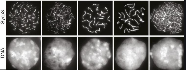
Immunocytochemistry/Immunofluorescence: SCP3/SYCP3 Antibody [NB300-232] - Chromosome spread for sycp3. ICC/IF image submitted by a verified customer review.![Immunohistochemistry-Paraffin: SCP3/SYCP3 Antibody - BSA Free [NB300-232]](https://resources.rndsystems.com/images/products/SCP3-SYCP3-Antibody-Immunohistochemistry-Paraffin-NB300-232-img0011.jpg "Immunohistochemistry-Paraffin: SCP3/SYCP3 Antibody - BSA Free [NB300-232]")
Immunohistochemistry-Paraffin: SCP3/SYCP3 Antibody - BSA Free [NB300-232]
Immunohistochemistry-Paraffin: SCP3/SYCP3 Antibody [NB300-232] - IHC analysis of a formalin fixed and paraffin embedded tissue section of Mouse testis using SCP3 antibody at 1:1000 dilution. The signal was detected using HRP-DAB detection method and the nuclei were counterstained with hematoxylin. The antibody generated a specific signal of SCP3 in the spermatogonial cells and the spermatocytes. The signal was strongest in the nuclei of spermatogonial cells.![Immunocytochemistry/ Immunofluorescence: SCP3/SYCP3 Antibody - BSA Free [NB300-232]](https://resources.rndsystems.com/images/products/SCP3-SYCP3-Antibody-Immunocytochemistry-Immunofluorescence-NB300-232-img0016.jpg "Immunocytochemistry/ Immunofluorescence: SCP3/SYCP3 Antibody - BSA Free [NB300-232]")
Immunocytochemistry/ Immunofluorescence: SCP3/SYCP3 Antibody - BSA Free [NB300-232]
SCP3-SYCP3-Antibody-Immunocytochemistry-Immunofluorescence-NB300-232-img0016.jpg![Immunocytochemistry/ Immunofluorescence: SCP3/SYCP3 Antibody - BSA Free [NB300-232]](https://resources.rndsystems.com/images/products/SCP3-SYCP3-Antibody-Immunocytochemistry-Immunofluorescence-NB300-232-img0018.jpg "Immunocytochemistry/ Immunofluorescence: SCP3/SYCP3 Antibody - BSA Free [NB300-232]")
Immunocytochemistry/ Immunofluorescence: SCP3/SYCP3 Antibody - BSA Free [NB300-232]
SCP3-SYCP3-Antibody-Immunocytochemistry-Immunofluorescence-NB300-232-img0018.jpg![Western Blot: SCP3/SYCP3 AntibodyBSA Free [NB300-232]](https://resources.rndsystems.com/images/products/SCP3-SYCP3-Antibody-Western-Blot-NB300-232-img0012.jpg "Western Blot: SCP3/SYCP3 AntibodyBSA Free [NB300-232]")
Western Blot: SCP3/SYCP3 AntibodyBSA Free [NB300-232]
Western Blot: SCP3/SYCP3 Antibody [NB300-232] - SCP3 Antibody [NB300-232] - Analysis of SCP3 in mouse testis protein.![Immunohistochemistry-Paraffin: SCP3/SYCP3 Antibody - BSA Free [NB300-232]](https://resources.rndsystems.com/images/products/SCP3-SYCP3-Antibody-Immunohistochemistry-Paraffin-NB300-232-img0008.jpg "Immunohistochemistry-Paraffin: SCP3/SYCP3 Antibody - BSA Free [NB300-232]")
Immunohistochemistry-Paraffin: SCP3/SYCP3 Antibody - BSA Free [NB300-232]
Immunohistochemistry-Paraffin: SCP3/SYCP3 Antibody [NB300-232] - Analysis of SCP3 in human testis using DAB with hematoxylin counterstain.![Immunocytochemistry/ Immunofluorescence: SCP3/SYCP3 Antibody - BSA Free [NB300-232]](https://resources.rndsystems.com/images/products/SCP3-SYCP3-Antibody-Immunocytochemistry-Immunofluorescence-NB300-232-img0010.jpg "Immunocytochemistry/ Immunofluorescence: SCP3/SYCP3 Antibody - BSA Free [NB300-232]")
Immunocytochemistry/ Immunofluorescence: SCP3/SYCP3 Antibody - BSA Free [NB300-232]
Immunocytochemistry/Immunofluorescence: SCP3/SYCP3 Antibody [NB300-232] - SCP3 Antibody [NB300-232] - SCP3 labeled in mouse pachytene preparation (red), using NB300-232 SCP3 antibody. CDK2 staining, near teleomeres, is also present (green).![Immunocytochemistry/ Immunofluorescence: SCP3/SYCP3 Antibody - BSA Free [NB300-232]](https://resources.rndsystems.com/images/products/SCP3-SYCP3-Antibody-Immunocytochemistry-Immunofluorescence-NB300-232-img0017.jpg "Immunocytochemistry/ Immunofluorescence: SCP3/SYCP3 Antibody - BSA Free [NB300-232]")
Immunocytochemistry/ Immunofluorescence: SCP3/SYCP3 Antibody - BSA Free [NB300-232]
SCP3-SYCP3-Antibody-Immunocytochemistry-Immunofluorescence-NB300-232-img0017.jpg![Immunohistochemistry-Paraffin: SCP3/SYCP3 Antibody - BSA Free [NB300-232]](https://resources.rndsystems.com/images/products/SCP3-SYCP3-Antibody-Immunohistochemistry-Paraffin-NB300-232-img0009.jpg "Immunohistochemistry-Paraffin: SCP3/SYCP3 Antibody - BSA Free [NB300-232]")
Immunohistochemistry-Paraffin: SCP3/SYCP3 Antibody - BSA Free [NB300-232]
Immunohistochemistry-Paraffin: SCP3/SYCP3 Antibody [NB300-232] - IHC-P analysis of formalin fixed paraffin embedded tissue section of mouse testes using SCP3 antibody at 1:200 dilution. Specific staining may be seen in the spermatogonial cells and the primary spermatocytes.
Immunocytochemistry/ Immunofluorescence: SCP3/SYCP3 Antibody - BSA Free [NB300-232] -
UHRF1 deficiency resulted in impaired meiotic recombination & defective pachynema.a Double immunofluorescence of SYCP3 (green) & DMC1 (red) in testicular spread preparations. b, c The number of DMC1 foci in zygotene stage (b) & pachytene stage (c). d Immunostaining for SYCP3 (red) & gamma H2AX (green). e The percentage of abnormal gamma H2AX foci in the pachytene stage. f Immunostaining for SYCP3 (red) & MLH1 (green). g The number of MLH1 foci in pachynema. h Immunostaining for SYCP3 (red) & H1t (green). i The percentage of spermatocytes with H1T staining. ***p ≤ 0.001; *p ≤ 0.05. Scale bar, 5 μm in a, d, f, h. Image collected & cropped by CiteAb from the following publication (https://pubmed.ncbi.nlm.nih.gov/32081844), licensed under a CC-BY license. Not internally tested by Novus Biologicals.
Immunocytochemistry/ Immunofluorescence: SCP3/SYCP3 Antibody - BSA Free [NB300-232] -
Immunocytochemistry/ Immunofluorescence: SCP3/SYCP3 Antibody - BSA Free [NB300-232] - Effects of MLT on HR in DEHP-exposed fetal oocytes. (A)The immunofluorescence with Sycp3 (red) & RAD51 (green) in fetal oocytes. (B) The percentages of more, less & none staining of RAD51 in all MPI stages (control: 32.48 ± 2.54%, 67.52 ± 2.54%; DEHP: 59.83 ± 7.44%, 40.17 ± 7.44%; DEHP+MLT: 33.13 ± 2.79%, 66.87 ± 2.79%). (C) The percentages of more, less & none staining of RAD51 in pachytene & diplotene oocytes (control: 36.27 ± 8.02%, 19.09 ± 1.03%, 44.64 ± 8.08%; DEHP: 44.24 ± 2.98%, 22.50 ± 4.28%, 33.26 ± 1.30%; DEHP+MLT: 32.97 ± 4.27%, 22.00 ± 1.03%, 45.03 ± 4.76%). The results were presented as mean ± SEM. *P < 0.05, ** P < 0.01. Image collected & cropped by CiteAb from the following publication (https://pubmed.ncbi.nlm.nih.gov/30591620), licensed under a CC-BY license. Not internally tested by Novus Biologicals.
Immunocytochemistry/ Immunofluorescence: SCP3/SYCP3 Antibody - BSA Free [NB300-232] -
Immunocytochemistry/ Immunofluorescence: SCP3/SYCP3 Antibody - BSA Free [NB300-232] - UHRF1 deficiency resulted in impaired meiotic recombination & defective pachynema.a Double immunofluorescence of SYCP3 (green) & DMC1 (red) in testicular spread preparations. b, c The number of DMC1 foci in zygotene stage (b) & pachytene stage (c). d Immunostaining for SYCP3 (red) & gamma H2AX (green). e The percentage of abnormal gamma H2AX foci in the pachytene stage. f Immunostaining for SYCP3 (red) & MLH1 (green). g The number of MLH1 foci in pachynema. h Immunostaining for SYCP3 (red) & H1t (green). i The percentage of spermatocytes with H1T staining. ***p ≤ 0.001; *p ≤ 0.05. Scale bar, 5 μm in a, d, f, h. Image collected & cropped by CiteAb from the following publication (https://pubmed.ncbi.nlm.nih.gov/32081844), licensed under a CC-BY license. Not internally tested by Novus Biologicals.
Western Blot: SCP3/SYCP3 Antibody - BSA Free [NB300-232] -
Western Blot: SCP3/SYCP3 Antibody - BSA Free [NB300-232] - Effects of MLT on meiotic progression & DSBs in DEHP-exposed fetal ovaries. (A) Morphology of 12.5 dpc ovaries cultured for 6 days in control, DEHP & DEHP+MLT group in vitro. (B) Western blot analyses of the expression of Sycp3 & gamma H2afx protein in control, DEHP, & DEHP+MLT groups. (C) Relative expression level of genes Sycp3 & Trp53 in control, DEHP & DEHP+MLT groups. The results were presented as mean ± SEM. *P < 0.05, ** P < 0.01. Image collected & cropped by CiteAb from the following publication (https://pubmed.ncbi.nlm.nih.gov/30591620), licensed under a CC-BY license. Not internally tested by Novus Biologicals.
Immunocytochemistry/ Immunofluorescence: SCP3/SYCP3 Antibody - BSA Free [NB300-232] -
Immunocytochemistry/ Immunofluorescence: SCP3/SYCP3 Antibody - BSA Free [NB300-232] - Dynamic expression of germ cell markers in human fetal testes. (A) Histological sections of human testes at W10, W12, W17 & W22, immunostained for the early germ cell marker (nuclear) POU5F1 (red) & late germ cell marker (cytoplasmic) DDX4 (green). Inserts are magnifications of the dotted boxes. (B) Histological sections of human ovaries at W10, W12, W17 & W22, immunostained for the meiotic markers H2AFX (red) & SYCP3 (green). Note the presence of autofluorescent red blood cells. Scale bars are 200 µm & in the magnified inserts 20 µm. Image collected & cropped by CiteAb from the following publication (https://pubmed.ncbi.nlm.nih.gov/26834021), licensed under a CC-BY license. Not internally tested by Novus Biologicals.
Immunocytochemistry/ Immunofluorescence: SCP3/SYCP3 Antibody - BSA Free [NB300-232] -
Immunocytochemistry/ Immunofluorescence: SCP3/SYCP3 Antibody - BSA Free [NB300-232] - The effect of MLT on the formation of DSBs in fetal oocytes. (A)The immunofluorescence with Sycp3 (red) & gamma H2afx (green) in fetal oocytes. (B) The percentages of stronger, none & weaker gamma H2afx signal in oocyte in all MPI stages (control: 77.60 ± 5.04%, 22.40 ± 5.04%; DEHP: 87.24 ± 4.02%, 12.46 ± 4.02%; DEHP+MLT: 56.03 ± 9.52%, 43.97 ± 9.52%). (C) The percentages of stronger, weaker & none staining of gamma H2afx in pachytene & diplotene oocytes (control: 62.13 ± 3.37%, 35.92 ± 2.99%, 1.95 ± 0.78%; DEHP: 80.48 ± 1.53%, 17.41 ± 1.94%, 2.11 ± 0.78%; DEHP+MLT: 64.36 ± 2.39%, 35.64 ± 2.39%, 0.00 ± 0.00%). The results were presented as mean ± SEM. *P < 0.05, ** P < 0.01. Image collected & cropped by CiteAb from the following publication (https://pubmed.ncbi.nlm.nih.gov/30591620), licensed under a CC-BY license. Not internally tested by Novus Biologicals.
Immunocytochemistry/ Immunofluorescence: SCP3/SYCP3 Antibody - BSA Free [NB300-232] -
Immunocytochemistry/ Immunofluorescence: SCP3/SYCP3 Antibody - BSA Free [NB300-232] - Dose-dependent decrease or increase of the number of meiotic germ cells (SCP3, MLH1) & of gamma H2AX positive cell, respectively, in nicotine treated fetal ovaries cultured for 4 days. (A) Representative IF images of ovarian tissue sections for SCP3; (B) representative IF images of ovarian tissue sections for MLH1 & gamma H2AX; (C) Relative percentage of SCP3 & MLH1 positive cells of ovaries cultured without (control) & with 1mM or 10mM nicotine. All experiments were repeated at least three times. (*) & (**) indicate significant (P < 0.05) & highly significant (P < 0.01) difference, respectively. Image collected & cropped by CiteAb from the following publication (https://pubmed.ncbi.nlm.nih.gov/30001218), licensed under a CC-BY license. Not internally tested by Novus Biologicals.
Immunocytochemistry/ Immunofluorescence: SCP3/SYCP3 Antibody - BSA Free [NB300-232] -
Immunocytochemistry/ Immunofluorescence: SCP3/SYCP3 Antibody - BSA Free [NB300-232] - Effects of MLT on mismatch repair in DEHP-exposed fetal oocytes. (A)The immunofluorescence with Sycp3 (green) & MLH1 (red) in fetal oocytes. (B) The percentages of positive & negative MLH1 signal in oocytes (control: 23.63 ± 0.55%, 76.37 ± 0.55%; DEHP: 77.01 ± 4.41%, 22.99 ± 4.41%; DEHP+MLT: 55.04 ± 17.04%, 43.87 ± 17.08%). (C & D) The amounts of the MLH1 positive foci in pachytene (control: 7.91 ± 1.33; DEHP: 13.97 ± 0.95; DEHP+MLT: 6.74 ± 0.58) & diplotene (control: 12.11 ± 1.74; DEHP: 13.18 ± 1.16; DEHP+MLT: 7.58 ± 1.07) oocytes, respectively. The results were presented as mean ± SEM. *P < 0.05, ** P < 0.01. Image collected & cropped by CiteAb from the following publication (https://pubmed.ncbi.nlm.nih.gov/30591620), licensed under a CC-BY license. Not internally tested by Novus Biologicals.
Immunocytochemistry/ Immunofluorescence: SCP3/SYCP3 Antibody - BSA Free [NB300-232] -
Immunocytochemistry/ Immunofluorescence: SCP3/SYCP3 Antibody - BSA Free [NB300-232] - Hyper-5hmC resulted from UHRF1 deletion.a Venn diagram depicting reduced transcripts associated with 5hmC downregulation. b 5hmC level of meiosis prophase I spermatocytes (16 dpp). c 5hmC densities of all chromosomes. d The distribution of 5hmC density on the genome of spermatocytes. e Venn diagram depicting 5hmC peaks in Uhrf1f/f;Stra8-cre & Uhrf1f/f spermatocytes. f 5hmC densities was shown in the proximal promoter, TSS, & gene body regions of the DEGs. g 5hmC densities in TSSs of the total refgenes with different RPKMs. h The percentage of RNA polymerase II staining in pachynema. i Double immunofluorescence of testicular spread preparations, SYCP3 (red) & RNA polymerase II (green). Scale bar, 5 μm in i. Image collected & cropped by CiteAb from the following publication (https://pubmed.ncbi.nlm.nih.gov/32081844), licensed under a CC-BY license. Not internally tested by Novus Biologicals.
Immunocytochemistry/ Immunofluorescence: SCP3/SYCP3 Antibody - BSA Free [NB300-232] -
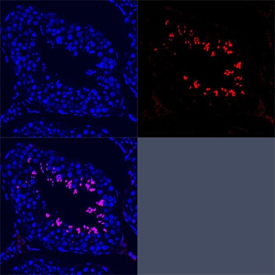
Immunocytochemistry/ Immunofluorescence: SCP3/SYCP3 Antibody - BSA Free [NB300-232] - UHRF1 deletion disrupted the meiotic progression & synaptonemal complex assembly.a Relative amounts of four spermatocyte populations (leptotene stage, zygotene stage, pachytene stage, & diplotene stage) during the prophase I in testes based on analyzing >600 spermatocytes in each stage. b, c The immunostaining of SYCP3 in the testicular sections (b) & surface-spread chromatin preparations of Uhrf1 deletion & control mice (c); d the percentage of spermatocytes with abnormal SYCP3 location. e Double immunofluorescence of testicular spread preparations of the adult mice, SYCP3 (green) & SYCP1 (red). f The percentage of spermatocytes with abnormal SYCP1 location. Lep leptotene, Zyg zygotene, Pac pachytene, Dip diplotene. Data are presented as mean ± SEM of three mice. ***p ≤ 0.001. Scale bar, 25 μm in b, 5 μm in c, e. Image collected & cropped by CiteAb from the following publication (https://pubmed.ncbi.nlm.nih.gov/32081844), licensed under a CC-BY license. Not internally tested by Novus Biologicals.
Immunocytochemistry/ Immunofluorescence: SCP3/SYCP3 Antibody - BSA Free [NB300-232] -
Immunocytochemistry/ Immunofluorescence: SCP3/SYCP3 Antibody - BSA Free [NB300-232] - Dynamic expression of germ cell markers in human fetal ovaries. (A) Histological sections of human ovaries at W10.5, W14, W17 & W21.5, immunostained for the early germ cell marker (nuclear) POU5F1 (red) & late germ cell marker (cytoplasmic) DDX4 (green). In zone 1, most germ cells are POU5F1+DDX4−/low; in zone 2 & 3, most germ cells are DDX4+. Several germ cells in zone 3 have developed into primordial follicles. Inserts are magnifications of the dotted boxes. (B) Histological sections of human ovaries at W10.5, W14, W17 & W21.5, immunostained for the meiotic markers H2AFX (red) & SYCP3 (green). Inserts are magnifications of the dotted boxes. Note the presence of autofluorescent red blood cells. Scale bars are 200 µm & in the magnified inserts 20 µm. Image collected & cropped by CiteAb from the following publication (https://pubmed.ncbi.nlm.nih.gov/26834021), licensed under a CC-BY license. Not internally tested by Novus Biologicals.
Immunocytochemistry/ Immunofluorescence: SCP3/SYCP3 Antibody - BSA Free [NB300-232] -
Immunocytochemistry/ Immunofluorescence: SCP3/SYCP3 Antibody - BSA Free [NB300-232] - CYP51 regulates the expression of the meiosis-specific cohesin subunits REC8 & STAG3. (A,B) RS21745 treatment decreased REC8 & STAG3 expression. Ovaries at 14.5 dpc were cultured with 10 μM RS21745 for 3 days in vitro. (A) Expression of the cohesin subunits following RS21745 treatment by qRT-PCR. All qRT-PCR values were normalized to beta -actin & were expressed as a relative ratio to the control; the means±s.e.m. of 3 values are shown. (B) Expression of REC8 & STAG3 following RS21745 treatment by western blotting GAPDH was used as internal reference. (C–F) Inhibition of CYP51 disturbed the distribution of both REC8 & STAG3 on the chromosomes. (C,E) In the control groups, REC8 & STAG3 expression was observed at high levels in zygotene oocytes. In the CYP51 inhibition groups, the cohesin signals were absent in zygotene cells. (D,F) The percentage of REC8-positive or STAG3-positive oocytes at the zygotene stage was recorded. Unidentified germ cells were not included in the analyses. Scale bars: 10 μm. The data are presented as the means±s.e.m. of 3–9 ovaries per group. Asterisk (*) denotes a statistically significant difference between the control & the treatment groups. ***P<0.001 (t-test). Image collected & cropped by CiteAb from the following publication (https://pubmed.ncbi.nlm.nih.gov/30420384), licensed under a CC-BY license. Not internally tested by Novus Biologicals.
Immunocytochemistry/ Immunofluorescence: SCP3/SYCP3 Antibody - BSA Free [NB300-232] -
Immunocytochemistry/ Immunofluorescence: SCP3/SYCP3 Antibody - BSA Free [NB300-232] - Induction of meiosis is compromised in the cortical germ cells of D17 embryos subject to oestrogen level alterations. (A) D17 (HH43) left gonad sections immunostained for gamma H2AX (green) & P63 (red); nuclei are counterstained with DAPI (blue). (B) Sections from the D17 (HH43) left gonad shown in A immunostained for gamma H2AX (green) & SYCP3 (red). ZW-WT, ZW wild type; ZW-Fa, ZW treated with fadrozole from D7-7.5 (HH31); ZW-E2, ZW treated with beta -oestradiol at D7-7.5; ZZ-E2, ZZ treated with beta -oestradiol at D7-7.5; ZW-Fa(sr), ZW, partially sex-reversed gonad, treated with fadrozole at D4; ZZ-E2(sr), ZZ, partially sex-reversed gonad, treated with beta -oestradiol at D4. All gonadal models have a cortical domain containing germ cells. In ZW-Fa ovary & ZW-E2 ovary, most cortical germ cells express SYCP3 like the ZW-WT control. In ZW-Fa(sr) ovotestis, very few germ cells express gamma H2AX & SYCP3 (orange dotted circled areas). In ZZ-E2(sr) ovotestis & ZZ-E2 testis overlain by a cortex, some cortical germ cells express gamma H2AX but none expresses SYCP3. White dotted line highlights the cortical domain borders. See Fig. S4 for the medullary structure of the ZZ-E2, ZW-Fa(sr) & ZZ-E2(sr) models. Image collected & cropped by CiteAb from the following publication (https://pubmed.ncbi.nlm.nih.gov/32001442), licensed under a CC-BY license. Not internally tested by Novus Biologicals.
Immunocytochemistry/ Immunofluorescence: SCP3/SYCP3 Antibody - BSA Free [NB300-232] -
Immunocytochemistry/ Immunofluorescence: SCP3/SYCP3 Antibody - BSA Free [NB300-232] - Exposure to DEHP impairs meiotic progression of oocytes from pachytene to diplotene. (a) Immunolabeling of the oocyte chromosomes with anti-SYCP3 antibody (red) & Hoechst 33342 (blue). (b) Effect of DEHP on meiotic progression of oocytes throughout prophase I stages; percentage of each group is presented as mean±SD. Control: 36.27±0.80% pachytene & 54.99±0.66% diplotene; 10 μM & 100 μM DEHP 57.79±4.22% & 56.62±6.62% pachytene & 39.79±4.22% & 36.62±6.62% diplotene, respectively. (c) Representative WB showing the effect of DEHP on the expression of germ cell (DAZL) & meiotic (STRA8 & SCP3) specific proteins. (d) Effect of DEHP on the levels of mRNA in the ovarian tissues of germ cell (Mvh & Dazl), meiotic (Stra8, Rec8, Scp1 & Scp3). All experiments were repeated at least three times independently. (* P<0.05; ** P<0.01) Image collected & cropped by CiteAb from the following publication (https://pubmed.ncbi.nlm.nih.gov/28771232), licensed under a CC-BY license. Not internally tested by Novus Biologicals.
Immunocytochemistry/ Immunofluorescence: SCP3/SYCP3 Antibody - BSA Free [NB300-232] -
Immunocytochemistry/ Immunofluorescence: SCP3/SYCP3 Antibody - BSA Free [NB300-232] - Loss of lamin C2 has no effect on meiotic telomere attachment.(A) 3D-preserved swab preparations showing wildtype (A) & knockout (A′) spermatocytes simultaneously labelled with anti-TRF1 & SUN1 antibodies. As in the wildtype, in lamin C2−/− spermatocytes virtual all telomeres appear to be attached to the NE as indicated by co-localisation of TRF1 & SUN1 signals. Scale bars 5 µm. (B) Quantifications of co-localised & non-co-localised TRF1/SUN1 signals (see A) revealed that ratios of co-localised to non-co-localised spots comparing wildtype & knockout spermatocytes show no significant difference (wildtype n = 33; lamin C2−/− n = 45; Pearson's Chi2 test p-value: 0.799). (C,D) Chromosome spread preparations of pachytene-like lamin C2−/− spermatocytes showing that all telomeres are associated with SUN1. In (C) TeloFISH & in (D) anti-SUN1 staining in co-localisation with SYCP3. Scale bars 10 µm. Image collected & cropped by CiteAb from the following publication (https://dx.plos.org/10.1371/journal.pgen.1003261), licensed under a CC-BY license. Not internally tested by Novus Biologicals.
Immunohistochemistry: SCP3/SYCP3 Antibody - BSA Free [NB300-232] -
Immunohistochemistry: SCP3/SYCP3 Antibody - BSA Free [NB300-232] - FANCM expression in human fetal ovaries.(A) Relative FANCM mRNA abundance was measured by RT-qPCR in human fetal ovaries from 5 to 32 weeks post-fertilization (wpf). (B) Germ cells (D2-40+) & somatic cells (D2-40-) were sorted from three ovaries ranging from 8 to 12 wpf & FANCM expression was measured. ACTB was used to normalize FANCM expression in all samples. Dots represent different ovaries & the mean is indicated by the line. (C) Immunohistochemistry of FANCM in human fetal & adult ovaries. Fetal ovaries at 8 & 22 wpf & adult ovaries were studied. FANCM positive cells appear in yellow/brown color (monoclonal FANCM CV5.1 antibody, Novus Biologicals, Abingdon, UK). Ovarian sections were counterstained with hematoxylin (blue staining). Oo, oogonia; Pa, oocyte at the pachytene stage of meiosis I, D, oocyte at the diplotene stage of meiosis I; Pr, oocyte in primordial follicle. (D) Co-staining in 22 wpf ovaries, for FANCM (purple) & DDX4 (brown) confirmed the germ cell identity of FANCM-positive cells (left). Successive staining for FANCM & SYCP3 in the same section (panels a & b). Negative control performed with non-immune mouse IgG (right). Scale bar: 10 μm. Image collected & cropped by CiteAb from the following publication (https://pubmed.ncbi.nlm.nih.gov/29231814), licensed under a CC-BY license. Not internally tested by Novus Biologicals.
Immunocytochemistry/ Immunofluorescence: SCP3/SYCP3 Antibody - BSA Free [NB300-232] -
Immunocytochemistry/ Immunofluorescence: SCP3/SYCP3 Antibody - BSA Free [NB300-232] - Loss of lamin C2 has no effect on meiotic telomere attachment.(A) 3D-preserved swab preparations showing wildtype (A) & knockout (A′) spermatocytes simultaneously labelled with anti-TRF1 & SUN1 antibodies. As in the wildtype, in lamin C2−/− spermatocytes virtual all telomeres appear to be attached to the NE as indicated by co-localisation of TRF1 & SUN1 signals. Scale bars 5 µm. (B) Quantifications of co-localised & non-co-localised TRF1/SUN1 signals (see A) revealed that ratios of co-localised to non-co-localised spots comparing wildtype & knockout spermatocytes show no significant difference (wildtype n = 33; lamin C2−/− n = 45; Pearson's Chi2 test p-value: 0.799). (C,D) Chromosome spread preparations of pachytene-like lamin C2−/− spermatocytes showing that all telomeres are associated with SUN1. In (C) TeloFISH & in (D) anti-SUN1 staining in co-localisation with SYCP3. Scale bars 10 µm. Image collected & cropped by CiteAb from the following publication (https://dx.plos.org/10.1371/journal.pgen.1003261), licensed under a CC-BY license. Not internally tested by Novus Biologicals.
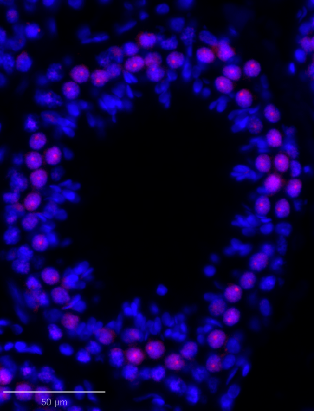
Immunohistochemistry-Paraffin: Rabbit Polyclonal SCP3/SYCP3 Antibody [NB300-232]
SYCP3 expression in adult male rhesus macaque testis seminiferous tubule. primary antibody - SYCP3 1:100.secondary antibody - donkey anti-rabbit 555.antigen retrieval buffer: 0.01M citrate (pH 6). Image from a verified customer review.Applications for SCP3/SYCP3 Antibody - BSA Free
Immunoblotting
Immunocytochemistry/ Immunofluorescence
Immunohistochemistry
Immunohistochemistry-Frozen
Immunohistochemistry-Paraffin
Immunoprecipitation
Western Blot
Reviewed Applications
Read 3 reviews rated 4.7 using NB300-232 in the following applications:
Formulation, Preparation, and Storage
Purification
Formulation
Format
Preservative
Concentration
Shipping
Stability & Storage
Background: SCP3/SYCP3
Long Name
Alternate Names
Gene Symbol
UniProt
Additional SCP3/SYCP3 Products
Product Documents for SCP3/SYCP3 Antibody - BSA Free
Certificate of Analysis
To download a Certificate of Analysis, please enter a lot or batch number in the search box below.
Product Specific Notices for SCP3/SYCP3 Antibody - BSA Free
This product is for research use only and is not approved for use in humans or in clinical diagnosis. Primary Antibodies are guaranteed for 1 year from date of receipt.
Citations for SCP3/SYCP3 Antibody - BSA Free
 Powered by Bioz
Customer Reviews for SCP3/SYCP3 Antibody - BSA Free (3)
Have you used SCP3/SYCP3 Antibody - BSA Free?
Submit a review and receive an Amazon gift card!
$25/€18/£15/$25CAN/¥2500 Yen for a review with an image
$10/€7/£6/$10CAN/¥1110 Yen for a review without an image
Submit a review
Customer Images
-
Application: Immunohistochemistry-ParaffinSample Tested: Testis tissueSpecies: Rhesus MacaqueVerified Customer | Posted 10/06/2025SYCP3 expression in adult male rhesus macaque testis seminiferous tubule. primary antibody - SYCP3 1:100 secondary antibody - donkey anti-rabbit 555 antigen retrieval buffer: 0.01M citrate (pH 6)Immunofluorescence
 Bio-Techne ResponseThis review was submitted through the legacy Novus Innovators Program, reflecting a new species or application tested on a primary antibody.
 -
Application: ImmunocytochemistrySample Tested: germ cellsSpecies: MouseVerified Customer | Posted 11/11/2019chromosome spread for sycp3
-
Application: Immunohistochemistry-ParaffinVerified Customer | Posted 07/12/2012
There are no reviews that match your criteria.
Protocols
View specific protocols for SCP3/SYCP3 Antibody - BSA Free (NB300-232):
Immunofluorescence Procedure
1. Freshly prepared slides are soaked in 1X ADB for 75 minutes.
2. Primary antibodies are added concurrently (SCP3 and CDK2).
3. The primary antibodies are incubated overnight in a hudid chamber (37 degrees Celcius).
4. The slides are washed for 40 minutes in 1X ADB.
5. The slides are detected with the appropriate secondary antibodies (RDAR for SCP1 and FDAM for CDK2).
6. The slides are incubated for 4 hours in a humid chamber (37 degrees Celcius).
7. The slides are washed for 20 minutes in 1X ADB, followed by 3 washes, 10 minutes each, in 1X PBS.
8. The slides are counterstained with DAPI.
9. Images are captured after allowing the slides to remain in the dark overnight at RT.
14. Dehydrate sections.
15. Mount coverslips.
*The above information is only intended as a guide. The researcher should determine what protocol best meets their needs. Please follow safe laboratory procedures.
Antigen Unmasking:
Bring slides to a boil in 10 mM sodium citrate buffer (pH 6.0) then maintain at a sub-boiling temperature for 10 minutes. Cool slides on bench-top for 30 minutes (keep slides in the sodium citrate buffer all the time).
Staining:
1. Wash sections in deionized water three times for 5 minutes each.
2. Wash sections in PBS for 5 minutes.
3. Block each section with 100-400 ul blocking solution (1% BSA in PBS) for 1 hour at room temperature.
4. Remove blocking solution and add 100-400 ul diluted primary antibody. Incubate overnight at 4 C.
5. Remove antibody solution and wash sections in wash buffer three times for 5 minutes each.
6. Add 100-400 ul HRP polymer conjugated secondary antibody. Incubate 30 minutes at room temperature.
7. Wash sections three times in wash buffer for 5 minutes each.
8. Add 100-400 ul DAB substrate to each section and monitor staining closely.
9. As soon as the sections develop, immerse slides in deionized water.
10. Counterstain sections in hematoxylin.
11. Wash sections in deionized water two times for 5 minutes each.
12. Dehydrate sections.
13. Mount coverslips.
1. Perform SDS-PAGE on samples to be analyzed, loading 10-25 ug of total protein per lane.
2. Transfer proteins to PVDF membrane according to the instructions provided by the manufacturer of the membrane and transfer apparatus.
3. Stain the membrane with Ponceau S (or similar product) to assess transfer success, and mark molecular weight standards where appropriate.
4. Rinse the blot TBS -0.05% Tween 20 (TBST).
5. Block the membrane in 5% Non-fat milk in TBST (blocking buffer) for at least 1 hour.
6. Wash the membrane in TBST three times for 10 minutes each.
7. Dilute primary antibody in blocking buffer and incubate overnight at 4C with gentle rocking.
8. Wash the membrane in TBST three times for 10 minutes each.
9. Incubate the membrane in diluted HRP conjugated secondary antibody in blocking buffer (as per manufacturer's instructions) for 1 hour at room temperature.
10. Wash the blot in TBST three times for 10 minutes each (this step can be repeated as required to reduce background).
11. Apply the detection reagent of choice in accordance with the manufacturers instructions.
Find general support by application which include: protocols, troubleshooting, illustrated assays, videos and webinars.
- Antigen Retrieval Protocol (PIER)
- Antigen Retrieval for Frozen Sections Protocol
- Appropriate Fixation of IHC/ICC Samples
- Cellular Response to Hypoxia Protocols
- Chromogenic IHC Staining of Formalin-Fixed Paraffin-Embedded (FFPE) Tissue Protocol
- Chromogenic Immunohistochemistry Staining of Frozen Tissue
- ClariTSA™ Fluorophore Kits
- Detection & Visualization of Antibody Binding
- Fluorescent IHC Staining of Frozen Tissue Protocol
- Graphic Protocol for Heat-induced Epitope Retrieval
- Graphic Protocol for the Preparation and Fluorescent IHC Staining of Frozen Tissue Sections
- Graphic Protocol for the Preparation and Fluorescent IHC Staining of Paraffin-embedded Tissue Sections
- Graphic Protocol for the Preparation of Gelatin-coated Slides for Histological Tissue Sections
- ICC Cell Smear Protocol for Suspension Cells
- ICC Immunocytochemistry Protocol Videos
- ICC for Adherent Cells
- IHC Sample Preparation (Frozen sections vs Paraffin)
- Immunocytochemistry (ICC) Protocol
- Immunocytochemistry Troubleshooting
- Immunofluorescence of Organoids Embedded in Cultrex Basement Membrane Extract
- Immunofluorescent IHC Staining of Formalin-Fixed Paraffin-Embedded (FFPE) Tissue Protocol
- Immunohistochemistry (IHC) and Immunocytochemistry (ICC) Protocols
- Immunohistochemistry Frozen Troubleshooting
- Immunohistochemistry Paraffin Troubleshooting
- Immunoprecipitation Protocol
- Preparing Samples for IHC/ICC Experiments
- Preventing Non-Specific Staining (Non-Specific Binding)
- Primary Antibody Selection & Optimization
- Protocol for Heat-Induced Epitope Retrieval (HIER)
- Protocol for Making a 4% Formaldehyde Solution in PBS
- Protocol for VisUCyte™ HRP Polymer Detection Reagent
- Protocol for the Fluorescent ICC Staining of Cell Smears - Graphic
- Protocol for the Fluorescent ICC Staining of Cultured Cells on Coverslips - Graphic
- Protocol for the Preparation & Fixation of Cells on Coverslips
- Protocol for the Preparation and Chromogenic IHC Staining of Frozen Tissue Sections
- Protocol for the Preparation and Chromogenic IHC Staining of Frozen Tissue Sections - Graphic
- Protocol for the Preparation and Chromogenic IHC Staining of Paraffin-embedded Tissue Sections
- Protocol for the Preparation and Chromogenic IHC Staining of Paraffin-embedded Tissue Sections - Graphic
- Protocol for the Preparation and Fluorescent ICC Staining of Cells on Coverslips
- Protocol for the Preparation and Fluorescent ICC Staining of Non-adherent Cells
- Protocol for the Preparation and Fluorescent ICC Staining of Stem Cells on Coverslips
- Protocol for the Preparation and Fluorescent IHC Staining of Frozen Tissue Sections
- Protocol for the Preparation and Fluorescent IHC Staining of Paraffin-embedded Tissue Sections
- Protocol for the Preparation of Gelatin-coated Slides for Histological Tissue Sections
- Protocol for the Preparation of a Cell Smear for Non-adherent Cell ICC - Graphic
- R&D Systems Quality Control Western Blot Protocol
- TUNEL and Active Caspase-3 Detection by IHC/ICC Protocol
- The Importance of IHC/ICC Controls
- Troubleshooting Guide: Immunohistochemistry
- Troubleshooting Guide: Western Blot Figures
- Western Blot Conditions
- Western Blot Protocol
- Western Blot Protocol for Cell Lysates
- Western Blot Troubleshooting
- Western Blot Troubleshooting Guide
- View all Protocols, Troubleshooting, Illustrated assays and Webinars
FAQs for SCP3/SYCP3 Antibody - BSA Free
-
Q: Could you tell us immunogen sequence of this antibody?
A:
The exact immunogen sequence for our product NB300-232 is proprietary. We can tell you that the immunogen falls between aa185-236 of the human SYCP3 protein (Uniprot Site).
-
Q: Is it a monoclonal antibody or polyclonal antibody?
A: This antibody is a rabbit polyclonal antibody.
-
Q: Was a permeabilization step performed for the mouse pachytene cells?
A: Our lab did perform a permeabilization step for this staining. The lab used 0.15% Triton-X in the fixation buffer. The tissues were then incubated for 2 hours.
-
Q: What was the dilution used for the CDK2 antibody in the ICC staining?
A: The antibody was used at a dilution of 1:500 for this image.
-
Q: What was the incubation time and temperature for this CDK2 antibody?
A: Primary incubation was performed at 4C overnight
-
Q: Could you tell us immunogen sequence of this antibody?
A:
The exact immunogen sequence for our product NB300-232 is proprietary. We can tell you that the immunogen falls between aa185-236 of the human SYCP3 protein (Uniprot Site).
-
Q: Is it a monoclonal antibody or polyclonal antibody?
A: This antibody is a rabbit polyclonal antibody.
-
Q: Was a permeabilization step performed for the mouse pachytene cells?
A: Our lab did perform a permeabilization step for this staining. The lab used 0.15% Triton-X in the fixation buffer. The tissues were then incubated for 2 hours.
-
Q: What was the dilution used for the CDK2 antibody in the ICC staining?
A: The antibody was used at a dilution of 1:500 for this image.
-
Q: What was the incubation time and temperature for this CDK2 antibody?
A: Primary incubation was performed at 4C overnight