SERCA2 ATPase Antibody (2A7-A1)
Novus Biologicals | Catalog # NB300-581
![Immunocytochemistry/ Immunofluorescence: SERCA2 ATPase Antibody (2A7-A1) [NB300-581]](https://resources.rndsystems.com/images/products/SERCA2-ATPase-Antibody-2A7-A1-Immunocytochemistry-Immunofluorescence-NB300-581-img0010.jpg "Immunocytochemistry/ Immunofluorescence: SERCA2 ATPase Antibody (2A7-A1) [NB300-581]")
Loading...
Key Product Details
Species Reactivity
Validated:
Human, Mouse, Rat, Porcine, Amphibian, Canine, Guinea Pig, Rabbit, Sheep
Cited:
Mouse, Rat, Porcine
Applications
Validated:
Immunohistochemistry, Immunohistochemistry-Paraffin, Immunohistochemistry-Frozen, Western Blot, Flow Cytometry, Immunocytochemistry/ Immunofluorescence, Simple Western, Immunoprecipitation
Cited:
Western Blot, Immunocytochemistry/ Immunofluorescence, Immunoprecipitation, IF/IHC
Label
Unconjugated
Antibody Source
Monoclonal Mouse IgG2A Clone # 2A7-A1
Loading...
Product Specifications
Immunogen
Purified canine cardiac sarcoplasmic reticulum vesicles.
Reactivity Notes
Sheep reactivity reported in scientific literature (PMID: 21289077). Amphibian reactivity reported in scientific literature (PMID: 12022876). Please note that this antibody is reactive to Mouse and derived from the same host, Mouse. Additional Mouse on Mouse blocking steps may be required for IHC and ICC experiments. Please contact Technical Support for more information.
Specificity
Detects sarcoplasmic or endoplasmic reticulum calcium (SERCA) 2 ATPase. NB300-581 recognizes the SERCA2a and SERCA2b isoforms identically. However, the antibody does not recognize the skeletal muscle (SERCA1) or housekeeping isoforms (SERCA3).
Clonality
Monoclonal
Host
Mouse
Isotype
IgG2A
Scientific Data Images for SERCA2 ATPase Antibody (2A7-A1)
Immunocytochemistry/ Immunofluorescence: SERCA2 ATPase Antibody (2A7-A1) [NB300-581]
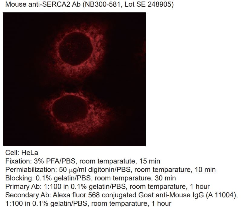
Immunocytochemistry/Immunofluorescence: SERCA2 ATPase Antibody (2A7-A1) [NB300-581] - HeLa cells. Primary antibody at 1:100, secondary antibody: goat-anti mouse IgG AlexaFluor 568 at 1:100. ICC/IF image submitted by a verified customer review.![Immunohistochemistry-Paraffin: SERCA2 ATPase Antibody (2A7-A1) [NB300-581]](https://resources.rndsystems.com/images/products/SERCA2-ATPase-Antibody-2A7-A1-Immunohistochemistry-Paraffin-NB300-581-img0008.jpg "Immunohistochemistry-Paraffin: SERCA2 ATPase Antibody (2A7-A1) [NB300-581]")
Immunohistochemistry-Paraffin: SERCA2 ATPase Antibody (2A7-A1) [NB300-581]
Immunohistochemistry-Paraffin: SERCA2 ATPase Antibody (2A7-A1) [NB300-581] - Both normal and cancer biopsies of deparaffinized Human skeletal muscle tissues.![Immunocytochemistry/ Immunofluorescence: SERCA2 ATPase Antibody (2A7-A1) [NB300-581]](https://resources.rndsystems.com/images/products/SERCA2-ATPase-Antibody-2A7-A1-Immunocytochemistry-Immunofluorescence-NB300-581-img0004.jpg "Immunocytochemistry/ Immunofluorescence: SERCA2 ATPase Antibody (2A7-A1) [NB300-581]")
Immunocytochemistry/ Immunofluorescence: SERCA2 ATPase Antibody (2A7-A1) [NB300-581]
Immunocytochemistry/Immunofluorescence: SERCA2 ATPase Antibody (2A7-A1) [NB300-581] - SERCA2 ATPase staining (green), F-Actin staining with Phalloidin (red) and nuclei with DAPI (blue) is shown. Cells were grown on chamber slides and fixed with formaldehyde prior to staining. Cells were probed without (control) or with or an antibody recognizing SERCA2 ATPase at a dilution of 1:100-1:200 over night at 4C, washed with PBS and incubated with a DyLight-488 conjugated.![Immunocytochemistry/ Immunofluorescence: SERCA2 ATPase Antibody (2A7-A1) [NB300-581]](https://resources.rndsystems.com/images/products/SERCA2-ATPase-Antibody-2A7-A1-Immunocytochemistry-Immunofluorescence-NB300-581-img0005.jpg "Immunocytochemistry/ Immunofluorescence: SERCA2 ATPase Antibody (2A7-A1) [NB300-581]")
Immunocytochemistry/ Immunofluorescence: SERCA2 ATPase Antibody (2A7-A1) [NB300-581]
Immunocytochemistry/Immunofluorescence: SERCA2 ATPase Antibody (2A7-A1) [NB300-581] - SERCA2 ATPase staining (green), F-Actin staining with Phalloidin (red) and nuclei with DAPI (blue) is shown. Cells were grown on chamber slides and fixed with formaldehyde prior to staining. Cells were probed without (control) or with or an antibody recognizing SERCA2 ATPase at a dilution of 1:100-1:200 over night at 4C, washed with PBS and incubated with a DyLight-488 conjugated.![Immunocytochemistry/ Immunofluorescence: SERCA2 ATPase Antibody (2A7-A1) [NB300-581]](https://resources.rndsystems.com/images/products/SERCA2-ATPase-Antibody-2A7-A1-Immunocytochemistry-Immunofluorescence-NB300-581-img0006.jpg "Immunocytochemistry/ Immunofluorescence: SERCA2 ATPase Antibody (2A7-A1) [NB300-581]")
Immunocytochemistry/ Immunofluorescence: SERCA2 ATPase Antibody (2A7-A1) [NB300-581]
Immunocytochemistry/Immunofluorescence: SERCA2 ATPase Antibody (2A7-A1) [NB300-581] - SERCA2 ATPase staining (green), F-Actin staining with Phalloidin (red) and nuclei with DAPI (blue) is shown. Cells were grown on chamber slides and fixed with formaldehyde prior to staining. Cells were probed without (control) or with or an antibody recognizing SERCA2 ATPase at a dilution of 1:100-1:200 over night at 4C, washed with PBS and incubated with a DyLight-488 conjugated.![Immunohistochemistry-Paraffin: SERCA2 ATPase Antibody (2A7-A1) [NB300-581]](https://resources.rndsystems.com/images/products/SERCA2-ATPase-Antibody-2A7-A1-Immunohistochemistry-Paraffin-NB300-581-img0007.jpg "Immunohistochemistry-Paraffin: SERCA2 ATPase Antibody (2A7-A1) [NB300-581]")
Immunohistochemistry-Paraffin: SERCA2 ATPase Antibody (2A7-A1) [NB300-581]
Immunohistochemistry-Paraffin: SERCA2 ATPase Antibody (2A7-A1) [NB300-581] - Both normal and cancer biopsies of deparaffinized Human tonsil tissues.![Immunohistochemistry-Paraffin: SERCA2 ATPase Antibody (2A7-A1) [NB300-581]](https://resources.rndsystems.com/images/products/SERCA2-ATPase-Antibody-2A7-A1-Immunohistochemistry-Paraffin-NB300-581-img0009.jpg "Immunohistochemistry-Paraffin: SERCA2 ATPase Antibody (2A7-A1) [NB300-581]")
Immunohistochemistry-Paraffin: SERCA2 ATPase Antibody (2A7-A1) [NB300-581]
Immunohistochemistry-Paraffin: SERCA2 ATPase Antibody (2A7-A1) [NB300-581] - Both normal and cancer biopsies of deparaffinized Human liver tissues.Applications for SERCA2 ATPase Antibody (2A7-A1)
Application
Recommended Usage
Flow Cytometry
1 ug / 10^6 cells
Immunocytochemistry/ Immunofluorescence
1:10 - 1:500
Immunohistochemistry
1:100
Immunohistochemistry-Frozen
1:100
Immunohistochemistry-Paraffin
1 ug/mL
Immunoprecipitation
1:10 - 1:500
Simple Western
1:10 - 1:250
Western Blot
1:1000
Application Notes
WB: Detects an approx. 110 kDa protein representing SERCA2 ATPase in rat cardiac tissue.
Reviewed Applications
Read 2 reviews rated 3 using NB300-581 in the following applications:
Flow Cytometry Panel Builder
Bio-Techne Knows Flow Cytometry
Save time and reduce costly mistakes by quickly finding compatible reagents using the Panel Builder Tool.
Advanced Features
- Spectra Viewer - Custom analysis of spectra from multiple fluorochromes
- Spillover Popups - Visualize the spectra of individual fluorochromes
- Antigen Density Selector - Match fluorochrome brightness with antigen density

Formulation, Preparation, and Storage
Purification
Unpurified
Formulation
Ascites
Preservative
0.05% Sodium Azide
Concentration
This product is unpurified. The exact concentration of antibody is not quantifiable.
Shipping
The product is shipped with polar packs. Upon receipt, store it immediately at the temperature recommended below.
Stability & Storage
Store at -20C. Avoid freeze-thaw cycles.
Background: SERCA2 ATPase
Long Name
Sarcoplasmic/endoplasmic reticulum calcium ATPase 2
Alternate Names
ATP2A2, ATP2B, Calcium pump 2, SERCA2, SR Ca(2+)-ATPase 2
Gene Symbol
ATP2A2
UniProt
Additional SERCA2 ATPase Products
Product Documents for SERCA2 ATPase Antibody (2A7-A1)
Certificate of Analysis
To download a Certificate of Analysis, please enter a lot or batch number in the search box below.
Product Specific Notices for SERCA2 ATPase Antibody (2A7-A1)
This product is for research use only and is not approved for use in humans or in clinical diagnosis. Primary Antibodies are guaranteed for 1 year from date of receipt.
Citations for SERCA2 ATPase Antibody (2A7-A1)
 Powered by Bioz
Customer Reviews for SERCA2 ATPase Antibody (2A7-A1) (2)
3 out of 5
2 Customer Ratings
Have you used SERCA2 ATPase Antibody (2A7-A1)?
Submit a review and receive an Amazon gift card!
$25/€18/£15/$25CAN/¥2500 Yen for a review with an image
$10/€7/£6/$10CAN/¥1110 Yen for a review without an image
Submit a review
Customer Images
Showing
1
-
2 of
2 reviews
Showing All
Filter By:
-
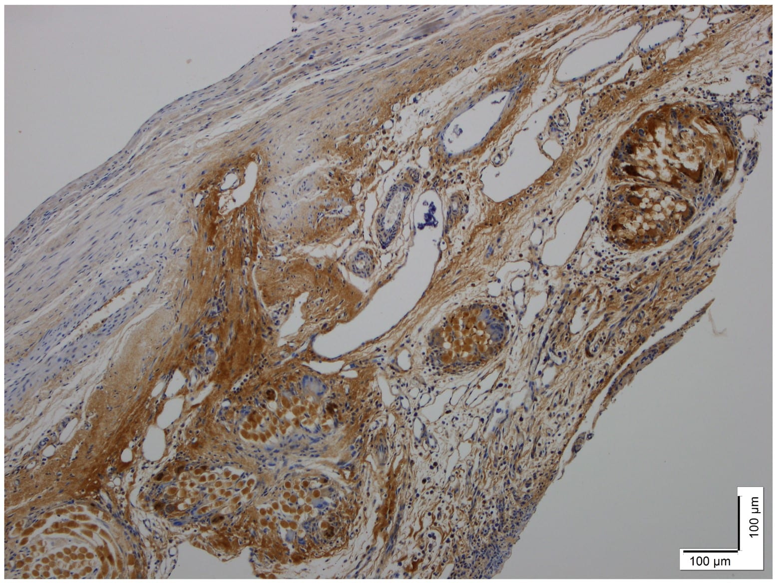
Application: Immunohistochemistry-ParaffinSample Tested: cardiac tissueSpecies: RatVerified Customer | Posted 12/16/2020Rat model of heart failure post-myocardial infarction. Non-specific staining in infarct region and healthy myocardium due to high cross-reactivity. 10X magnification1:900 dilution Nuclear counterstain

-
Application: ImmunocytochemistrySample Tested: hela cellSpecies: HumanVerified Customer | Posted 08/01/2018HeLa cell Primary Ab: 1:100 Secondary Ab: Alexa fluor 568 conjugated Goat anti-Mouse IgG 1:100
There are no reviews that match your criteria.
Protocols
Find general support by application which include: protocols, troubleshooting, illustrated assays, videos and webinars.
- 7-Amino Actinomycin D (7-AAD) Cell Viability Flow Cytometry Protocol
- Antigen Retrieval Protocol (PIER)
- Antigen Retrieval for Frozen Sections Protocol
- Appropriate Fixation of IHC/ICC Samples
- Cellular Response to Hypoxia Protocols
- Chromogenic IHC Staining of Formalin-Fixed Paraffin-Embedded (FFPE) Tissue Protocol
- Chromogenic Immunohistochemistry Staining of Frozen Tissue
- ClariTSA™ Fluorophore Kits
- Detection & Visualization of Antibody Binding
- Extracellular Membrane Flow Cytometry Protocol
- Flow Cytometry Protocol for Cell Surface Markers
- Flow Cytometry Protocol for Staining Membrane Associated Proteins
- Flow Cytometry Staining Protocols
- Flow Cytometry Troubleshooting Guide
- Fluorescent IHC Staining of Frozen Tissue Protocol
- Graphic Protocol for Heat-induced Epitope Retrieval
- Graphic Protocol for the Preparation and Fluorescent IHC Staining of Frozen Tissue Sections
- Graphic Protocol for the Preparation and Fluorescent IHC Staining of Paraffin-embedded Tissue Sections
- Graphic Protocol for the Preparation of Gelatin-coated Slides for Histological Tissue Sections
- ICC Cell Smear Protocol for Suspension Cells
- ICC Immunocytochemistry Protocol Videos
- ICC for Adherent Cells
- IHC Sample Preparation (Frozen sections vs Paraffin)
- Immunocytochemistry (ICC) Protocol
- Immunocytochemistry Troubleshooting
- Immunofluorescence of Organoids Embedded in Cultrex Basement Membrane Extract
- Immunofluorescent IHC Staining of Formalin-Fixed Paraffin-Embedded (FFPE) Tissue Protocol
- Immunohistochemistry (IHC) and Immunocytochemistry (ICC) Protocols
- Immunohistochemistry Frozen Troubleshooting
- Immunohistochemistry Paraffin Troubleshooting
- Immunoprecipitation Protocol
- Intracellular Flow Cytometry Protocol Using Alcohol (Methanol)
- Intracellular Flow Cytometry Protocol Using Detergents
- Intracellular Nuclear Staining Flow Cytometry Protocol Using Detergents
- Intracellular Staining Flow Cytometry Protocol Using Alcohol Permeabilization
- Intracellular Staining Flow Cytometry Protocol Using Detergents to Permeabilize Cells
- Preparing Samples for IHC/ICC Experiments
- Preventing Non-Specific Staining (Non-Specific Binding)
- Primary Antibody Selection & Optimization
- Propidium Iodide Cell Viability Flow Cytometry Protocol
- Protocol for Heat-Induced Epitope Retrieval (HIER)
- Protocol for Liperfluo
- Protocol for Making a 4% Formaldehyde Solution in PBS
- Protocol for VisUCyte™ HRP Polymer Detection Reagent
- Protocol for the Characterization of Human Th22 Cells
- Protocol for the Characterization of Human Th9 Cells
- Protocol for the Fluorescent ICC Staining of Cell Smears - Graphic
- Protocol for the Fluorescent ICC Staining of Cultured Cells on Coverslips - Graphic
- Protocol for the Preparation & Fixation of Cells on Coverslips
- Protocol for the Preparation and Chromogenic IHC Staining of Frozen Tissue Sections
- Protocol for the Preparation and Chromogenic IHC Staining of Frozen Tissue Sections - Graphic
- Protocol for the Preparation and Chromogenic IHC Staining of Paraffin-embedded Tissue Sections
- Protocol for the Preparation and Chromogenic IHC Staining of Paraffin-embedded Tissue Sections - Graphic
- Protocol for the Preparation and Fluorescent ICC Staining of Cells on Coverslips
- Protocol for the Preparation and Fluorescent ICC Staining of Non-adherent Cells
- Protocol for the Preparation and Fluorescent ICC Staining of Stem Cells on Coverslips
- Protocol for the Preparation and Fluorescent IHC Staining of Frozen Tissue Sections
- Protocol for the Preparation and Fluorescent IHC Staining of Paraffin-embedded Tissue Sections
- Protocol for the Preparation of Gelatin-coated Slides for Histological Tissue Sections
- Protocol for the Preparation of a Cell Smear for Non-adherent Cell ICC - Graphic
- Protocol: Annexin V and PI Staining by Flow Cytometry
- Protocol: Annexin V and PI Staining for Apoptosis by Flow Cytometry
- R&D Systems Quality Control Western Blot Protocol
- TUNEL and Active Caspase-3 Detection by IHC/ICC Protocol
- The Importance of IHC/ICC Controls
- Troubleshooting Guide: Fluorokine Flow Cytometry Kits
- Troubleshooting Guide: Immunohistochemistry
- Troubleshooting Guide: Western Blot Figures
- Western Blot Conditions
- Western Blot Protocol
- Western Blot Protocol for Cell Lysates
- Western Blot Troubleshooting
- Western Blot Troubleshooting Guide
- View all Protocols, Troubleshooting, Illustrated assays and Webinars
Loading...