SETD1A Antibody - BSA Free
Novus Biologicals | Catalog # NB100-558
![Western Blot: SETD1A Antibody [NB100-558]](https://resources.rndsystems.com/images/products/SETD1A-Antibody-Western-Blot-NB100-558-img0016.jpg "Western Blot: SETD1A Antibody [NB100-558]")
Key Product Details
Validated by
Independent Antibodies
Species Reactivity
Validated:
Human, Mouse
Cited:
Human
Applications
Validated:
Immunohistochemistry, Immunohistochemistry-Paraffin, Western Blot, Immunoprecipitation, Chromatin Immunoprecipitation (ChIP), Chromatin Immunoprecipitation Sequencing
Cited:
Chemotaxis
Label
Unconjugated
Antibody Source
Polyclonal Rabbit IgG
Format
BSA Free
Loading...
Product Specifications
Immunogen
A synthetic peptide which maps to a region between residues 1200 and 1250 of human hSET1 (KIAA0339) using the numbering given in TrEMBL entry O15047 (GeneID 9739).
Clonality
Polyclonal
Host
Rabbit
Isotype
IgG
Scientific Data Images for SETD1A Antibody - BSA Free
Western Blot: SETD1A Antibody [NB100-558]
Western Blot: SETD1A Antibody [NB100-558] - Detection of human hSET1 by western blot. Samples: Whole cell lysate (10 ug) from HEK293T, HeLa, Jurkat, and Hep-G2 cells prepared using NETN lysis buffer. Antibody: Affinity purified rabbit anti-hSET1 antibody A300-289A (lot A300-289A-6) used for WB at 0.04 ug/ml. Detection: Chemiluminescence with an exposure time of 75 seconds.![Immunohistochemistry-Paraffin: SETD1A Antibody [NB100-558]](https://resources.rndsystems.com/images/products/SETD1A-Antibody-Immunohistochemistry-Paraffin-NB100-558-img0017.jpg "Immunohistochemistry-Paraffin: SETD1A Antibody [NB100-558]")
Immunohistochemistry-Paraffin: SETD1A Antibody [NB100-558]
Immunohistochemistry-Paraffin: SETD1A Antibody [NB100-558] - Section of human ovarian carcinoma. Antibody: Affinity purified rabbit anti-hSET1 used at 1:5,000 (0.2 ug/ml). Secondary: HRP-conjugated goat anti-rabbit IgG. Substrate: DAB.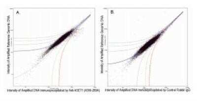
Chromatin Immunoprecipitation: SETD1A Antibody [NB100-558] - Chromatin Immunoprecipitation: SETD1A Antibody [NB100-558] - ChIP-chip scatter plot of anti-hSET1 (NB100-558) enriched DNA binding sites versus input reference DNA. A. 10 mcg of NB100-558 was used to immunoprecipitate chromatin from K562 cells per Ren et al (Genes Dev. 2002 16: 245-256). Immunoprecipitated DNA and reference DNA were amplified via ligation-mediated PCR and the products labeled with fluorescent dUTPs. The labeled ChIP and reference DNA were pooled, hybridized to a DNA microarray, and analyzed. Data points below the +3 SD curve (red line) represent significantly enriched binding sites. B. As a control, a similar experiment was performed using normal rabbit IgG. Compared to the anti-hSET1 ChIP, normal rabbit IgG showed little enrichment.
![Immunoprecipitation: SETD1A Antibody [NB100-558]](https://resources.rndsystems.com/images/products/SETD1A-Antibody-Immunoprecipitation-NB100-558-img0015.jpg "Immunoprecipitation: SETD1A Antibody [NB100-558]")
Immunoprecipitation: SETD1A Antibody [NB100-558]
Immunoprecipitation: SETD1A Antibody [NB100-558] - Detection of human hSET1 by western blot of immunoprecipitates. Samples: Whole cell lysate (1.0 mg per IP reaction; 20% of IP loaded) from HEK293T cells prepared using NETN lysis buffer. Antibodies: Affinity purified rabbit anti-hSET1 antibody NB100-558 (lot NB100-558-6) used for IP at 6 ug per reaction. hSET1 was also immunoprecipitated by a previous lot of this antibody (lot NB100-558-5) and another rabbit anti-hSET1 antibody. For blotting immunoprecipitated hSET1, the other antibody was used at 1:1000. Detection: Chemiluminescence with an exposure time of 30 seconds.Applications for SETD1A Antibody - BSA Free
Application
Recommended Usage
Chromatin Immunoprecipitation (ChIP)
10 ug
Immunohistochemistry
1:2000 - 1:10000
Immunohistochemistry-Paraffin
1:2000 - 1:10000
Immunoprecipitation
2-10 ug/mg lysate
Western Blot
1:10000 - 1:25000
Formulation, Preparation, and Storage
Purification
Immunogen affinity purified
Formulation
Tris-Citrate/Phosphate (pH 7.0 - 8.0)
Format
BSA Free
Preservative
0.09% Sodium Azide
Concentration
1.0 mg/ml
Shipping
The product is shipped with polar packs. Upon receipt, store it immediately at the temperature recommended below.
Stability & Storage
Store at 4C. Do not freeze.
Background: SETD1A
Alternate Names
histone-lysine N-methyltransferase SETD1A, hSET1A, KIAA0339EC 2.1.1.43, KMT2FSET1, Lysine N-methyltransferase 2F, SET domain containing 1A, SET domain-containing protein 1A, Set1, Set1/Ash2 histone methyltransferase complex subunit SET1, SET1A
Entrez Gene IDs
9739 (Human)
Gene Symbol
SETD1A
UniProt
Additional SETD1A Products
Product Documents for SETD1A Antibody - BSA Free
Certificate of Analysis
To download a Certificate of Analysis, please enter a lot or batch number in the search box below.
Product Specific Notices for SETD1A Antibody - BSA Free
This product is for research use only and is not approved for use in humans or in clinical diagnosis. Primary Antibodies are guaranteed for 1 year from date of receipt.
Citations for SETD1A Antibody - BSA Free
 Powered by Bioz
Customer Reviews for SETD1A Antibody - BSA Free
There are currently no reviews for this product. Be the first to review SETD1A Antibody - BSA Free and earn rewards!
Have you used SETD1A Antibody - BSA Free?
Submit a review and receive an Amazon gift card!
$25/€18/£15/$25CAN/¥2500 Yen for a review with an image
$10/€7/£6/$10CAN/¥1110 Yen for a review without an image
Submit a review
Protocols
Find general support by application which include: protocols, troubleshooting, illustrated assays, videos and webinars.
- Antigen Retrieval Protocol (PIER)
- Antigen Retrieval for Frozen Sections Protocol
- Appropriate Fixation of IHC/ICC Samples
- Cellular Response to Hypoxia Protocols
- ChIP Protocol Video
- Chromatin Immunoprecipitation (ChIP) Protocol
- Chromatin Immunoprecipitation Protocol
- Chromogenic IHC Staining of Formalin-Fixed Paraffin-Embedded (FFPE) Tissue Protocol
- Chromogenic Immunohistochemistry Staining of Frozen Tissue
- ClariTSA™ Fluorophore Kits
- Detection & Visualization of Antibody Binding
- Fluorescent IHC Staining of Frozen Tissue Protocol
- Graphic Protocol for Heat-induced Epitope Retrieval
- Graphic Protocol for the Preparation and Fluorescent IHC Staining of Frozen Tissue Sections
- Graphic Protocol for the Preparation and Fluorescent IHC Staining of Paraffin-embedded Tissue Sections
- Graphic Protocol for the Preparation of Gelatin-coated Slides for Histological Tissue Sections
- IHC Sample Preparation (Frozen sections vs Paraffin)
- Immunofluorescent IHC Staining of Formalin-Fixed Paraffin-Embedded (FFPE) Tissue Protocol
- Immunohistochemistry (IHC) and Immunocytochemistry (ICC) Protocols
- Immunohistochemistry Frozen Troubleshooting
- Immunohistochemistry Paraffin Troubleshooting
- Immunoprecipitation Protocol
- Preparing Samples for IHC/ICC Experiments
- Preventing Non-Specific Staining (Non-Specific Binding)
- Primary Antibody Selection & Optimization
- Protocol for Heat-Induced Epitope Retrieval (HIER)
- Protocol for Making a 4% Formaldehyde Solution in PBS
- Protocol for VisUCyte™ HRP Polymer Detection Reagent
- Protocol for the Preparation & Fixation of Cells on Coverslips
- Protocol for the Preparation and Chromogenic IHC Staining of Frozen Tissue Sections
- Protocol for the Preparation and Chromogenic IHC Staining of Frozen Tissue Sections - Graphic
- Protocol for the Preparation and Chromogenic IHC Staining of Paraffin-embedded Tissue Sections
- Protocol for the Preparation and Chromogenic IHC Staining of Paraffin-embedded Tissue Sections - Graphic
- Protocol for the Preparation and Fluorescent IHC Staining of Frozen Tissue Sections
- Protocol for the Preparation and Fluorescent IHC Staining of Paraffin-embedded Tissue Sections
- Protocol for the Preparation of Gelatin-coated Slides for Histological Tissue Sections
- R&D Systems Quality Control Western Blot Protocol
- TUNEL and Active Caspase-3 Detection by IHC/ICC Protocol
- The Importance of IHC/ICC Controls
- Troubleshooting Guide: Immunohistochemistry
- Troubleshooting Guide: Western Blot Figures
- Western Blot Conditions
- Western Blot Protocol
- Western Blot Protocol for Cell Lysates
- Western Blot Troubleshooting
- Western Blot Troubleshooting Guide
- View all Protocols, Troubleshooting, Illustrated assays and Webinars
Loading...