SETDB1 Antibody - BSA Free
Novus Biologicals | Catalog # NBP2-20322
![Immunocytochemistry: SETDB1 Antibody [NBP2-20322]](https://resources.rndsystems.com/images/products/SETDB1-Antibody-Immunocytochemistry-NBP2-20322-img0006.jpg "Immunocytochemistry: SETDB1 Antibody [NBP2-20322]")
Loading...
Key Product Details
Species Reactivity
Validated:
Human, Mouse
Cited:
Human
Predicted:
Bovine (94%), Rat (90%). Backed by our 100% Guarantee.
Applications
Validated:
Immunohistochemistry, Immunohistochemistry-Paraffin, Western Blot, Immunocytochemistry/ Immunofluorescence, Immunoprecipitation, Chromatin Immunoprecipitation (ChIP), Proximity Ligation Assay
Cited:
Western Blot, Immunocytochemistry/ Immunofluorescence, Immunoprecipitation, Chemotaxis, Proximity Ligation Assay
Label
Unconjugated
Antibody Source
Polyclonal Rabbit IgG
Format
BSA Free
Loading...
Product Specifications
Immunogen
Recombinant protein encompassing a sequence within the center region of human SETDB1. The exact sequence is proprietary.
Localization
Nucleus
Clonality
Polyclonal
Host
Rabbit
Isotype
IgG
Theoretical MW
143 kDa.
Disclaimer note: The observed molecular weight of the protein may vary from the listed predicted molecular weight due to post translational modifications, post translation cleavages, relative charges, and other experimental factors.
Disclaimer note: The observed molecular weight of the protein may vary from the listed predicted molecular weight due to post translational modifications, post translation cleavages, relative charges, and other experimental factors.
Scientific Data Images for SETDB1 Antibody - BSA Free
Immunocytochemistry: SETDB1 Antibody [NBP2-20322]
Immunocytochemistry: SETDB1 Antibody [NBP2-20322] - Paraformaldehyde Fixed Human Dermal Microvascular Endothelial Cells were immunostained with 1:100 dilution of pAb. Image submitted by a verified customer review.
Immunohistochemistry-Paraffin: SETDB1 Antibody [NBP2-20322] -
SETDB1 antibody [N2C1], Internal detects SETDB1 protein at nucleus by immunohistochemical analysis.Sample: Paraffin-embedded mouse testis.
SETDB1 stained by SETDB1 antibody [N2C1], Internal (NBP2-20322) diluted at 1:500.
Antigen Retrieval: Citrate buffer, pH 6.0, 15 min

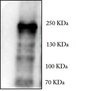
Western Blot: SETDB1 Antibody [NBP2-20322] -
HeLa whole cell and nuclear extracts (30 ug) were separated by 5% SDS-PAGE, and the membrane was blotted with SETDB1 antibody [N2C1], Internal (NBP2-20322) diluted at 1:1000. The HRP-conjugated anti-rabbit IgG antibody was used to detect the primary antibody.
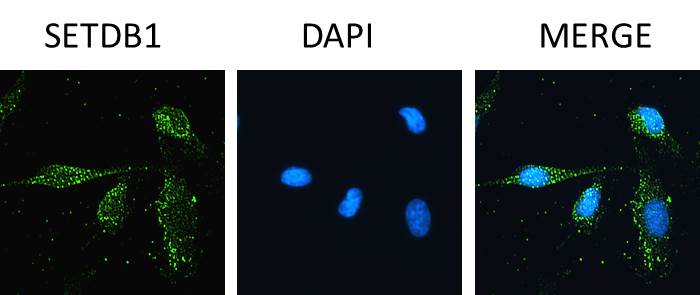
Immunocytochemistry/ Immunofluorescence: SETDB1 Antibody [NBP2-20322] -
SETDB1 antibody [N2C1], Internal detects SETDB1 protein at nucleus by immunofluorescent analysis.Sample: HeLa cells were fixed in 4% paraformaldehyde at RT for 15 min.
Green: SETDB1 stained by SETDB1 antibody [N2C1], Internal (NBP2-20322) diluted at 1:500.
Red: alpha Tubulin, a cytoskeleton marker, stained by alpha Tubulin antibody [GT114] diluted at 1:1000.
Scale bar= 10um.

Immunoprecipitation: SETDB1 Antibody [NBP2-20322] -
SETDB1 antibody [N2C1], Internal immunoprecipitates SETDB1 protein in IP experiments.IP samples: HeLa nuclear extract
A. Control with 4 ug of preimmune Rabbit IgG
B. Immunoprecipitation of SETDB1 protein by 4 ug SETDB1 antibody [N2C1], Internal (NBP2-20322)
5 % SDS-PAGE
The immunoprecipitated SETDB1 protein was detected by SETDB1 antibody [N2C1], Internal (NBP2-20322) diluted at 1:1000.
[EasyBlot anti-rabbit IgG was used as a secondary reagent]

Chromatin Immunoprecipitation: SETDB1 Antibody [NBP2-20322] -
Cross-linked ChIP was performed with HeLa chromatin extract and 5 ug of either control rabbit IgG or anti-SETDB1 antibody. The precipitated DNA was detected by PCR with primer set targeting to p53P2.
Western Blot: SETDB1 Antibody - BSA Free [NBP2-20322] -
Demonstration of IFI16’s interaction and recruitment of specific H3K9 MTases during de novo KSHV infection.(A) TIME cells either left uninfected or infected with KSHV for 6 or 24 hr were IPed with anti-IFI16 antibodies and western blotted for the indicated proteins. (B) To confirm IFI16’s interaction with H3K9 MTases, TIME cells were infected as in (A) and IPed with antibodies against the MTases and blotted for the corresponding MTase and IFI16. (C) 293 T cells lacking IFI16 transfected with control plasmid or His-IFI16 expressing plasmid for 72 hr were utilized for His-tag pulldown using HisPur cobalt resin. Inputs and elutions were blotted for the indicated proteins. (D) 293 T cells transfected with control plasmid, IFI16 expressing plasmid or LANA expressing plasmid for 72 hr were IPed with anti-IFI16 mAb or LANA mAb. Inputs and elutions were blotted for the indicated proteins. Image collected and cropped by CiteAb from the following open publication (https://pubmed.ncbi.nlm.nih.gov/31682228), licensed under a CC-BY license. Not internally tested by Novus Biologicals.
Western Blot: SETDB1 Antibody - BSA Free [NBP2-20322] -
Demonstration of IFI16’s interaction with specific H3K9 MTases in KSHV latently infected PEL (BCBL-1 and BC-3) cells and in uninfected control BJAB cells.(A) Nuclear fractions were isolated from latently infected cells and uninfected BJAB cells and treated with Benzonase. IPs were performed using anti-IFI16 mAb and LANA mAb and WBs were performed. (B) To confirm IFI16’s and LANA’s interaction with H3K9 MTases, IPs were done with Abs against the H3K9 MTases and blotted for the corresponding MTase, IFI16, LANA and HP1 alpha (heterochromatin protein 1 alpha ). Image collected and cropped by CiteAb from the following open publication (https://pubmed.ncbi.nlm.nih.gov/31682228), licensed under a CC-BY license. Not internally tested by Novus Biologicals.
Western Blot: SETDB1 Antibody - BSA Free [NBP2-20322] -
Demonstration of IFI16’s interaction and recruitment of specific H3K9 MTases during de novo KSHV infection.(A) TIME cells either left uninfected or infected with KSHV for 6 or 24 hr were IPed with anti-IFI16 antibodies and western blotted for the indicated proteins. (B) To confirm IFI16’s interaction with H3K9 MTases, TIME cells were infected as in (A) and IPed with antibodies against the MTases and blotted for the corresponding MTase and IFI16. (C) 293 T cells lacking IFI16 transfected with control plasmid or His-IFI16 expressing plasmid for 72 hr were utilized for His-tag pulldown using HisPur cobalt resin. Inputs and elutions were blotted for the indicated proteins. (D) 293 T cells transfected with control plasmid, IFI16 expressing plasmid or LANA expressing plasmid for 72 hr were IPed with anti-IFI16 mAb or LANA mAb. Inputs and elutions were blotted for the indicated proteins. Image collected and cropped by CiteAb from the following open publication (https://pubmed.ncbi.nlm.nih.gov/31682228), licensed under a CC-BY license. Not internally tested by Novus Biologicals.
Western Blot: SETDB1 Antibody - BSA Free [NBP2-20322] -
Effect of A366 on KSHV life cycle and the demonstration of IFI16’s association with cellular H3K9 methyltransferase(s) (H3K9 MTase) and recruitment of various H3K9 MTases to the KSHV genome during de novo infection.(A) MTT cell viability assay of BCBL-1 cells treated with the H3K9me3 specific chemical inhibitor A366 at different concentrations and different time points. (B) q-RT PCR (two-step, sybr Green) of KSHV mRNAs in BCBL-1 cells treated for 72 hr with either vehicle control DMSO or A366 (10 uM and 100 uM). (C) WB of different H3 methylations and IFI16 after A366 treatment of BCBL-1 cells. (D) H3K9 methyltransferase activity (ng/h/mg) assay. TIME cells were infected with KSHV for 6 or 24 hr followed by isolation of nuclear fraction, benzonase treatment and IP with anti-IFI16 or control IgG in the presence of benzonase using the catch and release method. Elution was performed under non-denaturing conditions to keep the associated H3K9 methyltransferase active. H3K9 methyltransferase activity was assayed in the eluate (Materials and methods). *, p<0.05; **, p<0.01; ***,<0.001; unpaired t-test. (E) TIME cells were infected with KSHV genome labeled with EdU or unlabeled control KSHV (100 DNA copies/cell) for 24 hr followed by EdU-KSHV genome pulldown using Click chemistry. The inputs and eluates were blotted for different H3K9 MTases. (F) TIME cells were infected with EdU-labeled KSHV as in (D) and stained using the Click-iT EdU Alexa Fluor 594 Imaging Kit (red). Subsequently, IFA was performed against different H3K9 MTases and colocalization of the IFA signal (green) with KSHV EdU-genome staining (red) resulting in yellow was evaluated (enlarged image, white arrows). Image collected and cropped by CiteAb from the following open publication (https://pubmed.ncbi.nlm.nih.gov/31682228), licensed under a CC-BY license. Not internally tested by Novus Biologicals.
Western Blot: SETDB1 Antibody - BSA Free [NBP2-20322] -
Demonstration of IFI16’s interaction with specific H3K9 MTases in KSHV latently infected PEL (BCBL-1 and BC-3) cells and in uninfected control BJAB cells.(A) Nuclear fractions were isolated from latently infected cells and uninfected BJAB cells and treated with Benzonase. IPs were performed using anti-IFI16 mAb and LANA mAb and WBs were performed. (B) To confirm IFI16’s and LANA’s interaction with H3K9 MTases, IPs were done with Abs against the H3K9 MTases and blotted for the corresponding MTase, IFI16, LANA and HP1 alpha (heterochromatin protein 1 alpha ). Image collected and cropped by CiteAb from the following open publication (https://pubmed.ncbi.nlm.nih.gov/31682228), licensed under a CC-BY license. Not internally tested by Novus Biologicals.
Western Blot: SETDB1 Antibody - BSA Free [NBP2-20322] -
Demonstration of IFI16’s interaction and recruitment of specific H3K9 MTases during de novo KSHV infection.(A) TIME cells either left uninfected or infected with KSHV for 6 or 24 hr were IPed with anti-IFI16 antibodies and western blotted for the indicated proteins. (B) To confirm IFI16’s interaction with H3K9 MTases, TIME cells were infected as in (A) and IPed with antibodies against the MTases and blotted for the corresponding MTase and IFI16. (C) 293 T cells lacking IFI16 transfected with control plasmid or His-IFI16 expressing plasmid for 72 hr were utilized for His-tag pulldown using HisPur cobalt resin. Inputs and elutions were blotted for the indicated proteins. (D) 293 T cells transfected with control plasmid, IFI16 expressing plasmid or LANA expressing plasmid for 72 hr were IPed with anti-IFI16 mAb or LANA mAb. Inputs and elutions were blotted for the indicated proteins. Image collected and cropped by CiteAb from the following open publication (https://pubmed.ncbi.nlm.nih.gov/31682228), licensed under a CC-BY license. Not internally tested by Novus Biologicals.Applications for SETDB1 Antibody - BSA Free
Application
Recommended Usage
Chromatin Immunoprecipitation (ChIP)
Assay dependent
Immunocytochemistry/ Immunofluorescence
Assay dependent
Immunohistochemistry
Assay dependent
Immunohistochemistry-Paraffin
Assay dependent
Immunoprecipitation
1:100-1:500
Proximity Ligation Assay
Reported in scientific literature (PMID:31682228)
Western Blot
1:500-1:3000
Reviewed Applications
Read 4 reviews rated 3.8 using NBP2-20322 in the following applications:
Formulation, Preparation, and Storage
Purification
Antigen Affinity-purified
Formulation
PBS, 20% Glycerol
Format
BSA Free
Preservative
0.025% Proclin 300
Concentration
Concentrations vary lot to lot. See vial label for concentration. If unlisted please contact technical services.
Shipping
The product is shipped with polar packs. Upon receipt, store it immediately at the temperature recommended below.
Stability & Storage
Aliquot and store at -20C or -80C. Avoid freeze-thaw cycles.
Background: SETDB1
Alternate Names
ERG-associated protein with SET domain, ESETEC 2.1.1.43, H3-K9-HMTase 4, Histone H3-K9 methyltransferase 4, histone-lysine N-methyltransferase SETDB1, histone-lysine N-methyltransferase, H3lysine-9 specific 4, KG1T, KIAA0067H3-K9-HMTase4, KMT1EERG-associated protein with a SET domain, ESET, Lysine N-methyltransferase 1E, SET domain bifurcated 1, SET domain, bifurcated 1
Gene Symbol
SETDB1
UniProt
Additional SETDB1 Products
Product Documents for SETDB1 Antibody - BSA Free
Certificate of Analysis
To download a Certificate of Analysis, please enter a lot or batch number in the search box below.
Product Specific Notices for SETDB1 Antibody - BSA Free
This product is for research use only and is not approved for use in humans or in clinical diagnosis. Primary Antibodies are guaranteed for 1 year from date of receipt.
⚠ WARNING: This product can expose you to chemicals including mercury, which is known to the State of California to cause reproductive toxicity with developmental effects. For more information go to www.P65Warnings.ca.gov.Citations for SETDB1 Antibody - BSA Free
 Powered by Bioz
Customer Reviews for SETDB1 Antibody - BSA Free (4)
3.8 out of 5
4 Customer Ratings
Have you used SETDB1 Antibody - BSA Free?
Submit a review and receive an Amazon gift card!
$25/€18/£15/$25CAN/¥2500 Yen for a review with an image
$10/€7/£6/$10CAN/¥1110 Yen for a review without an image
Submit a review
Customer Images

Showing
1
-
4 of
4 reviews
Showing All
Filter By:
-
Application: Western BlotSample Tested: 293TSpecies: HumanVerified Customer | Posted 06/18/2018293T whole cell lysate (50 ug) was subjected WB with NBP2-20322 (1:2000).

-
Application: ImmunocytochemistrySample Tested: Human Dermal Microvascular Endothelial Cells - Paraformaldehyde FixedSpecies: HumanVerified Customer | Posted 07/12/2017Paraformaldehyde Fixed Human Dermal Microvascular Endothelial Cells were immunostained with 1:100 dilution of NBP2-20322.
-
Application: ImmunoprecipitationSample Tested: Human Dermal Microvascular Endothelial whole cell lysateSpecies: HumanVerified Customer | Posted 04/20/2017
-
Application: Western BlotSample Tested: Human Dermal Microvascular Endothelial whole cell lysateSpecies: HumanVerified Customer | Posted 04/19/2017
There are no reviews that match your criteria.
Protocols
Find general support by application which include: protocols, troubleshooting, illustrated assays, videos and webinars.
- Antigen Retrieval Protocol (PIER)
- Antigen Retrieval for Frozen Sections Protocol
- Appropriate Fixation of IHC/ICC Samples
- Cellular Response to Hypoxia Protocols
- ChIP Protocol Video
- Chromatin Immunoprecipitation (ChIP) Protocol
- Chromatin Immunoprecipitation Protocol
- Chromogenic IHC Staining of Formalin-Fixed Paraffin-Embedded (FFPE) Tissue Protocol
- Chromogenic Immunohistochemistry Staining of Frozen Tissue
- ClariTSA™ Fluorophore Kits
- Detection & Visualization of Antibody Binding
- Fluorescent IHC Staining of Frozen Tissue Protocol
- Graphic Protocol for Heat-induced Epitope Retrieval
- Graphic Protocol for the Preparation and Fluorescent IHC Staining of Frozen Tissue Sections
- Graphic Protocol for the Preparation and Fluorescent IHC Staining of Paraffin-embedded Tissue Sections
- Graphic Protocol for the Preparation of Gelatin-coated Slides for Histological Tissue Sections
- ICC Cell Smear Protocol for Suspension Cells
- ICC Immunocytochemistry Protocol Videos
- ICC for Adherent Cells
- IHC Sample Preparation (Frozen sections vs Paraffin)
- Immunocytochemistry (ICC) Protocol
- Immunocytochemistry Troubleshooting
- Immunofluorescence of Organoids Embedded in Cultrex Basement Membrane Extract
- Immunofluorescent IHC Staining of Formalin-Fixed Paraffin-Embedded (FFPE) Tissue Protocol
- Immunohistochemistry (IHC) and Immunocytochemistry (ICC) Protocols
- Immunohistochemistry Frozen Troubleshooting
- Immunohistochemistry Paraffin Troubleshooting
- Immunoprecipitation Protocol
- Preparing Samples for IHC/ICC Experiments
- Preventing Non-Specific Staining (Non-Specific Binding)
- Primary Antibody Selection & Optimization
- Protocol for Heat-Induced Epitope Retrieval (HIER)
- Protocol for Making a 4% Formaldehyde Solution in PBS
- Protocol for VisUCyte™ HRP Polymer Detection Reagent
- Protocol for the Fluorescent ICC Staining of Cell Smears - Graphic
- Protocol for the Fluorescent ICC Staining of Cultured Cells on Coverslips - Graphic
- Protocol for the Preparation & Fixation of Cells on Coverslips
- Protocol for the Preparation and Chromogenic IHC Staining of Frozen Tissue Sections
- Protocol for the Preparation and Chromogenic IHC Staining of Frozen Tissue Sections - Graphic
- Protocol for the Preparation and Chromogenic IHC Staining of Paraffin-embedded Tissue Sections
- Protocol for the Preparation and Chromogenic IHC Staining of Paraffin-embedded Tissue Sections - Graphic
- Protocol for the Preparation and Fluorescent ICC Staining of Cells on Coverslips
- Protocol for the Preparation and Fluorescent ICC Staining of Non-adherent Cells
- Protocol for the Preparation and Fluorescent ICC Staining of Stem Cells on Coverslips
- Protocol for the Preparation and Fluorescent IHC Staining of Frozen Tissue Sections
- Protocol for the Preparation and Fluorescent IHC Staining of Paraffin-embedded Tissue Sections
- Protocol for the Preparation of Gelatin-coated Slides for Histological Tissue Sections
- Protocol for the Preparation of a Cell Smear for Non-adherent Cell ICC - Graphic
- R&D Systems Quality Control Western Blot Protocol
- TUNEL and Active Caspase-3 Detection by IHC/ICC Protocol
- The Importance of IHC/ICC Controls
- Troubleshooting Guide: Immunohistochemistry
- Troubleshooting Guide: Western Blot Figures
- Western Blot Conditions
- Western Blot Protocol
- Western Blot Protocol for Cell Lysates
- Western Blot Troubleshooting
- Western Blot Troubleshooting Guide
- View all Protocols, Troubleshooting, Illustrated assays and Webinars
Loading...