![Western Blot: SETDB2 Antibody [NB100-1137]](https://resources.rndsystems.com/images/products/SETDB2-Antibody-Western-Blot-NB100-1137-img0010.jpg "Western Blot: SETDB2 Antibody [NB100-1137]")
Loading...
Key Product Details
Species Reactivity
Validated:
Human
Cited:
Human
Predicted:
Bovine (100%), Canine (100%), Mouse (100%), Porcine (100%), Rat (100%). Backed by our 100% Guarantee.
Applications
Validated:
Western Blot, Peptide ELISA, Immunocytochemistry/ Immunofluorescence, Immunoprecipitation, Chromatin Immunoprecipitation (ChIP)
Cited:
Western Blot, Immunocytochemistry/ Immunofluorescence, Immunoprecipitation, Chemotaxis
Label
Unconjugated
Antibody Source
Polyclonal Goat IgG
Loading...
Product Specifications
Immunogen
Peptide with sequence GEKNGDAKTFWME-C corresponding to N-Terminus according to NP_114121.2, NP_001153780.1.
Specificity
This antibody is expected to recognize both reported isoforms (NP_114121.2; NP_001153780.1).
Clonality
Polyclonal
Host
Goat
Isotype
IgG
Scientific Data Images for SETDB2 Antibody
Western Blot: SETDB2 Antibody [NB100-1137]
Western Blot: SETDB2 Antibody [NB100-1137] - Staining of Nuclear HeLa lysate with antibody at 0.3 ug/mL (35 ug protein in RIPA buffer). Detected by chemiluminescence.![Immunocytochemistry/ Immunofluorescence: SETDB2 Antibody [NB100-1137]](https://resources.rndsystems.com/images/products/SETDB2-Antibody-Immunocytochemistry-Immunofluorescence-NB100-1137-img0007.jpg "Immunocytochemistry/ Immunofluorescence: SETDB2 Antibody [NB100-1137]")
Immunocytochemistry/ Immunofluorescence: SETDB2 Antibody [NB100-1137]
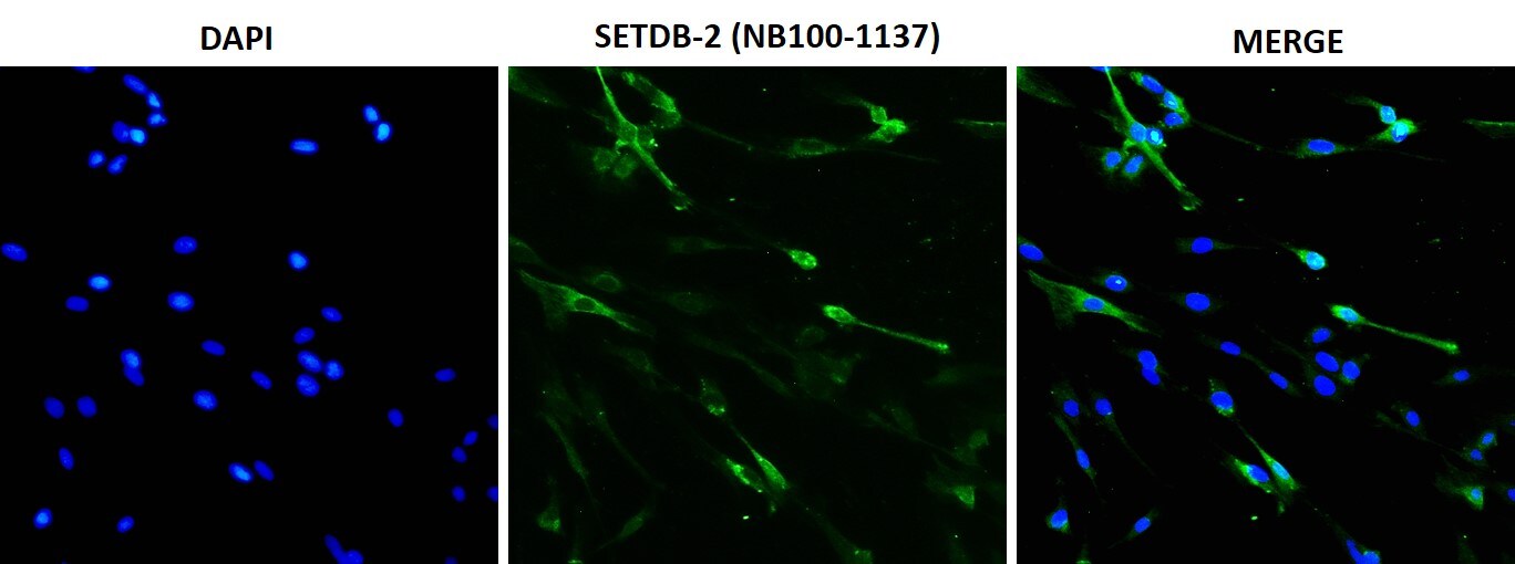
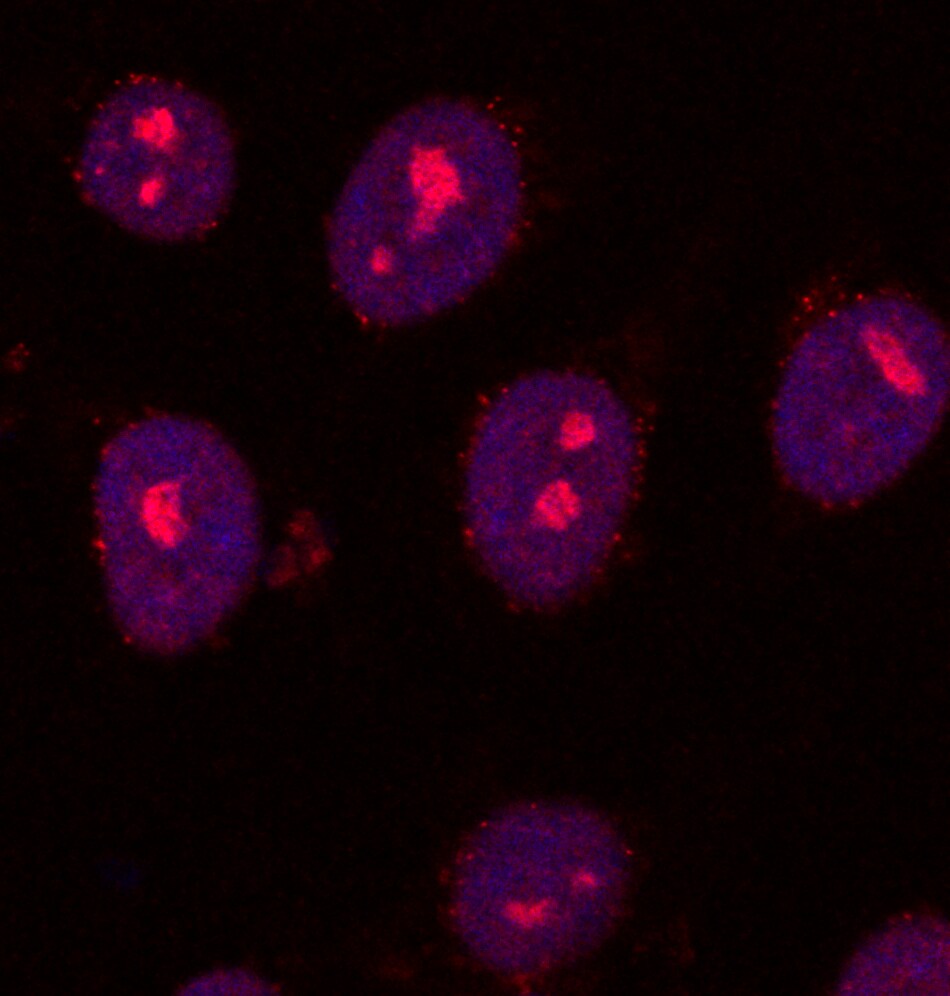
Immunocytochemistry/Immunofluorescence: SETDB2 Antibody [NB100-1137] - Human dermal microvascular endothelial cells were fixed with 4% paraformaldehyde, permeabilized with 0.2% TritonX-100 and immunostained with NB100-1137 (1:100 dilution). Secondary antibody conjugated to Alexafluor 488. Image submitted by a verified customer review.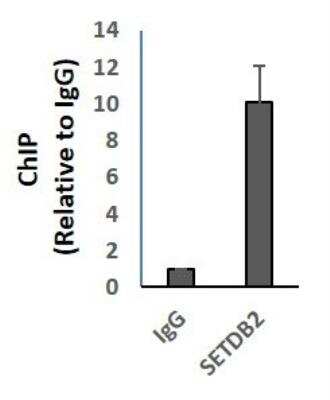
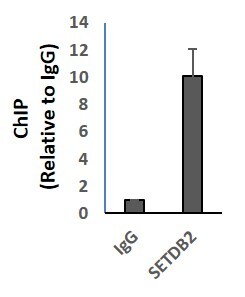
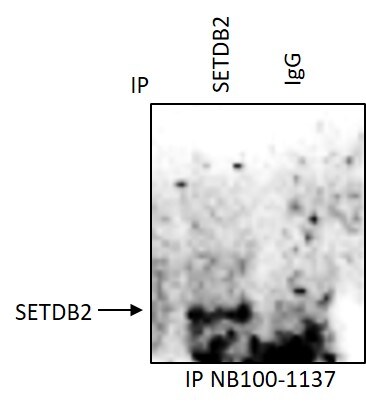
Chromatin Immunoprecipitation (ChIP): SETDB2 Antibody [NB100-1137] - Human B-cell nuclear extract (500 ug). Gene of interest promoter primers were used for amplification to detect ChIP efficiency to IgG control. Image submitted by a verified customer review.
![Western Blot: SETDB2 Antibody [NB100-1137]](https://resources.rndsystems.com/images/products/SETDB2-Antibody-Western-Blot-NB100-1137-img0005.jpg "Western Blot: SETDB2 Antibody [NB100-1137]")
Western Blot: SETDB2 Antibody [NB100-1137]
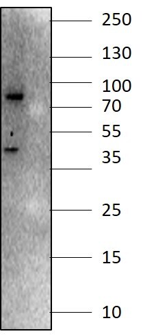
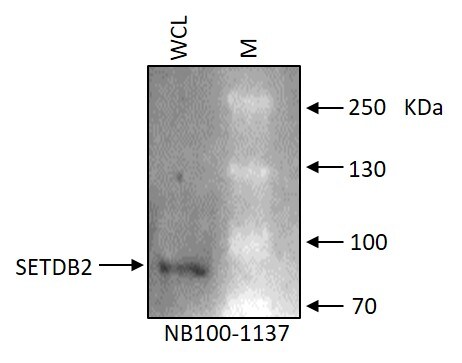
Western Blot: SETDB2 Antibody [NB100-1137] - RL human non-Hodgkin's lymphoma B cell line whole cell lysate (WCL). Single band around correct MW is specific to the target protein SETDB2. Image submitted by a verified customer review.![Immunocytochemistry/ Immunofluorescence: SETDB2 Antibody [NB100-1137]](https://resources.rndsystems.com/images/products/SETDB2-Antibody-Chromatin-Immunoprecipitation-NB100-1137-img0006.jpg "Immunocytochemistry/ Immunofluorescence: SETDB2 Antibody [NB100-1137]")
Immunocytochemistry/ Immunofluorescence: SETDB2 Antibody [NB100-1137]
Immunocytochemistry/Immunofluorescence: SETDB2 Antibody [NB100-1137] - SETDB2 in human induced Pluripotent Stem Cells (iPSCs) using anti-SETDB2 antibody. Image submitted by a verified customer review.
Western Blot: SETDB2 Antibody [NB100-1137] -
Demonstration of IFI16’s interaction with specific H3K9 MTases in KSHV latently infected PEL (BCBL-1 and BC-3) cells and in uninfected control BJAB cells.(A) Nuclear fractions were isolated from latently infected cells and uninfected BJAB cells and treated with Benzonase. IPs were performed using anti-IFI16 mAb and LANA mAb and WBs were performed. (B) To confirm IFI16’s and LANA’s interaction with H3K9 MTases, IPs were done with Abs against the H3K9 MTases and blotted for the corresponding MTase, IFI16, LANA and HP1 alpha (heterochromatin protein 1 alpha ). Image collected and cropped by CiteAb from the following open publication (https://pubmed.ncbi.nlm.nih.gov/31682228), licensed under a CC-BY license. Not internally tested by Novus Biologicals.
Western Blot: SETDB2 Antibody [NB100-1137] -
Demonstration of IFI16’s interaction and recruitment of specific H3K9 MTases during de novo KSHV infection.(A) TIME cells either left uninfected or infected with KSHV for 6 or 24 hr were IPed with anti-IFI16 antibodies and western blotted for the indicated proteins. (B) To confirm IFI16’s interaction with H3K9 MTases, TIME cells were infected as in (A) and IPed with antibodies against the MTases and blotted for the corresponding MTase and IFI16. (C) 293 T cells lacking IFI16 transfected with control plasmid or His-IFI16 expressing plasmid for 72 hr were utilized for His-tag pulldown using HisPur cobalt resin. Inputs and elutions were blotted for the indicated proteins. (D) 293 T cells transfected with control plasmid, IFI16 expressing plasmid or LANA expressing plasmid for 72 hr were IPed with anti-IFI16 mAb or LANA mAb. Inputs and elutions were blotted for the indicated proteins. Image collected and cropped by CiteAb from the following open publication (https://pubmed.ncbi.nlm.nih.gov/31682228), licensed under a CC-BY license. Not internally tested by Novus Biologicals.
Western Blot: SETDB2 Antibody [NB100-1137] -
Demonstration of IFI16’s interaction with specific H3K9 MTases in KSHV latently infected PEL (BCBL-1 and BC-3) cells and in uninfected control BJAB cells.(A) Nuclear fractions were isolated from latently infected cells and uninfected BJAB cells and treated with Benzonase. IPs were performed using anti-IFI16 mAb and LANA mAb and WBs were performed. (B) To confirm IFI16’s and LANA’s interaction with H3K9 MTases, IPs were done with Abs against the H3K9 MTases and blotted for the corresponding MTase, IFI16, LANA and HP1 alpha (heterochromatin protein 1 alpha ). Image collected and cropped by CiteAb from the following open publication (https://pubmed.ncbi.nlm.nih.gov/31682228), licensed under a CC-BY license. Not internally tested by Novus Biologicals.
Western Blot: SETDB2 Antibody [NB100-1137] -
Effect of A366 on KSHV life cycle and the demonstration of IFI16’s association with cellular H3K9 methyltransferase(s) (H3K9 MTase) and recruitment of various H3K9 MTases to the KSHV genome during de novo infection.(A) MTT cell viability assay of BCBL-1 cells treated with the H3K9me3 specific chemical inhibitor A366 at different concentrations and different time points. (B) q-RT PCR (two-step, sybr Green) of KSHV mRNAs in BCBL-1 cells treated for 72 hr with either vehicle control DMSO or A366 (10 uM and 100 uM). (C) WB of different H3 methylations and IFI16 after A366 treatment of BCBL-1 cells. (D) H3K9 methyltransferase activity (ng/h/mg) assay. TIME cells were infected with KSHV for 6 or 24 hr followed by isolation of nuclear fraction, benzonase treatment and IP with anti-IFI16 or control IgG in the presence of benzonase using the catch and release method. Elution was performed under non-denaturing conditions to keep the associated H3K9 methyltransferase active. H3K9 methyltransferase activity was assayed in the eluate (Materials and methods). *, p<0.05; **, p<0.01; ***,<0.001; unpaired t-test. (E) TIME cells were infected with KSHV genome labeled with EdU or unlabeled control KSHV (100 DNA copies/cell) for 24 hr followed by EdU-KSHV genome pulldown using Click chemistry. The inputs and eluates were blotted for different H3K9 MTases. (F) TIME cells were infected with EdU-labeled KSHV as in (D) and stained using the Click-iT EdU Alexa Fluor 594 Imaging Kit (red). Subsequently, IFA was performed against different H3K9 MTases and colocalization of the IFA signal (green) with KSHV EdU-genome staining (red) resulting in yellow was evaluated (enlarged image, white arrows). Image collected and cropped by CiteAb from the following open publication (https://pubmed.ncbi.nlm.nih.gov/31682228), licensed under a CC-BY license. Not internally tested by Novus Biologicals.
Western Blot: SETDB2 Antibody [NB100-1137] -
Demonstration of IFI16’s interaction and recruitment of specific H3K9 MTases during de novo KSHV infection.(A) TIME cells either left uninfected or infected with KSHV for 6 or 24 hr were IPed with anti-IFI16 antibodies and western blotted for the indicated proteins. (B) To confirm IFI16’s interaction with H3K9 MTases, TIME cells were infected as in (A) and IPed with antibodies against the MTases and blotted for the corresponding MTase and IFI16. (C) 293 T cells lacking IFI16 transfected with control plasmid or His-IFI16 expressing plasmid for 72 hr were utilized for His-tag pulldown using HisPur cobalt resin. Inputs and elutions were blotted for the indicated proteins. (D) 293 T cells transfected with control plasmid, IFI16 expressing plasmid or LANA expressing plasmid for 72 hr were IPed with anti-IFI16 mAb or LANA mAb. Inputs and elutions were blotted for the indicated proteins. Image collected and cropped by CiteAb from the following open publication (https://pubmed.ncbi.nlm.nih.gov/31682228), licensed under a CC-BY license. Not internally tested by Novus Biologicals.Applications for SETDB2 Antibody
Application
Recommended Usage
Peptide ELISA
Detection limit 1:1000
Western Blot
1 - 3 ug/mL
Application Notes
Approx 85 kDa band observed in nuclear lysates of cell line HeLa (calculated MW of 81.9 kDa according to NP_114121.2). Not suitable on mouse heart and rat heart lysates. This SETDB2 antibody is validated for WB, CHIP, ICC/IF IP from verified customer reviews.
Reviewed Applications
Read 6 reviews rated 4 using NB100-1137 in the following applications:
Formulation, Preparation, and Storage
Purification
Immunogen affinity purified
Formulation
Tris saline (20 mM Tris pH 7.3, 150 mM NaCl), 0.5% BSA
Preservative
0.02% Sodium Azide
Concentration
0.5 mg/ml
Shipping
The product is shipped with polar packs. Upon receipt, store it immediately at the temperature recommended below.
Stability & Storage
Store at -20C. Avoid freeze-thaw cycles.
Background: SETDB2
Alternate Names
C13orf4, chromosome 13 open reading frame 4, chronic lymphocytic leukemia deletion region 8, Chronic lymphocytic leukemia deletion region gene 8 protein, CLLD8DKFZp761J1217, DKFZp586I0123, EC 2.1.1.43, histone-lysine N-methyltransferase SETDB2, KMT1FCLLL8, Lysine N-methyltransferase 1F, SET domain bifurcated 2, SET domain, bifurcated 2
Entrez Gene IDs
83852 (Human)
Gene Symbol
SETDB2
UniProt
Additional SETDB2 Products
Product Documents for SETDB2 Antibody
Certificate of Analysis
To download a Certificate of Analysis, please enter a lot or batch number in the search box below.
Product Specific Notices for SETDB2 Antibody
This product is for research use only and is not approved for use in humans or in clinical diagnosis. Primary Antibodies are guaranteed for 1 year from date of receipt.
Citations for SETDB2 Antibody
 Powered by Bioz
Customer Reviews for SETDB2 Antibody (6)
4 out of 5
6 Customer Ratings
Have you used SETDB2 Antibody?
Submit a review and receive an Amazon gift card!
$25/€18/£15/$25CAN/¥2500 Yen for a review with an image
$10/€7/£6/$10CAN/¥1110 Yen for a review without an image
Submit a review
Customer Images
Showing
1
-
5 of
6 reviews
Showing All
Filter By:
-
Application: Western BlotSample Tested: THP1 cell lineSpecies: HumanVerified Customer | Posted 07/18/2018Thp1 whole cell lysate was immunobloted with SETDB2 antibody.

-
Application: Chromatin ImmunoprecipitationSample Tested: B cellsSpecies: HumanVerified Customer | Posted 06/22/2018ChIP was performed with 500 ug of Nuclear extract using 3 ug of NB100-1137. Gene of interest promoter primers was used to detect ChIP efficiency and compared to ChIP using control IgG.
-
Application: ImmunocytochemistrySample Tested: Human Dermal Microvascular Endothelial cellsSpecies: HumanVerified Customer | Posted 06/18/2018Human Dermal Microvascular Endothelial cells were fixed with 4% paraformaldehyde, permeabilized with 0.2% TritonX-100 and immunostained with NB100-1137 (1:100 dilution). Secondary antibody conjugated to Alexafluor 488 was used.
-
Application: ImmunoprecipitationSample Tested: RL human non-Hodgkin's lymphoma B cell lineSpecies: HumanVerified Customer | Posted 05/16/2018IP was performed on human non-Hodgkin's lymphoma B cell line with NB100-1137 and IgG as indicated. WB was performed with NB100-1137.
-
Application: Western BlotSample Tested: RL human non-Hodgkin's lymphoma B cell lineSpecies: HumanVerified Customer | Posted 01/04/2018WCL, whole cell lysate.
-
Application: ImmunocytochemistrySample Tested: Induced pluripotent stem cellsSpecies: HumanVerified Customer | Posted 02/25/2016iPS cells, nuclear staining
There are no reviews that match your criteria.
Protocols
Find general support by application which include: protocols, troubleshooting, illustrated assays, videos and webinars.
- Appropriate Fixation of IHC/ICC Samples
- Cellular Response to Hypoxia Protocols
- ChIP Protocol Video
- Chromatin Immunoprecipitation (ChIP) Protocol
- Chromatin Immunoprecipitation Protocol
- ClariTSA™ Fluorophore Kits
- Detection & Visualization of Antibody Binding
- ELISA Sample Preparation & Collection Guide
- ELISA Troubleshooting Guide
- How to Run an R&D Systems DuoSet ELISA
- How to Run an R&D Systems Quantikine ELISA
- How to Run an R&D Systems Quantikine™ QuicKit™ ELISA
- ICC Cell Smear Protocol for Suspension Cells
- ICC Immunocytochemistry Protocol Videos
- ICC for Adherent Cells
- Immunocytochemistry (ICC) Protocol
- Immunocytochemistry Troubleshooting
- Immunofluorescence of Organoids Embedded in Cultrex Basement Membrane Extract
- Immunohistochemistry (IHC) and Immunocytochemistry (ICC) Protocols
- Immunoprecipitation Protocol
- Preparing Samples for IHC/ICC Experiments
- Preventing Non-Specific Staining (Non-Specific Binding)
- Primary Antibody Selection & Optimization
- Protocol for VisUCyte™ HRP Polymer Detection Reagent
- Protocol for the Fluorescent ICC Staining of Cell Smears - Graphic
- Protocol for the Fluorescent ICC Staining of Cultured Cells on Coverslips - Graphic
- Protocol for the Preparation and Fluorescent ICC Staining of Cells on Coverslips
- Protocol for the Preparation and Fluorescent ICC Staining of Non-adherent Cells
- Protocol for the Preparation and Fluorescent ICC Staining of Stem Cells on Coverslips
- Protocol for the Preparation of a Cell Smear for Non-adherent Cell ICC - Graphic
- Quantikine HS ELISA Kit Assay Principle, Alkaline Phosphatase
- Quantikine HS ELISA Kit Principle, Streptavidin-HRP Polymer
- R&D Systems Quality Control Western Blot Protocol
- Sandwich ELISA (Colorimetric) – Biotin/Streptavidin Detection Protocol
- Sandwich ELISA (Colorimetric) – Direct Detection Protocol
- TUNEL and Active Caspase-3 Detection by IHC/ICC Protocol
- The Importance of IHC/ICC Controls
- Troubleshooting Guide: ELISA
- Troubleshooting Guide: Western Blot Figures
- Western Blot Conditions
- Western Blot Protocol
- Western Blot Protocol for Cell Lysates
- Western Blot Troubleshooting
- Western Blot Troubleshooting Guide
- View all Protocols, Troubleshooting, Illustrated assays and Webinars
Loading...