SFRS4 Antibody - BSA Free
Novus Biologicals | Catalog # NBP2-04144
![Western Blot: SFRS4 Antibody [NBP2-04144]](https://resources.rndsystems.com/images/products/SFRS4-Antibody-Western-Blot-NBP2-04144-img0006.jpg "Western Blot: SFRS4 Antibody [NBP2-04144]")
Key Product Details
Validated by
Biological Validation
Species Reactivity
Validated:
Human
Cited:
Human
Applications
Validated:
Immunohistochemistry, Immunohistochemistry-Paraffin, Western Blot, Immunoprecipitation
Cited:
Western Blot
Label
Unconjugated
Antibody Source
Polyclonal Rabbit IgG
Format
BSA Free
Loading...
Product Specifications
Immunogen
The immunogen recognized by this antibody maps to a region between residue 400 and 450 of human Pre-mRNA-Splicing Factor SRP75 using the numbering given in entry NP_005617.2 (Gene ID 6429).
Clonality
Polyclonal
Host
Rabbit
Isotype
IgG
Scientific Data Images for SFRS4 Antibody - BSA Free
Western Blot: SFRS4 Antibody [NBP2-04144]
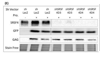
Western Blot: SFRS4 Antibody [NBP2-04144] - J-Lat cells treated with sh vectors targeting SR proteins were induced with Prostratin over 24 hours. Cells were harvested for protein using RIPA. Blots for SRSF4, GFP, HIV p24 GAG protein, and the stain free gel are shown. The J-LAt cell line used had an inducibe HIV construct that when induced would produce GFP as well as HIV proteins such as p24 (GAG). The sh vector was used to knock down the protein. Western blot image submitted by a verified customer review.![Immunohistochemistry-Paraffin: SFRS4 Antibody [NBP2-04144]](https://resources.rndsystems.com/images/products/SFRS4-Antibody-Immunohistochemistry-NBP2-04144-img0003.jpg "Immunohistochemistry-Paraffin: SFRS4 Antibody [NBP2-04144]")
Immunohistochemistry-Paraffin: SFRS4 Antibody [NBP2-04144]
Immunohistochemistry-Paraffin: SFRS4 Antibody [NBP2-04144] - Sections of human lung carcinoma. Antibody: Affinity purified rabbit anti- SRp75 used at a dilution of 1:1000 (1 ug/mL). Detection: DAB. Counterstain: Hematoxylin (blue).![Western Blot: SFRS4 Antibody [NBP2-04144]](https://resources.rndsystems.com/images/products/SFRS4-Antibody-Western-Blot-NBP2-04144-img0004.jpg "Western Blot: SFRS4 Antibody [NBP2-04144]")
Western Blot: SFRS4 Antibody [NBP2-04144]
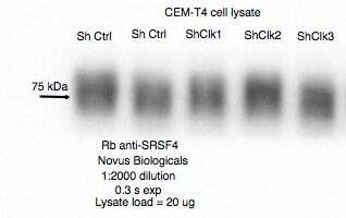
Western Blot: SFRS4 Antibody [NBP2-04144] - CEM-T4 cell lysate. Western blot image submitted by a verified customer review.![Immunoprecipitation: SFRS4 Antibody [NBP2-04144]](https://resources.rndsystems.com/images/products/SFRS4-Antibody-Immunoprecipitation-NBP2-04144-img0001.jpg "Immunoprecipitation: SFRS4 Antibody [NBP2-04144]")
Immunoprecipitation: SFRS4 Antibody [NBP2-04144]
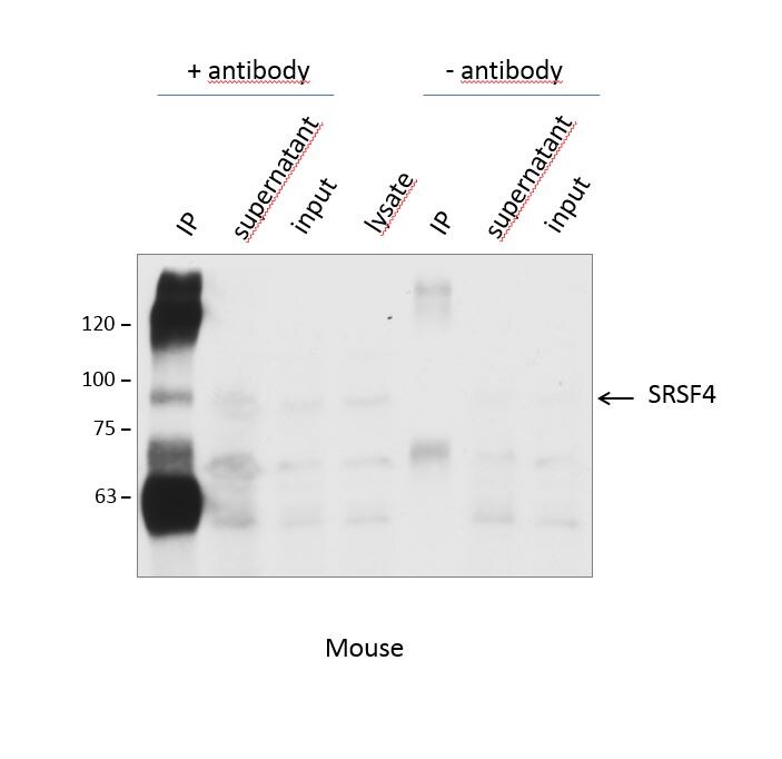
Immunoprecipitation: SFRS4 Antibody [NBP2-04144] - Samples: Whole cell lysate from HeLa (15 and 50 ug for WB; 1 mg for IP, 20% of IP loaded), 293T (T; 50 ug) and Jurkat (J; 50 ug) cells. Antibodies: Affinity purified rabbit anti-SRp75 antibody NBP2-04144 used for WB at 0.1 ug/mL (A) and 1 ug/mL (B) and used for IP at 6 ug/mg lysate. SRp75 was also immunoprecipitated by rabbit anti-SRp75 antibody NBP2-04143, which recognizes a downstream epitope. Detection: Chemiluminescence with exposure times of 3 minutes (A) and 3 seconds (B).Applications for SFRS4 Antibody - BSA Free
Application
Recommended Usage
Immunohistochemistry
1:500 to 1:2000
Immunohistochemistry-Paraffin
1:500 to 1:2000
Immunoprecipitation
2-10 ug/ml lysate
Western Blot
1:2000-1:10000
Application Notes
Epitope retrieval with citrate buffer pH 6.0 is recommended for FFPE tissue sections.
Reviewed Applications
Read 3 reviews rated 4.7 using NBP2-04144 in the following applications:
Formulation, Preparation, and Storage
Purification
Immunogen affinity purified
Formulation
Tris-Citrate/Phosphate (pH 7.0 - 8.0)
Format
BSA Free
Preservative
0.09% Sodium Azide
Concentration
1.0 mg/ml
Shipping
The product is shipped with polar packs. Upon receipt, store it immediately at the temperature recommended below.
Stability & Storage
Store at 4C. Do not freeze.
Background: SFRS4
Alternate Names
Pre-mRNA-splicing factor SRP75, serine/arginine-rich splicing factor 4, SFRS4, Splicing factor, arginine/serine-rich 4SRP001LB, SRP75SR splicing factor 4
Gene Symbol
SRSF4
UniProt
Additional SFRS4 Products
Product Documents for SFRS4 Antibody - BSA Free
Certificate of Analysis
To download a Certificate of Analysis, please enter a lot or batch number in the search box below.
Product Specific Notices for SFRS4 Antibody - BSA Free
This product is for research use only and is not approved for use in humans or in clinical diagnosis. Primary Antibodies are guaranteed for 1 year from date of receipt.
Citations for SFRS4 Antibody - BSA Free
 Powered by Bioz
Customer Reviews for SFRS4 Antibody - BSA Free (3)
4.7 out of 5
3 Customer Ratings
Have you used SFRS4 Antibody - BSA Free?
Submit a review and receive an Amazon gift card!
$25/€18/£15/$25CAN/¥2500 Yen for a review with an image
$10/€7/£6/$10CAN/¥1110 Yen for a review without an image
Submit a review
Customer Images
Showing
1
-
3 of
3 reviews
Showing All
Filter By:
-
Application: Western BlotSample Tested: Cell lysate from Jurkat cells grown in RPMI medium, Hela whole cell lysate and Human primary macrophagesSpecies: HumanVerified Customer | Posted 07/24/2020J-Lat cells treated with sh vectors targeting SR proteins were induced with Prostratin over 24 hours. Cells were harvested for protein using RIPA. Blots for SRSF4, GFP, HIV p24 GAG protein, and the stain free gel are shown.The J-LAt cell line used had an inducibe HIV construct that when induced would produce GFP as well as HIV proteins such as p24 (GAG). The sh vector was used to knock down the protein.

-
Application: Western BlotSample Tested: HeLa Cell lysateSpecies: HumanVerified Customer | Posted 06/09/2018Rb anti-SRSF4 in CEM-T4 cell lysate
-
Application: ImmunoprecipitationSample Tested: P19 cell line (nucleus and cytoplasm)Species: MouseVerified Customer | Posted 11/10/2015IP analysis of SFRS4 in P19 cells
There are no reviews that match your criteria.
Protocols
Find general support by application which include: protocols, troubleshooting, illustrated assays, videos and webinars.
- Antigen Retrieval Protocol (PIER)
- Antigen Retrieval for Frozen Sections Protocol
- Appropriate Fixation of IHC/ICC Samples
- Cellular Response to Hypoxia Protocols
- Chromogenic IHC Staining of Formalin-Fixed Paraffin-Embedded (FFPE) Tissue Protocol
- Chromogenic Immunohistochemistry Staining of Frozen Tissue
- ClariTSA™ Fluorophore Kits
- Detection & Visualization of Antibody Binding
- Fluorescent IHC Staining of Frozen Tissue Protocol
- Graphic Protocol for Heat-induced Epitope Retrieval
- Graphic Protocol for the Preparation and Fluorescent IHC Staining of Frozen Tissue Sections
- Graphic Protocol for the Preparation and Fluorescent IHC Staining of Paraffin-embedded Tissue Sections
- Graphic Protocol for the Preparation of Gelatin-coated Slides for Histological Tissue Sections
- IHC Sample Preparation (Frozen sections vs Paraffin)
- Immunofluorescent IHC Staining of Formalin-Fixed Paraffin-Embedded (FFPE) Tissue Protocol
- Immunohistochemistry (IHC) and Immunocytochemistry (ICC) Protocols
- Immunohistochemistry Frozen Troubleshooting
- Immunohistochemistry Paraffin Troubleshooting
- Immunoprecipitation Protocol
- Preparing Samples for IHC/ICC Experiments
- Preventing Non-Specific Staining (Non-Specific Binding)
- Primary Antibody Selection & Optimization
- Protocol for Heat-Induced Epitope Retrieval (HIER)
- Protocol for Making a 4% Formaldehyde Solution in PBS
- Protocol for VisUCyte™ HRP Polymer Detection Reagent
- Protocol for the Preparation & Fixation of Cells on Coverslips
- Protocol for the Preparation and Chromogenic IHC Staining of Frozen Tissue Sections
- Protocol for the Preparation and Chromogenic IHC Staining of Frozen Tissue Sections - Graphic
- Protocol for the Preparation and Chromogenic IHC Staining of Paraffin-embedded Tissue Sections
- Protocol for the Preparation and Chromogenic IHC Staining of Paraffin-embedded Tissue Sections - Graphic
- Protocol for the Preparation and Fluorescent IHC Staining of Frozen Tissue Sections
- Protocol for the Preparation and Fluorescent IHC Staining of Paraffin-embedded Tissue Sections
- Protocol for the Preparation of Gelatin-coated Slides for Histological Tissue Sections
- R&D Systems Quality Control Western Blot Protocol
- TUNEL and Active Caspase-3 Detection by IHC/ICC Protocol
- The Importance of IHC/ICC Controls
- Troubleshooting Guide: Immunohistochemistry
- Troubleshooting Guide: Western Blot Figures
- Western Blot Conditions
- Western Blot Protocol
- Western Blot Protocol for Cell Lysates
- Western Blot Troubleshooting
- Western Blot Troubleshooting Guide
- View all Protocols, Troubleshooting, Illustrated assays and Webinars
Loading...