SLC34A1 Antibody (10B1.3E9) - BSA Free
Novus Biologicals | Catalog # NBP2-42216
Key Product Details
Species Reactivity
Validated:
Human, Mouse
Cited:
Human, Mouse
Applications
Validated:
Immunohistochemistry, Immunohistochemistry-Paraffin, Western Blot, Flow Cytometry, Immunocytochemistry/ Immunofluorescence, Immunoprecipitation
Cited:
Western Blot, Immunoprecipitation, IF/IHC
Label
Unconjugated
Antibody Source
Monoclonal Mouse IgG1 kappa Clone # 10B1.3E9
Format
BSA Free
Loading...
Product Specifications
Immunogen
Partial synthetic peptide made to an internal portion of the human SLC34A1 protein (between amino acids 25-150) [UniProt Q06495]
Localization
Membrane-Cytoplasmic
Marker
Tubulointerstitium Marker
Clonality
Monoclonal
Host
Mouse
Isotype
IgG1 kappa
Theoretical MW
68.9 kDa.
Disclaimer note: The observed molecular weight of the protein may vary from the listed predicted molecular weight due to post translational modifications, post translation cleavages, relative charges, and other experimental factors.
Disclaimer note: The observed molecular weight of the protein may vary from the listed predicted molecular weight due to post translational modifications, post translation cleavages, relative charges, and other experimental factors.
Scientific Data Images for SLC34A1 Antibody (10B1.3E9) - BSA Free
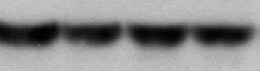
![Western Blot: SLC34A1 Antibody (10B1.3E9)BSA Free [NBP2-42216]](https://resources.rndsystems.com/images/products/SLC34A1-Antibody-10B1-3E9-Western-Blot-NBP2-42216-img0001.jpg "Western Blot: SLC34A1 Antibody (10B1.3E9)BSA Free [NBP2-42216]")
Western Blot: SLC34A1 Antibody (10B1.3E9)BSA Free [NBP2-42216]
Western Blot: SLC34A1 Antibody (10B1.3E9) [NBP2-42216] - WB detection of SLC34A1 /NPTIIa protein in a lysate of mouse thymus using SLC34A1 clone 10B1.3E9 at its 1:500 dilution. The antibody detected a single specific band at ~80 kDa representing the glycosylated form of SLC34A1 protein.![Immunocytochemistry/ Immunofluorescence: SLC34A1 Antibody (10B1.3E9) - BSA Free [NBP2-42216]](https://resources.rndsystems.com/images/products/SLC34A1-Antibody-10B1-3E9-Immunocytochemistry-Immunofluorescence-NBP2-42216-img0007.jpg "Immunocytochemistry/ Immunofluorescence: SLC34A1 Antibody (10B1.3E9) - BSA Free [NBP2-42216]")
Immunocytochemistry/ Immunofluorescence: SLC34A1 Antibody (10B1.3E9) - BSA Free [NBP2-42216]
Immunocytochemistry/Immunofluorescence: SLC34A1 Antibody (10B1.3E9) [NBP2-42216] - Hek293 cells were fixed for 10 minutes using 10% formalin and then permeabilized using 1X TBS + 0.5% Triton-X100. The cells were incubated with hSLC34A1 (10B1.3E9) at a 1:50 dilution overnight at 4 degrees Celsius and detected with Dylight 488 (Green). Actin was detected with Phalloidin 568 (Red). Nuclei were detected with DAPI (Blue). Cells were imaged using a 40X objective.![Immunohistochemistry-Paraffin: SLC34A1 Antibody (10B1.3E9) - BSA Free [NBP2-42216]](https://resources.rndsystems.com/images/products/SLC34A1-Antibody-10B1-3E9-Immunohistochemistry-Paraffin-NBP2-42216-img0006.jpg "Immunohistochemistry-Paraffin: SLC34A1 Antibody (10B1.3E9) - BSA Free [NBP2-42216]")
Immunohistochemistry-Paraffin: SLC34A1 Antibody (10B1.3E9) - BSA Free [NBP2-42216]
Immunohistochemistry-Paraffin: SLC34A1 Antibody (10B1.3E9) [NBP2-42216] - IHC analysis of a formalin-fixed and paraffin-embedded tissue section of human normal kidney using SLC34A1 antibody (clone 10B1.3E9) at 1:75 dilution. The epithelial cells of various renal ducts and tubules depicted very nice membrane-cytoplasmic SLC34A1 immunostaining while the Bowman's capsule and the nuclei of cells were largely negative for SLC34A1 protein.![Flow Cytometry: SLC34A1 Antibody (10B1.3E9) - BSA Free [NBP2-42216]](https://resources.rndsystems.com/images/products/SLC34A1-Antibody-10B1-3E9-Flow-Cytometry-NBP2-42216-img0011.jpg "Flow Cytometry: SLC34A1 Antibody (10B1.3E9) - BSA Free [NBP2-42216]")
Flow Cytometry: SLC34A1 Antibody (10B1.3E9) - BSA Free [NBP2-42216]
Flow Cytometry: SLC34A1 Antibody (10B1.3E9) [NBP2-42216] - An intracellular stain was performed on Hek293 cells with SLC34A1 Antibody [10B1.3E9] NBP2-42216PCP (blue) and a matched isotype control (orange). Cells were fixed with 4% PFA and then permeablized with 0.1% saponin. Cells were incubated in an antibody dilution of 10 ug/mL for 30 minutes at room temperature. Both antibodies were conjugated to PerCP.![Flow Cytometry: SLC34A1 Antibody (10B1.3E9) - BSA Free [NBP2-42216]](https://resources.rndsystems.com/images/products/SLC34A1-Antibody-10B1-3E9-Flow-Cytometry-NBP2-42216-img0002.jpg "Flow Cytometry: SLC34A1 Antibody (10B1.3E9) - BSA Free [NBP2-42216]")
Flow Cytometry: SLC34A1 Antibody (10B1.3E9) - BSA Free [NBP2-42216]
Flow Cytometry: SLC34A1 Antibody (10B1.3E9) [NBP2-42216] - FLOW detection of SLC34A1 protein on HEK293 cells - After fixation and permeabilization, 2 x 10^6 cells/ml were stained using SLC34A1 antibody (clone 10B1.3E9) at 1:1000 dilution. Signal was developped using GtxMs dylight 488 secondary (blue peak). Shown with secondary control (orange peak). Data was acquired on BD FACSCalibur.![Flow Cytometry: SLC34A1 Antibody (10B1.3E9) - BSA Free [NBP2-42216]](https://resources.rndsystems.com/images/products/SLC34A1-Antibody-10B1-3E9-Flow-Cytometry-NBP2-42216-img0008.jpg "Flow Cytometry: SLC34A1 Antibody (10B1.3E9) - BSA Free [NBP2-42216]")
Flow Cytometry: SLC34A1 Antibody (10B1.3E9) - BSA Free [NBP2-42216]
Flow Cytometry: SLC34A1 Antibody (10B1.3E9) [NBP2-42216] - An intracellular stain was performed on Hek293 cells with SLC34A1 Antibody [10B1.3E9] NBP2-42216AF647 (blue) and a matched isotype control (orange). Cells were fixed with 4% PFA and then permeabilized with 0.1% saponin. Cells were incubated in an antibody dilution of 5 ug/mL for 30 minutes at room temperature. Both antibodies were conjugated to Alexa Fluor 647.![Flow Cytometry: SLC34A1 Antibody (10B1.3E9) - BSA Free [NBP2-42216]](https://resources.rndsystems.com/images/products/SLC34A1-Antibody-10B1-3E9-Flow-Cytometry-NBP2-42216-img0009.jpg "Flow Cytometry: SLC34A1 Antibody (10B1.3E9) - BSA Free [NBP2-42216]")
Flow Cytometry: SLC34A1 Antibody (10B1.3E9) - BSA Free [NBP2-42216]
Flow Cytometry: SLC34A1 Antibody (10B1.3E9) [NBP2-42216] - An intracellular stain was performed on Hek293 cells with SLC34A1 Antibody [10B1.3E9] NBP2-42216APC (blue) and a matched isotype control (orange). Cells were fixed with 4% PFA and then permeabilized with 0.1% saponin. Cells were incubated in an antibody dilution of 1 ug/mL for 30 minutes at room temperature. Both antibodies were conjugated to Allophycocyanin.![Flow Cytometry: SLC34A1 Antibody (10B1.3E9) - BSA Free [NBP2-42216]](https://resources.rndsystems.com/images/products/SLC34A1-Antibody-10B1-3E9-Flow-Cytometry-NBP2-42216-img0010.jpg "Flow Cytometry: SLC34A1 Antibody (10B1.3E9) - BSA Free [NBP2-42216]")
Flow Cytometry: SLC34A1 Antibody (10B1.3E9) - BSA Free [NBP2-42216]
Flow Cytometry: SLC34A1 Antibody (10B1.3E9) [NBP2-42216] - An intracellular stain was performed on Hek293 cells with SLC34A1 Antibody [10B1.3E9] NBP2-42216PE (blue) and a matched isotype control (orange). Cells were fixed with 4% PFA and then permeabilized with 0.1% saponin. Cells were incubated in an antibody dilution of 2.5 ug/mL for 30 minutes at room temperature. Both antibodies were conjugated to Phycoerythrin.Applications for SLC34A1 Antibody (10B1.3E9) - BSA Free
Application
Recommended Usage
Flow Cytometry
1:1000
Immunocytochemistry/ Immunofluorescence
1:50
Immunohistochemistry
1:50-1:100
Immunohistochemistry-Paraffin
1:50-1:100
Immunoprecipitation
reported in scientific literature (PMID 35307350)
Western Blot
1:500
Application Notes
SLC34A1 is a 639 amino acids long protein with predicted molecular weight of 68.9 kDa, however, because of glycosylation, the processed protein may show up at higher than predicted molecular weight position on Western blot.
Reviewed Applications
Read 1 review rated 5 using NBP2-42216 in the following applications:
Flow Cytometry Panel Builder
Bio-Techne Knows Flow Cytometry
Save time and reduce costly mistakes by quickly finding compatible reagents using the Panel Builder Tool.
Advanced Features
- Spectra Viewer - Custom analysis of spectra from multiple fluorochromes
- Spillover Popups - Visualize the spectra of individual fluorochromes
- Antigen Density Selector - Match fluorochrome brightness with antigen density

Formulation, Preparation, and Storage
Purification
Protein G purified
Formulation
PBS
Format
BSA Free
Preservative
0.02% Sodium Azide
Concentration
1.0 mg/ml
Shipping
The product is shipped with polar packs. Upon receipt, store it immediately at the temperature recommended below.
Stability & Storage
Store at -20C. Avoid freeze-thaw cycles.
Background: SLC34A1
Alternate Names
FRTS2, Na(+)/Pi cotransporter 2A, Na(+)-dependent phosphate cotransporter 2A, naPi-2a, NaPi-3, NPT2NPHLOP1, NPTIIa, renal sodium-dependent phosphate transporter, SLC11, SLC17A2Na+-phosphate cotransporter type II, sodium/phosphate co-transporter, Sodium/phosphate cotransporter 2A, sodium-dependent phosphate transport protein 2A, Sodium-phosphate transport protein 2A, solute carrier family 17 (sodium phosphate), member 2, solute carrier family 34 (sodium phosphate), member 1, Solute carrier family 34 member 1
Entrez Gene IDs
6569 (Human)
Gene Symbol
SLC34A1
UniProt
Additional SLC34A1 Products
Product Documents for SLC34A1 Antibody (10B1.3E9) - BSA Free
Certificate of Analysis
To download a Certificate of Analysis, please enter a lot or batch number in the search box below.
Product Specific Notices for SLC34A1 Antibody (10B1.3E9) - BSA Free
This product is for research use only and is not approved for use in humans or in clinical diagnosis. Primary Antibodies are guaranteed for 1 year from date of receipt.
Citations for SLC34A1 Antibody (10B1.3E9) - BSA Free
 Powered by Bioz
Customer Reviews for SLC34A1 Antibody (10B1.3E9) - BSA Free (1)
5 out of 5
1 Customer Rating
Have you used SLC34A1 Antibody (10B1.3E9) - BSA Free?
Submit a review and receive an Amazon gift card!
$25/€18/£15/$25CAN/¥2500 Yen for a review with an image
$10/€7/£6/$10CAN/¥1110 Yen for a review without an image
Submit a review
Customer Images
Showing
1
-
1 of
1 review
Showing All
Filter By:
-
Application: Western BlotSample Tested: Kidney tissueSpecies: MouseVerified Customer | Posted 08/17/2021SLC34A1 Antibody in kidney

There are no reviews that match your criteria.
Protocols
View specific protocols for SLC34A1 Antibody (10B1.3E9) - BSA Free (NBP2-42216):
Immunocytochemistry Protocol
Culture cells to appropriate density in 35 mm culture dishes or 6-well plates.
1. Remove culture medium and add 10% formalin to the dish. Fix at room temperature for 30 minutes.
2. Remove the formalin and add ice cold methanol. Incubate for 5-10 minutes.
3. Remove methanol and add washing solution (i.e. PBS). Be sure to not let the specimen dry out. Wash three times for 10 minutes.
4. To block nonspecific antibody binding incubate in 10% normal goat serum from 1 hour to overnight at room temperature.
5. Add primary antibody at appropriate dilution and incubate at room temperature from 2 hours to overnight at room temperature.
6. Remove primary antibody and replace with washing solution. Wash three times for 10 minutes.
7. Add secondary antibody at appropriate dilution. Incubate for 1 hour at room temperature.
8. Remove antibody and replace with wash solution, then wash for 10 minutes. Add Hoechst 33258 to wash solution at 1:25,0000 and incubate for 10 minutes. Wash a third time for 10 minutes.
9. Cells can be viewed directly after washing. The plates can also be stored in PBS containing Azide covered in Parafilm (TM). Cells can also be cover-slipped using Fluoromount, with appropriate sealing.
*The above information is only intended as a guide. The researcher should determine what protocol best meets their needs. Please follow safe laboratory procedures.
Reagents needed:
a. Washing Buffer: Tris Buffer Saline with 0.01% of tween 20).
b. Blocking Buffer: 5% skimmed milk powder in washing buffer).
c. Secondary antibody, Horseradish peroxidase conjugated.
d. Chemiluminescent solution (SuperSignal WestPicoTM, Pierce).
Western blot Method:
1. Perform SDS-PAGE using PVDF membrane. Cut into strips.
2. Activate strips with methanol by dipping them into methanol for 5 min.
3. Discard the methanol and take fresh methanol to repeat step b.
4. Let the strips dry, and then add blocking solution and incubate at RT in a shaker for 30-45 minutes.
5. Dilute primary antibody in blocking buffer. Incubate the number of strips required with the diluted primary antibody at room temperature for 2 hours in a shaker.
6. Wash strips two times with washing buffer at 30 minutes intervals.
7. Dilute HRP conjugated secondary antibody in blocking buffer. Add diluted secondary antibody to the membrane strips and incubate for exactly 1 hour while shaking at RT.
8. Wash the strips with washing buffer for 2-3 hours with 3 to 4 changes on a shaker. This helps in reducing the back ground staining.
9. Prepare the chemiluminescent solution (SuperSignal WestPicoTM) by mixing solution A and Solution B at 1:1. Mix well. Soak the strip in the chemiluminescent solution; keep for 3-5 minutes under constant shaking.
10. Expose the membrane to a sheet of film and develop.
Find general support by application which include: protocols, troubleshooting, illustrated assays, videos and webinars.
- 7-Amino Actinomycin D (7-AAD) Cell Viability Flow Cytometry Protocol
- Antigen Retrieval Protocol (PIER)
- Antigen Retrieval for Frozen Sections Protocol
- Appropriate Fixation of IHC/ICC Samples
- Cellular Response to Hypoxia Protocols
- Chromogenic IHC Staining of Formalin-Fixed Paraffin-Embedded (FFPE) Tissue Protocol
- Chromogenic Immunohistochemistry Staining of Frozen Tissue
- ClariTSA™ Fluorophore Kits
- Detection & Visualization of Antibody Binding
- Extracellular Membrane Flow Cytometry Protocol
- Flow Cytometry Protocol for Cell Surface Markers
- Flow Cytometry Protocol for Staining Membrane Associated Proteins
- Flow Cytometry Staining Protocols
- Flow Cytometry Troubleshooting Guide
- Fluorescent IHC Staining of Frozen Tissue Protocol
- Graphic Protocol for Heat-induced Epitope Retrieval
- Graphic Protocol for the Preparation and Fluorescent IHC Staining of Frozen Tissue Sections
- Graphic Protocol for the Preparation and Fluorescent IHC Staining of Paraffin-embedded Tissue Sections
- Graphic Protocol for the Preparation of Gelatin-coated Slides for Histological Tissue Sections
- ICC Cell Smear Protocol for Suspension Cells
- ICC Immunocytochemistry Protocol Videos
- ICC for Adherent Cells
- IHC Sample Preparation (Frozen sections vs Paraffin)
- Immunocytochemistry (ICC) Protocol
- Immunocytochemistry Troubleshooting
- Immunofluorescence of Organoids Embedded in Cultrex Basement Membrane Extract
- Immunofluorescent IHC Staining of Formalin-Fixed Paraffin-Embedded (FFPE) Tissue Protocol
- Immunohistochemistry (IHC) and Immunocytochemistry (ICC) Protocols
- Immunohistochemistry Frozen Troubleshooting
- Immunohistochemistry Paraffin Troubleshooting
- Immunoprecipitation Protocol
- Intracellular Flow Cytometry Protocol Using Alcohol (Methanol)
- Intracellular Flow Cytometry Protocol Using Detergents
- Intracellular Nuclear Staining Flow Cytometry Protocol Using Detergents
- Intracellular Staining Flow Cytometry Protocol Using Alcohol Permeabilization
- Intracellular Staining Flow Cytometry Protocol Using Detergents to Permeabilize Cells
- Preparing Samples for IHC/ICC Experiments
- Preventing Non-Specific Staining (Non-Specific Binding)
- Primary Antibody Selection & Optimization
- Propidium Iodide Cell Viability Flow Cytometry Protocol
- Protocol for Heat-Induced Epitope Retrieval (HIER)
- Protocol for Liperfluo
- Protocol for Making a 4% Formaldehyde Solution in PBS
- Protocol for VisUCyte™ HRP Polymer Detection Reagent
- Protocol for the Characterization of Human Th22 Cells
- Protocol for the Characterization of Human Th9 Cells
- Protocol for the Fluorescent ICC Staining of Cell Smears - Graphic
- Protocol for the Fluorescent ICC Staining of Cultured Cells on Coverslips - Graphic
- Protocol for the Preparation & Fixation of Cells on Coverslips
- Protocol for the Preparation and Chromogenic IHC Staining of Frozen Tissue Sections
- Protocol for the Preparation and Chromogenic IHC Staining of Frozen Tissue Sections - Graphic
- Protocol for the Preparation and Chromogenic IHC Staining of Paraffin-embedded Tissue Sections
- Protocol for the Preparation and Chromogenic IHC Staining of Paraffin-embedded Tissue Sections - Graphic
- Protocol for the Preparation and Fluorescent ICC Staining of Cells on Coverslips
- Protocol for the Preparation and Fluorescent ICC Staining of Non-adherent Cells
- Protocol for the Preparation and Fluorescent ICC Staining of Stem Cells on Coverslips
- Protocol for the Preparation and Fluorescent IHC Staining of Frozen Tissue Sections
- Protocol for the Preparation and Fluorescent IHC Staining of Paraffin-embedded Tissue Sections
- Protocol for the Preparation of Gelatin-coated Slides for Histological Tissue Sections
- Protocol for the Preparation of a Cell Smear for Non-adherent Cell ICC - Graphic
- Protocol: Annexin V and PI Staining by Flow Cytometry
- Protocol: Annexin V and PI Staining for Apoptosis by Flow Cytometry
- R&D Systems Quality Control Western Blot Protocol
- TUNEL and Active Caspase-3 Detection by IHC/ICC Protocol
- The Importance of IHC/ICC Controls
- Troubleshooting Guide: Fluorokine Flow Cytometry Kits
- Troubleshooting Guide: Immunohistochemistry
- Troubleshooting Guide: Western Blot Figures
- Western Blot Conditions
- Western Blot Protocol
- Western Blot Protocol for Cell Lysates
- Western Blot Troubleshooting
- Western Blot Troubleshooting Guide
- View all Protocols, Troubleshooting, Illustrated assays and Webinars
Loading...