Sodium Potassium ATPase Alpha 1 Antibody (464.6) - BSA Free
Novus Biologicals | Catalog # NB300-146
Key Product Details
Species Reactivity
Validated:
Cited:
Applications
Validated:
Cited:
Label
Antibody Source
Format
Product Specifications
Immunogen
Reactivity Notes
Marker
Specificity
Clonality
Host
Isotype
Theoretical MW
Disclaimer note: The observed molecular weight of the protein may vary from the listed predicted molecular weight due to post translational modifications, post translation cleavages, relative charges, and other experimental factors.
Scientific Data Images for Sodium Potassium ATPase Alpha 1 Antibody (464.6) - BSA Free
![Western Blot: Sodium Potassium ATPase Alpha 1 Antibody (464.6) [NB300-146]](https://resources.rndsystems.com/images/products/Sodium-Potassium-ATPase-Alpha-1-Antibody-464-6-Western-Blot-NB300-146-img0022.jpg "Western Blot: Sodium Potassium ATPase Alpha 1 Antibody (464.6) [NB300-146]")
Western Blot: Sodium Potassium ATPase Alpha 1 Antibody (464.6) [NB300-146]
Sodium-Potassium-ATPase-Alpha-1-Antibody-464-6-Western-Blot-NB300-146-img0022.jpg![Immunocytochemistry/ Immunofluorescence: Sodium Potassium ATPase Alpha 1 Antibody (464.6) [NB300-146]](https://resources.rndsystems.com/images/products/Sodium-Potassium-ATPase-Alpha-1-Antibody-464-6-Immunocytochemistry-Immunofluorescence-NB300-146-img0023.jpg "Immunocytochemistry/ Immunofluorescence: Sodium Potassium ATPase Alpha 1 Antibody (464.6) [NB300-146]")
Immunocytochemistry/ Immunofluorescence: Sodium Potassium ATPase Alpha 1 Antibody (464.6) [NB300-146]
Sodium-Potassium-ATPase-Alpha-1-Antibody-464-6-Immunocytochemistry-Immunofluorescence-NB300-146-img0023.jpg![Immunohistochemistry: Sodium Potassium ATPase Alpha 1 Antibody (464.6) [NB300-146]](https://resources.rndsystems.com/images/products/Sodium-Potassium-ATPase-Alpha-1-Antibody-464-6-Immunohistochemistry-NB300-146-img0012.jpg "Immunohistochemistry: Sodium Potassium ATPase Alpha 1 Antibody (464.6) [NB300-146]")
Immunohistochemistry: Sodium Potassium ATPase Alpha 1 Antibody (464.6) [NB300-146]
Immunohistochemistry: Sodium Potassium ATPase Alpha 1 Antibody (464.6) [NB300-146] - Staining of human enodmetrial glands within the uterus using NB300-146. Note the absence of staining in the surrounding myometrial smooth muscle.![Flow Cytometry: Sodium Potassium ATPase Alpha 1 Antibody (464.6) [NB300-146]](https://resources.rndsystems.com/images/products/Sodium-Potassium-ATPase-Alpha-1-Antibody-464-6-Flow-Cytometry-NB300-146-img0027.jpg "Flow Cytometry: Sodium Potassium ATPase Alpha 1 Antibody (464.6) [NB300-146]")
Flow Cytometry: Sodium Potassium ATPase Alpha 1 Antibody (464.6) [NB300-146]
Flow Cytometry: Sodium Potassium ATPase Alpha 1 Antibody (464.6) [NB300-146] - An intracellular stain was performed on NIH3T3 cells with Sodium Potassium ATPase Alpha 1 Antibody (464.6) NB300-146 (blue) and a matched isotype control MAB002 (orange). Cells were fixed with 4% PFA and then permeabilized with 0.1% saponin. Cells were incubated in an antibody dilution of 1 ug/mL for 30 minutes at room temperature, followed by Mouse IgG (H+L) Cross-Adsorbed Secondary Antibody, Dylight 550 (84540, Thermo Fisher).![Western Blot: Sodium Potassium ATPase Alpha 1 Antibody (464.6) [NB300-146]](https://resources.rndsystems.com/images/products/Sodium-Potassium-ATPase-Alpha-1-Antibody-464-6-Western-Blot-NB300-146-img0013.jpg "Western Blot: Sodium Potassium ATPase Alpha 1 Antibody (464.6) [NB300-146]")
Western Blot: Sodium Potassium ATPase Alpha 1 Antibody (464.6) [NB300-146]
Western Blot: Sodium Potassium ATPase Alpha 1 Antibody (464.6) [NB300-146] - Analysis detecting Na, K-ATPase (alpha) in porcine proximal tubule protein.![Immunocytochemistry/ Immunofluorescence: Sodium Potassium ATPase Alpha 1 Antibody (464.6) [NB300-146]](https://resources.rndsystems.com/images/products/Sodium-Potassium-ATPase-Alpha-1-Antibody-464-6-Immunocytochemistry-Immunofluorescence-NB300-146-img0014.jpg "Immunocytochemistry/ Immunofluorescence: Sodium Potassium ATPase Alpha 1 Antibody (464.6) [NB300-146]")
Immunocytochemistry/ Immunofluorescence: Sodium Potassium ATPase Alpha 1 Antibody (464.6) [NB300-146]
Immunocytochemistry/Immunofluorescence: Sodium Potassium ATPase Alpha 1 Antibody (464.6) [NB300-146] - Hek293 cells were fixed for 10 minutes using 10% formalin and then permeabilized for 5 minutes using 1X TBS + 0.5% Triton X-100. The cells were incubated with anti-Sodium Potassium ATPase Alpha 1 (464.6) at 10 ug/ml overnight at 4C and detected with an anti-mouse DyLight 488 (Green) at a 1:500 dilution. Actin was detected with Phalloidin 568 (Red) at a 1:200 dilution. Nuclei were counterstained with DAPI (Blue). Cells were imaged using a 40X objective.![Immunocytochemistry/ Immunofluorescence: Sodium Potassium ATPase Alpha 1 Antibody (464.6) [NB300-146]](https://resources.rndsystems.com/images/products/Sodium-Potassium-ATPase-Alpha-1-Antibody-464-6-Immunocytochemistry-Immunofluorescence-NB300-146-img0015.jpg "Immunocytochemistry/ Immunofluorescence: Sodium Potassium ATPase Alpha 1 Antibody (464.6) [NB300-146]")
Immunocytochemistry/ Immunofluorescence: Sodium Potassium ATPase Alpha 1 Antibody (464.6) [NB300-146]
Immunocytochemistry/Immunofluorescence: Sodium Potassium ATPase Alpha 1 Antibody (464.6) [NB300-146] - HeLa cells were fixed for 10 minutes using 10% formalin and then permeabilized for 5 minutes using 1X TBS + 0.5% Triton X-100. The cells were incubated with anti-Sodium Potassium ATPase Alpha 1 (464.6) at 10 ug/ml overnight at 4C and detected with an anti-mouse DyLight 488 (Green) at a 1:500 dilution. Actin was detected with Phalloidin 568 (Red) at a 1:200 dilution. Nuclei were counterstained with DAPI (Blue). Cells were imaged using a 40X objective.![Immunocytochemistry/ Immunofluorescence: Sodium Potassium ATPase Alpha 1 Antibody (464.6) [NB300-146]](https://resources.rndsystems.com/images/products/Sodium-Potassium-ATPase-Alpha-1-Antibody-464-6-Immunocytochemistry-Immunofluorescence-NB300-146-img0007.jpg "Immunocytochemistry/ Immunofluorescence: Sodium Potassium ATPase Alpha 1 Antibody (464.6) [NB300-146]")
Immunocytochemistry/ Immunofluorescence: Sodium Potassium ATPase Alpha 1 Antibody (464.6) [NB300-146]
Immunocytochemistry/Immunofluorescence: Sodium Potassium ATPase Alpha 1 Antibody (464.6) [NB300-146] - Detection of ATPA1 (Green) in HepG2 cells using NB300-146. Nuclei (Blue) were counterstained using Hoechst 33258.![Immunocytochemistry/ Immunofluorescence: Sodium Potassium ATPase Alpha 1 Antibody (464.6) [NB300-146]](https://resources.rndsystems.com/images/products/Sodium-Potassium-ATPase-Alpha-1-Antibody-464-6-Immunocytochemistry-Immunofluorescence-NB300-146-img0019.jpg "Immunocytochemistry/ Immunofluorescence: Sodium Potassium ATPase Alpha 1 Antibody (464.6) [NB300-146]")
Immunocytochemistry/ Immunofluorescence: Sodium Potassium ATPase Alpha 1 Antibody (464.6) [NB300-146]
Sodium-Potassium-ATPase-Alpha-1-Antibody-464-6-Immunocytochemistry-Immunofluorescence-NB300-146-img0019.jpg![Immunocytochemistry/ Immunofluorescence: Sodium Potassium ATPase Alpha 1 Antibody (464.6) [NB300-146]](https://resources.rndsystems.com/images/products/Sodium-Potassium-ATPase-Alpha-1-Antibody-464-6-Immunocytochemistry-Immunofluorescence-NB300-146-img0020.jpg "Immunocytochemistry/ Immunofluorescence: Sodium Potassium ATPase Alpha 1 Antibody (464.6) [NB300-146]")
Immunocytochemistry/ Immunofluorescence: Sodium Potassium ATPase Alpha 1 Antibody (464.6) [NB300-146]
Sodium-Potassium-ATPase-Alpha-1-Antibody-464-6-Immunocytochemistry-Immunofluorescence-NB300-146-img0020.jpg![Flow Cytometry: Sodium Potassium ATPase Alpha 1 Antibody (464.6) [NB300-146]](https://resources.rndsystems.com/images/products/Sodium-Potassium-ATPase-Alpha-1-Antibody-464-6-Flow-Cytometry-NB300-146-img0016.jpg "Flow Cytometry: Sodium Potassium ATPase Alpha 1 Antibody (464.6) [NB300-146]")
Flow Cytometry: Sodium Potassium ATPase Alpha 1 Antibody (464.6) [NB300-146]
Flow Cytometry: Sodium Potassium ATPase Alpha 1 Antibody (464.6) [NB300-146] - An intracellular stain was performed on A549 cells with NB300-146 and a matched isotype control. Cells were fixed with 4% PFA and then permeablized with 0.1% saponin. Cells were incubated in an antibody dilution of 1 ug/mL for 30 minutes at room temperature, followed by mouse F(ab)2 IgG (H+L) APC-conjugated secondary antibody (F0101B, R&D Systems).![Flow Cytometry: Sodium Potassium ATPase Alpha 1 Antibody (464.6) [NB300-146]](https://resources.rndsystems.com/images/products/Sodium-Potassium-ATPase-Alpha-1-Antibody-464-6-Flow-Cytometry-NB300-146-img0017.jpg "Flow Cytometry: Sodium Potassium ATPase Alpha 1 Antibody (464.6) [NB300-146]")
Flow Cytometry: Sodium Potassium ATPase Alpha 1 Antibody (464.6) [NB300-146]
Flow Cytometry: Sodium Potassium ATPase Alpha 1 Antibody (464.6) [NB300-146] - An intracellular stain was performed on A549 cells with NBP2-61137AF647 (Blue) and a matched isotype control (Orange). Cells were fixed with 4% PFA and then permeabilized with 0.1% saponin. Cells were incubated in an antibody dilution of 2.5 ug/mL for 30 minutes at room temperature. Both antibodies were conjugated to Alexa Fluor 647. Image using the Alexa Fluor 647 form of this antibody.![Flow Cytometry: Sodium Potassium ATPase Alpha 1 Antibody (464.6) [NB300-146]](https://resources.rndsystems.com/images/products/Sodium-Potassium-ATPase-Alpha-1-Antibody-464-6-Flow-Cytometry-NB300-146-img0018.jpg "Flow Cytometry: Sodium Potassium ATPase Alpha 1 Antibody (464.6) [NB300-146]")
Flow Cytometry: Sodium Potassium ATPase Alpha 1 Antibody (464.6) [NB300-146]
Flow Cytometry: Sodium Potassium ATPase Alpha 1 Antibody (464.6) [NB300-146] - An intracellular stain was performed on A549 cells with - Purified NBP2-61137 and a matched isotype control. Cells were fixed with 4% PFA and then permeablized with 0.1% saponin. Cells were incubated in an antibody dilution of 1 ug/mL for 30 minutes at room temperature, followed by mouse F(ab)2 IgG (H+L) APC-conjugated secondary antibody (F0101B, R&D Systems). Image using the Purified form of this antibody.![Flow Cytometry: Sodium Potassium ATPase Alpha 1 Antibody (464.6) [NB300-146]](https://resources.rndsystems.com/images/products/Sodium-Potassium-ATPase-Alpha-1-Antibody-464-6-Flow-Cytometry-NB300-146-img0025.jpg "Flow Cytometry: Sodium Potassium ATPase Alpha 1 Antibody (464.6) [NB300-146]")
Flow Cytometry: Sodium Potassium ATPase Alpha 1 Antibody (464.6) [NB300-146]
Flow Cytometry: Sodium Potassium ATPase Alpha 1 Antibody (464.6) [NB300-146] - An intracellular stain was performed on RCC4 cells with Sodium Potassium ATPase Alpha 1 Antibody (464.6) NB300-146 (blue) and a matched isotype control MAB002 (orange). Cells were fixed with 4% PFA and then permeabilized with 0.1% saponin. Cells were incubated in an antibody dilution of 1 ug/mL for 30 minutes at room temperature, followed by Mouse IgG (H+L) Cross-Adsorbed Secondary Antibody, Dylight 550 (84540, Thermo Fisher).![Flow Cytometry: Sodium Potassium ATPase Alpha 1 Antibody (464.6) [NB300-146]](https://resources.rndsystems.com/images/products/Sodium-Potassium-ATPase-Alpha-1-Antibody-464-6-Flow-Cytometry-NB300-146-img0026.jpg "Flow Cytometry: Sodium Potassium ATPase Alpha 1 Antibody (464.6) [NB300-146]")
Flow Cytometry: Sodium Potassium ATPase Alpha 1 Antibody (464.6) [NB300-146]
Flow Cytometry: Sodium Potassium ATPase Alpha 1 Antibody (464.6) [NB300-146] - An intracellular stain was performed on SK-MEL-28 cells with Sodium Potassium ATPase Alpha 1 Antibody (464.6) NB300-146 (blue) and a matched isotype control MAB002 (orange). Cells were fixed with 4% PFA and then permeabilized with 0.1% saponin. Cells were incubated in an antibody dilution of 1 ug/mL for 30 minutes at room temperature, followed by Mouse IgG (H+L) Cross-Adsorbed Secondary Antibody, Dylight 550 (84540, Thermo Fisher).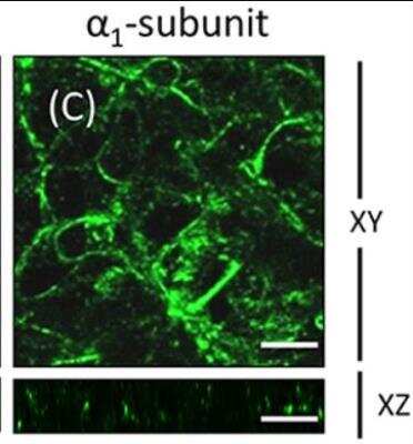
 [NB300-146] -")
Immunocytochemistry/ Immunofluorescence: Sodium Potassium ATPase Alpha 1 Antibody (464.6) [NB300-146] -
Immunocytochemistry/ Immunofluorescence: Sodium Potassium ATPase Alpha 1 Antibody (464.6) [NB300-146] - The cellular model of the blood-CSF barrier.(A) Schematic representation of a choroidal villus & of the experimental set up illustrating the two-chamber culture device. Left: the choroidal epithelium which forms the actual tight barrier controlling access into the CSF [18] delimits a stroma, in which the fenestrated vessels lacking a typical blood-brain barrier phenotype, express P-selectin [4]. Right: The epithelial cell monolayer grown on the lower side of the filter separates the upper chamber corresponding to the blood/stromal or basolateral space, from the bottom chamber representing the CSF or apical compartment (adapted from [18,41]. (B & C) Immunofluorescent staining of occludin (B) & claudin 1/3 (C) showing a typical intercellular distribution of the tight junction proteins in the confluent inverted monolayers of choroidal epithelial cells. (D) Immunofluorescent staining of Na+K+ ATPase & ABCC1, showing the expected respective apical & basolateral membrane localization in the choroidal epithelial cells. The left image is a close up of a single cell to better appreciate the polarity of distribution of the 2 proteins. Nuclei appear in blue. Arrows show the lateral cellular membranes best seen in the z direction by confocal analysis, arrowheads show the basal labeling of ABCC1. Image collected & cropped by CiteAb from the following publication (https://dx.plos.org/10.1371/journal.pone.0150945), licensed under a CC-BY license. Not internally tested by Novus Biologicals. [NB300-146] -")
Western Blot: Sodium Potassium ATPase Alpha 1 Antibody (464.6) [NB300-146] -
Western Blot: Sodium Potassium ATPase Alpha 1 Antibody (464.6) [NB300-146] - Endothelial-mesenchymal transformation (EMT) of RCECs using specific endothelial-mesenchymal transformation medium (SEMTM). (A) Immunohistochemistry of the fibroblastic markers alpha -SMA & vimentin were characteristic of fibroblasts in EMT-RCECs compared to the hexogonal morphology of Fresh RCECs (Scale bar upper panel = 100 µm, Scale bar middle & bottom panel = 50 µm). (B) RT-PCR showed that both Fresh RCECs & EMT-RCECs expressed alpha -Sma. Original gel is shown in Supplemental Fig. S2. However, qRT-PCR in the lower panel shows alpha -Sma was significantly upregulated in EMT-RCECs. (C) Western blots showed that Fresh RCECs & EMT-RCECs expressed Vimentin, Atp1a1, a marker of differentiated endothelial cells, & b-actin. Original gel & blots are shown in Supplemental Figs S3, 4. Semiquantitative analysis shows significant upregulation of the protein levels of Vimentin (p = 0.048) & downregulation of the protein level of Atp1a1 (p = 0.0043). Data expressed as mean ± SD of three replicate experiments. Student’s t test (*p < 0.05, **p < 0.01). (n = 3). RCECs: rabbit corneal endothelial cells, alpha -SMA: smooth muscle alpha -actin. Atp1a1: Na,K‐ATPase alpha ‐subunit. Image collected & cropped by CiteAb from the following publication (https://pubmed.ncbi.nlm.nih.gov/30442918), licensed under a CC-BY license. Not internally tested by Novus Biologicals. [NB300-146] -")
Western Blot: Sodium Potassium ATPase Alpha 1 Antibody (464.6) [NB300-146] -
Western Blot: Sodium Potassium ATPase Alpha 1 Antibody (464.6) [NB300-146] - SDS–PAGE western blot validation of the increased abundance of Annexin V, Na/K ATPase & alpha -tubulin in the EVs derived from TIS Cal51 cells compared with EVs derived from non-senescent non-treated controls. EVs isolated from TIS & control cells were immunoblotted to validate the protein expression of annexin V, Na/K ATPase & alpha -tubulin from the MS data. CD63 & HSP70 were used as loading controls. Densitometry was completed using Image J software (Fiji). All values are expressed as the mean of three independent experiments±s.e.m. The limit of ±1 skewness was set as normal distribution. Skewed data were transformed to Y=Log(Y) to fit a normal distribution. Results were analysed using the Student’s t-test with significant differences having a *P⩽0.05, **P⩽0.01, ***P⩽0.001. Image collected & cropped by CiteAb from the following publication (https://www.nature.com/articles/oncsis201782), licensed under a CC-BY license. Not internally tested by Novus Biologicals. [NB300-146] -")
Immunocytochemistry/ Immunofluorescence: Sodium Potassium ATPase Alpha 1 Antibody (464.6) [NB300-146] -
Immunocytochemistry/ Immunofluorescence: Sodium Potassium ATPase Alpha 1 Antibody (464.6) [NB300-146] - ARPE-19 cells cultured on transwell inserts for 4 weeks are not completely polarized. ARPE-19 cells were cultured up to 4 weeks on transwell inserts covered with laminin. The immunofluorescence image in (A) shows actin localization using rhodamine phalloidin. The cells are flat with stress fibers & very little circumferential actin microfilament bundles. The expression of CD147, a RPE marker, was detected using a specific antibody in the apical membrane domain (B). The expression of Na+, K+-ATPase using anti-alpha 1(C) & anti-beta 1 antibodies (D) was observed mostly at the basolateral membrane. Immunofluorescence detection with anti-human beta 2 antibody revealed a very weak signal (E). Scale bar: 10 μm. Image collected & cropped by CiteAb from the following publication (http://journal.frontiersin.org/article/10.3389/fphys.2016.00450/full), licensed under a CC-BY license. Not internally tested by Novus Biologicals.Applications for Sodium Potassium ATPase Alpha 1 Antibody (464.6) - BSA Free
Flow Cytometry
Immunocytochemistry/ Immunofluorescence
Immunohistochemistry
Immunohistochemistry-Frozen
Immunohistochemistry-Paraffin
Immunoprecipitation
Western Blot
Reviewed Applications
Read 4 reviews rated 4.3 using NB300-146 in the following applications:
Flow Cytometry Panel Builder
Bio-Techne Knows Flow Cytometry
Save time and reduce costly mistakes by quickly finding compatible reagents using the Panel Builder Tool.
Advanced Features
- Spectra Viewer - Custom analysis of spectra from multiple fluorochromes
- Spillover Popups - Visualize the spectra of individual fluorochromes
- Antigen Density Selector - Match fluorochrome brightness with antigen density

Formulation, Preparation, and Storage
Purification
Formulation
Format
Preservative
Concentration
Shipping
Stability & Storage
Background: Sodium Potassium ATPase Alpha 1
Long Name
Alternate Names
Gene Symbol
UniProt
Additional Sodium Potassium ATPase Alpha 1 Products
Product Documents for Sodium Potassium ATPase Alpha 1 Antibody (464.6) - BSA Free
Certificate of Analysis
To download a Certificate of Analysis, please enter a lot or batch number in the search box below.
Product Specific Notices for Sodium Potassium ATPase Alpha 1 Antibody (464.6) - BSA Free
This product is for research use only and is not approved for use in humans or in clinical diagnosis. Primary Antibodies are guaranteed for 1 year from date of receipt.
Citations for Sodium Potassium ATPase Alpha 1 Antibody (464.6) - BSA Free
 Powered by Bioz
Customer Reviews for Sodium Potassium ATPase Alpha 1 Antibody (464.6) - BSA Free (4)
Have you used Sodium Potassium ATPase Alpha 1 Antibody (464.6) - BSA Free?
Submit a review and receive an Amazon gift card!
$25/€18/£15/$25CAN/¥2500 Yen for a review with an image
$10/€7/£6/$10CAN/¥1110 Yen for a review without an image
Submit a review
Customer Images

-
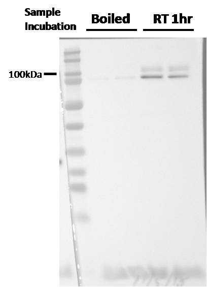
Application: Western BlotSample Tested: Human microglia cell lysateSpecies: HumanVerified Customer | Posted 02/08/2019NOT boiling the samples gives a much better signal.

-
Application: Western BlotSample Tested: Human cancer cellSpecies: HumanVerified Customer | Posted 08/22/2015
-
Application: Western BlotSample Tested: Human Postmortem prefrontal cortex tissueSpecies: HumanVerified Customer | Posted 08/21/2012
-
Application: Western BlotSample Tested: U87MG whole cell lysateSpecies: HumanVerified Customer | Posted 12/02/2010
There are no reviews that match your criteria.
Protocols
View specific protocols for Sodium Potassium ATPase Alpha 1 Antibody (464.6) - BSA Free (NB300-146):
Sample Preparation.
1. Grow cells to 60-85% confluency. Flow cytometry requires between 2 x 10⁵ and 1 x 10⁶ cells for optimal performance.
2. If cells are adherent, harvest gently by washing once with staining buffer and then scraping. Avoid using trypsin as this can disrupt certain epitopes of interest. If enzymatic harvest is required, use Accutase, Collagenase, or TrypLE Express for a less damaging option.
3. Reserve 100 uL for counting, then transfer cell volume into a 50 mL conical tube and centrifuge for 8 minutes at 400 RCF.
a. Count cells using a hemocytometer and a 1:1 trypan blue exclusion stain to determine cell viability before starting the flow protocol. If cells appear blue, do not proceed.
4. Re-suspend cells to a concentration of 1 x 10⁶ cells/mL in staining buffer (NBP2-26247).
5. Aliquot out 100 uL samples in accordance with your experimental samples.
Tip: When cell surface and intracellular staining are required in the same sample, it is advisable that the cell surface staining be performed first since the fixation and permeabilization steps might reduce the availability of surface antigens.
Intracellular Staining.
Tip: When performing intracellular staining, it is important to use appropriate fixation and permeabilization reagents based upon the target and its subcellular location. Generally, our Intracellular Flow Assay Kit (NBP2-29450) is a good place to start as it contains an optimized combination of reagents for intracellular staining as well as an inhibitor of intracellular protein transport (necessary if staining secreted proteins). Certain targets may require more gentle or transient permeabilization protocols such as the commonly employed methanol or saponin-based methods.
Protocol for Cytoplasmic Targets:
1. Fix the cells by adding 100 uL fixation solution (such as 4% PFA) to each sample for 10-15 minutes.
2. Permeabilize cells by adding 100 uL of a permeabilization buffer to every 1 x 10⁶ cells present in the sample. Mix well and incubate at room temperature for 15 minutes.
a. For cytoplasmic targets, use a gentle permeabilization solution such as 1X PBS + 0.5% Saponin or 1X PBS + 0.5% Tween-20.
b. To maintain the permeabilized state throughout your experiment, use staining buffer + 0.1% of the permeabilization reagent (i.e. 0.1% Tween-20 or 0.1% Saponin).
3. Following the 15 minute incubation, add 2 mL of the staining buffer + 0.1% permeabilizer to each sample.
4. Centrifuge for 1 minute at 400 RCF.
5. Discard supernatant and re-suspend in 100 uL of staining buffer + 0.1% permeabilizer.
6. Add appropriate amount of each antibody (eg. 1 test or 1 ug per sample, as experimentally determined).
7. Mix well and incubate at room temperature for 30 minutes- 1 hour. Gently mix samples every 10-15 minutes.
8. Following the primary/conjugate incubation, add 1-2 mL/sample of staining buffer +0.1% permeabilizer and centrifuge for 1 minute at 400 RCF.
9. Wash twice by re-suspending cells in staining buffer (2 mL for tubes or 200 uL for wells) and centrifuging at 400 RCF for 5 minutes. Discard supernatant.
10. Add appropriate amount of secondary antibody (as experimentally determined) to each sample.
11. Incubate at room temperature in dark for 20 minutes.
12. Add 1-2 mL of staining buffer and centrifuge at 400 RCF for 1 minute and discard supernatant.
13. Wash twice by re-suspending cells in staining buffer (2 mL for tubes or 200 uL for wells) and centrifuging at 400 RCF for 5 minutes. Discard supernatant.
14. Resuspend in an appropriate volume of staining buffer (usually 500 uL per sample) and proceed with analysis on your flow cytometer.
Culture cells to appropriate density in 35 mm culture dishes or 6-well plates.
1. Remove culture medium and wash the cells briefly in PBS. Add 10% formalin to the dish and fix at room temperature for 10 minutes.
2. Remove the formalin and wash the cells in PBS.
3. Permeablize the cells with 0.1% Triton X100 or other suitable detergent for 10 min.
4. Remove the permeablization buffer and wash three times for 10 minutes each in PBS. Be sure to not let the specimen dry out.
5. To block nonspecific antibody binding, incubate in 10% normal goat serum from 1 hour to overnight at room temperature.
6. Add primary antibody at appropriate dilution and incubate overnight at 4C.
7. Remove primary antibody and replace with PBS. Wash three times for 10 minutes each.
8. Add secondary antibody at appropriate dilution. Incubate for 1 hour at room temperature.
9. Remove secondary antibody and replace with PBS. Wash three times for 10 minutes each.
10. Counter stain DNA with DAPi if required.
Antigen Unmasking:
Bring slides to a boil in 10 mM sodium citrate buffer (pH 6.0) then maintain at a sub-boiling temperature for 10 minutes. Cool slides on bench-top for 30 minutes (keep slides in the sodium citrate buffer at all times).
Staining:
1. Wash sections in deionized water three times for 5 minutes each.
2. Wash sections in PBS for 5 minutes.
3. Block each section with 100-400 ul blocking solution (1% BSA in PBS) for 1 hour at room temperature.
4. Remove blocking solution and add 100-400 ul diluted primary antibody. Incubate overnight at 4 C.
5. Remove antibody solution and wash sections in wash buffer three times for 5 minutes each.
6. Add 100-400 ul HRP polymer conjugated secondary antibody. Incubate 30 minutes at room temperature.
7. Wash sections three times in wash buffer for 5 minutes each.
8. Add 100-400 ul DAB substrate to each section and monitor staining closely.
9. As soon as the sections develop, immerse slides in deionized water.
10. Counterstain sections in hematoxylin.
11. Wash sections in deionized water two times for 5 minutes each.
12. Dehydrate sections.
13. Mount coverslips.
Find general support by application which include: protocols, troubleshooting, illustrated assays, videos and webinars.
- 7-Amino Actinomycin D (7-AAD) Cell Viability Flow Cytometry Protocol
- Antigen Retrieval Protocol (PIER)
- Antigen Retrieval for Frozen Sections Protocol
- Appropriate Fixation of IHC/ICC Samples
- Cellular Response to Hypoxia Protocols
- Chromogenic IHC Staining of Formalin-Fixed Paraffin-Embedded (FFPE) Tissue Protocol
- Chromogenic Immunohistochemistry Staining of Frozen Tissue
- ClariTSA™ Fluorophore Kits
- Detection & Visualization of Antibody Binding
- Extracellular Membrane Flow Cytometry Protocol
- Flow Cytometry Protocol for Cell Surface Markers
- Flow Cytometry Protocol for Staining Membrane Associated Proteins
- Flow Cytometry Staining Protocols
- Flow Cytometry Troubleshooting Guide
- Fluorescent IHC Staining of Frozen Tissue Protocol
- Graphic Protocol for Heat-induced Epitope Retrieval
- Graphic Protocol for the Preparation and Fluorescent IHC Staining of Frozen Tissue Sections
- Graphic Protocol for the Preparation and Fluorescent IHC Staining of Paraffin-embedded Tissue Sections
- Graphic Protocol for the Preparation of Gelatin-coated Slides for Histological Tissue Sections
- ICC Cell Smear Protocol for Suspension Cells
- ICC Immunocytochemistry Protocol Videos
- ICC for Adherent Cells
- IHC Sample Preparation (Frozen sections vs Paraffin)
- Immunocytochemistry (ICC) Protocol
- Immunocytochemistry Troubleshooting
- Immunofluorescence of Organoids Embedded in Cultrex Basement Membrane Extract
- Immunofluorescent IHC Staining of Formalin-Fixed Paraffin-Embedded (FFPE) Tissue Protocol
- Immunohistochemistry (IHC) and Immunocytochemistry (ICC) Protocols
- Immunohistochemistry Frozen Troubleshooting
- Immunohistochemistry Paraffin Troubleshooting
- Immunoprecipitation Protocol
- Intracellular Flow Cytometry Protocol Using Alcohol (Methanol)
- Intracellular Flow Cytometry Protocol Using Detergents
- Intracellular Nuclear Staining Flow Cytometry Protocol Using Detergents
- Intracellular Staining Flow Cytometry Protocol Using Alcohol Permeabilization
- Intracellular Staining Flow Cytometry Protocol Using Detergents to Permeabilize Cells
- Preparing Samples for IHC/ICC Experiments
- Preventing Non-Specific Staining (Non-Specific Binding)
- Primary Antibody Selection & Optimization
- Propidium Iodide Cell Viability Flow Cytometry Protocol
- Protocol for Heat-Induced Epitope Retrieval (HIER)
- Protocol for Liperfluo
- Protocol for Making a 4% Formaldehyde Solution in PBS
- Protocol for VisUCyte™ HRP Polymer Detection Reagent
- Protocol for the Characterization of Human Th22 Cells
- Protocol for the Characterization of Human Th9 Cells
- Protocol for the Fluorescent ICC Staining of Cell Smears - Graphic
- Protocol for the Fluorescent ICC Staining of Cultured Cells on Coverslips - Graphic
- Protocol for the Preparation & Fixation of Cells on Coverslips
- Protocol for the Preparation and Chromogenic IHC Staining of Frozen Tissue Sections
- Protocol for the Preparation and Chromogenic IHC Staining of Frozen Tissue Sections - Graphic
- Protocol for the Preparation and Chromogenic IHC Staining of Paraffin-embedded Tissue Sections
- Protocol for the Preparation and Chromogenic IHC Staining of Paraffin-embedded Tissue Sections - Graphic
- Protocol for the Preparation and Fluorescent ICC Staining of Cells on Coverslips
- Protocol for the Preparation and Fluorescent ICC Staining of Non-adherent Cells
- Protocol for the Preparation and Fluorescent ICC Staining of Stem Cells on Coverslips
- Protocol for the Preparation and Fluorescent IHC Staining of Frozen Tissue Sections
- Protocol for the Preparation and Fluorescent IHC Staining of Paraffin-embedded Tissue Sections
- Protocol for the Preparation of Gelatin-coated Slides for Histological Tissue Sections
- Protocol for the Preparation of a Cell Smear for Non-adherent Cell ICC - Graphic
- Protocol: Annexin V and PI Staining by Flow Cytometry
- Protocol: Annexin V and PI Staining for Apoptosis by Flow Cytometry
- R&D Systems Quality Control Western Blot Protocol
- TUNEL and Active Caspase-3 Detection by IHC/ICC Protocol
- The Importance of IHC/ICC Controls
- Troubleshooting Guide: Fluorokine Flow Cytometry Kits
- Troubleshooting Guide: Immunohistochemistry
- Troubleshooting Guide: Western Blot Figures
- Western Blot Conditions
- Western Blot Protocol
- Western Blot Protocol for Cell Lysates
- Western Blot Troubleshooting
- Western Blot Troubleshooting Guide
- View all Protocols, Troubleshooting, Illustrated assays and Webinars
FAQs for Sodium Potassium ATPase Alpha 1 Antibody (464.6) - BSA Free
-
Q: I am looking forward to pick up a plasma membrane marker. I have gastric cancer cell line and I am preparing PM out of that. To just check the quality of prep, I need a plasma membrane marker.
A:
Our sodium potassium ATPase and plasma membrane products may be of use to you. Please contact us at technical@novusbio.com with any additional questions.
-
Q: I would like to know whether it is possible to dilute this antibody in 50% glycerol final, and keep the aliquots at -20 degrees Celsius?
A: As far as the storage conditions are concerned, since this antibody is supplied as ascites and may contain proteases, it has to be frozen immediately at -20 or -80. From a technical perspective, adding glycerol as a cryoprotectant to avoid freeze/thaw damage should be acceptable; however, we suggest to store our antibodies as recommended in the datasheet which in this case it is better to aliquot the antibody in small volumes and store it at -20 or -80. We do supply some antibodies that we recommend adding glycerol but NB300-146 has not been tested in this storage condition.
-
Q: Was the epitope characterized?
A: We unfortunately have not mapped the epitope or binding region for this product yet. Our lab will always include 0.1% Tween-20 in their diluent and wash buffers for ICC staining, so I would recommend using this detergent for your customers staining. If they want to try first without the detergent, that is fine as well.
-
Q: I am looking forward to pick up a plasma membrane marker. I have gastric cancer cell line and I am preparing PM out of that. To just check the quality of prep, I need a plasma membrane marker.
A:
Our sodium potassium ATPase and plasma membrane products may be of use to you. Please contact us at technical@novusbio.com with any additional questions.
-
Q: I would like to know whether it is possible to dilute this antibody in 50% glycerol final, and keep the aliquots at -20 degrees Celsius?
A: As far as the storage conditions are concerned, since this antibody is supplied as ascites and may contain proteases, it has to be frozen immediately at -20 or -80. From a technical perspective, adding glycerol as a cryoprotectant to avoid freeze/thaw damage should be acceptable; however, we suggest to store our antibodies as recommended in the datasheet which in this case it is better to aliquot the antibody in small volumes and store it at -20 or -80. We do supply some antibodies that we recommend adding glycerol but NB300-146 has not been tested in this storage condition.
-
Q: Was the epitope characterized?
A: We unfortunately have not mapped the epitope or binding region for this product yet. Our lab will always include 0.1% Tween-20 in their diluent and wash buffers for ICC staining, so I would recommend using this detergent for your customers staining. If they want to try first without the detergent, that is fine as well.