SSEA-3 Antibody (MC-631) - BSA Free
Novus Biologicals | Catalog # NB100-1832
![Immunocytochemistry/ Immunofluorescence: SSEA-3 Antibody (MC-631) [NB100-1832]](https://resources.rndsystems.com/images/products/SSEA-3-Antibody-MC-631-Immunocytochemistry-Immunofluorescence-NB100-1832-img0016.jpg "Immunocytochemistry/ Immunofluorescence: SSEA-3 Antibody (MC-631) [NB100-1832]")
Key Product Details
Species Reactivity
Validated:
Cited:
Applications
Validated:
Cited:
Label
Antibody Source
Format
Product Specifications
Immunogen
Reactivity Notes
Marker
Specificity
Clonality
Host
Isotype
Scientific Data Images for SSEA-3 Antibody (MC-631) - BSA Free
Immunocytochemistry/ Immunofluorescence: SSEA-3 Antibody (MC-631) [NB100-1832]
SSEA-3-Antibody-MC-631-Immunocytochemistry-Immunofluorescence-NB100-1832-img0016.jpg![Flow Cytometry: SSEA-3 Antibody (MC-631) [NB100-1832]](https://resources.rndsystems.com/images/products/SSEA-3-Antibody-MC-631-Flow-Cytometry-NB100-1832-img0013.jpg "Flow Cytometry: SSEA-3 Antibody (MC-631) [NB100-1832]")
Flow Cytometry: SSEA-3 Antibody (MC-631) [NB100-1832]
Flow Cytometry: SSEA-3 Antibody (MC-631) [NB100-1832] - A surface stain was performed on Jurkat cells with SSEA-3 Antibody (MC-631) NB100-1832AF647 (blue) and a matched isotype control (orange). Cells were incubated in an antibody dilution of 5 ug/mL for 20 minutes at room temperature. Both antibodies were conjugated to Alexa Fluor 647.![Immunocytochemistry/ Immunofluorescence: SSEA-3 Antibody (MC-631) [NB100-1832]](https://resources.rndsystems.com/images/products/SSEA-3-Antibody-MC-631-Immunocytochemistry-Immunofluorescence-NB100-1832-img0012.jpg "Immunocytochemistry/ Immunofluorescence: SSEA-3 Antibody (MC-631) [NB100-1832]")
Immunocytochemistry/ Immunofluorescence: SSEA-3 Antibody (MC-631) [NB100-1832]
Immunocytochemistry/Immunofluorescence: SSEA-3 Antibody (MC-631) [NB100-1832] - Analysis of human embryonic stem cells stained with SSEA-3 antibody detected with Alexa Fluor 488 anti-rat IgM secondary antibody. - BSA Free [NB100-1832] -")
Immunocytochemistry/ Immunofluorescence: SSEA-3 Antibody (MC-631) - BSA Free [NB100-1832] -
PSC characterisation. (A) Human PSC-1 characterized for A) typical undifferentiated colony morphology in phase contrast image and (B) expression of pluripotency markers Nanog, OCT-3/4, SSEA-3, SSEA-4, TRA-1-60, and TRA-1-81, and lack of expression of early differentiation marker SSEA-1 after immunofluorescence staining. Corresponding nuclei stains with DAPI shown. (C) Cells showed normal female (46, XX) karyotype after 28 passages in total (9 passages in feeder-free culture). The results of the KaryoLite BoBs assay are shown as signal relative to karyotypically normal female (/F, red) and male (/M, blue) genomic DNA used as a reference (equal to 1) for each of the probes covering both p and q arms of all chromosomes. Software threshold for changes shown as a green lines and deviations in red. (D) Pluripotency shown after spontaneous differentiation in vitro as expression of markers for mesoderm, endoderm, and ectoderm. All scale bars 200 um. Image collected and cropped by CiteAb from the following open publication (https://pubmed.ncbi.nlm.nih.gov/30341351), licensed under a CC-BY license. Not internally tested by Novus Biologicals.Applications for SSEA-3 Antibody (MC-631) - BSA Free
Flow Cytometry
Immunocytochemistry/ Immunofluorescence
Immunohistochemistry
Immunohistochemistry-Frozen
Immunohistochemistry-Paraffin
Immunoprecipitation
Western Blot
Reviewed Applications
Read 1 review rated 5 using NB100-1832 in the following applications:
Flow Cytometry Panel Builder
Bio-Techne Knows Flow Cytometry
Save time and reduce costly mistakes by quickly finding compatible reagents using the Panel Builder Tool.
Advanced Features
- Spectra Viewer - Custom analysis of spectra from multiple fluorochromes
- Spillover Popups - Visualize the spectra of individual fluorochromes
- Antigen Density Selector - Match fluorochrome brightness with antigen density

Formulation, Preparation, and Storage
Purification
Formulation
Format
Preservative
Concentration
Shipping
Stability & Storage
Background: SSEA-3
Long Name
Alternate Names
Additional SSEA-3 Products
Product Documents for SSEA-3 Antibody (MC-631) - BSA Free
Certificate of Analysis
To download a Certificate of Analysis, please enter a lot or batch number in the search box below.
Product Specific Notices for SSEA-3 Antibody (MC-631) - BSA Free
This product is for research use only and is not approved for use in humans or in clinical diagnosis. Primary Antibodies are guaranteed for 1 year from date of receipt.
Citations for SSEA-3 Antibody (MC-631) - BSA Free
 Powered by Bioz
Customer Reviews for SSEA-3 Antibody (MC-631) - BSA Free (1)
Have you used SSEA-3 Antibody (MC-631) - BSA Free?
Submit a review and receive an Amazon gift card!
$25/€18/£15/$25CAN/¥2500 Yen for a review with an image
$10/€7/£6/$10CAN/¥1110 Yen for a review without an image
Submit a review
Customer Images
-
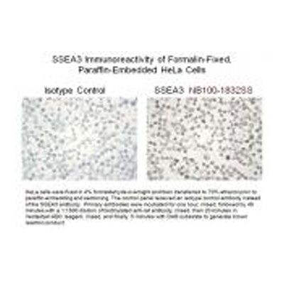
Application: Immunohistochemistry-ParaffinSample Tested: HeLa cellsSpecies: HumanVerified Customer | Posted 08/07/2010

There are no reviews that match your criteria.
Protocols
View specific protocols for SSEA-3 Antibody (MC-631) - BSA Free (NB100-1832):
Immunocytochemistry Protocol
Culture cells to appropriate density in 35 mm culture dishes or 6-well plates.
1. Remove culture medium and add 10% formalin to the dish. Fix at room temperature for 30 minutes.
2. Remove the formalin and add ice cold methanol. Incubate for 5-10 minutes.
3. Remove methanol and add washing solution (i.e. PBS). Be sure to not let the specimen dry out. Wash three times for 10 minutes.
4. To block nonspecific antibody binding incubate in 10% normal goat serum from 1 hour to overnight at room temperature.
5. Add primary antibody at appropriate dilution and incubate at room temperature from 2 hours to overnight at room temperature.
6. Remove primary antibody and replace with washing solution. Wash three times for 10 minutes.
7. Add secondary antibody at appropriate dilution. Incubate for 1 hour at room temperature.
8. Remove antibody and replace with wash solution, then wash for 10 minutes. Add Hoechst 33258 to wash solution at 1:25,0000 and incubate for 10 minutes. Wash a third time for 10 minutes.
9. Cells can be viewed directly after washing. The plates can also be stored in PBS containing Azide covered in Parafilm (TM). Cells can also be cover-slipped using Fluoromount, with appropriate sealing.
*The above information is only intended as a guide. The researcher should determine what protocol best meets their needs. Please follow safe laboratory procedures.
Find general support by application which include: protocols, troubleshooting, illustrated assays, videos and webinars.
- 7-Amino Actinomycin D (7-AAD) Cell Viability Flow Cytometry Protocol
- Antigen Retrieval Protocol (PIER)
- Antigen Retrieval for Frozen Sections Protocol
- Appropriate Fixation of IHC/ICC Samples
- Cellular Response to Hypoxia Protocols
- Chromogenic IHC Staining of Formalin-Fixed Paraffin-Embedded (FFPE) Tissue Protocol
- Chromogenic Immunohistochemistry Staining of Frozen Tissue
- ClariTSA™ Fluorophore Kits
- Detection & Visualization of Antibody Binding
- Extracellular Membrane Flow Cytometry Protocol
- Flow Cytometry Protocol for Cell Surface Markers
- Flow Cytometry Protocol for Staining Membrane Associated Proteins
- Flow Cytometry Staining Protocols
- Flow Cytometry Troubleshooting Guide
- Fluorescent IHC Staining of Frozen Tissue Protocol
- Graphic Protocol for Heat-induced Epitope Retrieval
- Graphic Protocol for the Preparation and Fluorescent IHC Staining of Frozen Tissue Sections
- Graphic Protocol for the Preparation and Fluorescent IHC Staining of Paraffin-embedded Tissue Sections
- Graphic Protocol for the Preparation of Gelatin-coated Slides for Histological Tissue Sections
- ICC Cell Smear Protocol for Suspension Cells
- ICC Immunocytochemistry Protocol Videos
- ICC for Adherent Cells
- IHC Sample Preparation (Frozen sections vs Paraffin)
- Immunocytochemistry (ICC) Protocol
- Immunocytochemistry Troubleshooting
- Immunofluorescence of Organoids Embedded in Cultrex Basement Membrane Extract
- Immunofluorescent IHC Staining of Formalin-Fixed Paraffin-Embedded (FFPE) Tissue Protocol
- Immunohistochemistry (IHC) and Immunocytochemistry (ICC) Protocols
- Immunohistochemistry Frozen Troubleshooting
- Immunohistochemistry Paraffin Troubleshooting
- Immunoprecipitation Protocol
- Intracellular Flow Cytometry Protocol Using Alcohol (Methanol)
- Intracellular Flow Cytometry Protocol Using Detergents
- Intracellular Nuclear Staining Flow Cytometry Protocol Using Detergents
- Intracellular Staining Flow Cytometry Protocol Using Alcohol Permeabilization
- Intracellular Staining Flow Cytometry Protocol Using Detergents to Permeabilize Cells
- Preparing Samples for IHC/ICC Experiments
- Preventing Non-Specific Staining (Non-Specific Binding)
- Primary Antibody Selection & Optimization
- Propidium Iodide Cell Viability Flow Cytometry Protocol
- Protocol for Heat-Induced Epitope Retrieval (HIER)
- Protocol for Liperfluo
- Protocol for Making a 4% Formaldehyde Solution in PBS
- Protocol for VisUCyte™ HRP Polymer Detection Reagent
- Protocol for the Characterization of Human Th22 Cells
- Protocol for the Characterization of Human Th9 Cells
- Protocol for the Fluorescent ICC Staining of Cell Smears - Graphic
- Protocol for the Fluorescent ICC Staining of Cultured Cells on Coverslips - Graphic
- Protocol for the Preparation & Fixation of Cells on Coverslips
- Protocol for the Preparation and Chromogenic IHC Staining of Frozen Tissue Sections
- Protocol for the Preparation and Chromogenic IHC Staining of Frozen Tissue Sections - Graphic
- Protocol for the Preparation and Chromogenic IHC Staining of Paraffin-embedded Tissue Sections
- Protocol for the Preparation and Chromogenic IHC Staining of Paraffin-embedded Tissue Sections - Graphic
- Protocol for the Preparation and Fluorescent ICC Staining of Cells on Coverslips
- Protocol for the Preparation and Fluorescent ICC Staining of Non-adherent Cells
- Protocol for the Preparation and Fluorescent ICC Staining of Stem Cells on Coverslips
- Protocol for the Preparation and Fluorescent IHC Staining of Frozen Tissue Sections
- Protocol for the Preparation and Fluorescent IHC Staining of Paraffin-embedded Tissue Sections
- Protocol for the Preparation of Gelatin-coated Slides for Histological Tissue Sections
- Protocol for the Preparation of a Cell Smear for Non-adherent Cell ICC - Graphic
- Protocol: Annexin V and PI Staining by Flow Cytometry
- Protocol: Annexin V and PI Staining for Apoptosis by Flow Cytometry
- R&D Systems Quality Control Western Blot Protocol
- TUNEL and Active Caspase-3 Detection by IHC/ICC Protocol
- The Importance of IHC/ICC Controls
- Troubleshooting Guide: Fluorokine Flow Cytometry Kits
- Troubleshooting Guide: Immunohistochemistry
- Troubleshooting Guide: Western Blot Figures
- Western Blot Conditions
- Western Blot Protocol
- Western Blot Protocol for Cell Lysates
- Western Blot Troubleshooting
- Western Blot Troubleshooting Guide
- View all Protocols, Troubleshooting, Illustrated assays and Webinars
FAQs for SSEA-3 Antibody (MC-631) - BSA Free
-
Q: Could you please let us know the accession number of the antigen protein for SSEA3 Antibody (Cat. No. NB100-1832)?
A: SSEA3 is a globoseries carbohydrate antigen found on glycolipids as well as glycopeptides on cellular surface of embryonic stem cells. Because it is not a protein in reality, it is not supposed to have an accession number. NB100-1832 was generated in rat host using murine 4-8 cell stage embryos as immunogen and this antibody specifically detects the SSEA3 antigen in various immunoassays.