![Western Blot: SSR2 Antibody (31G6)BSA Free [NBP2-42647]](https://resources.rndsystems.com/images/products/SSR2-Antibody-31G6-Western-Blot-NBP2-42647-img0001.jpg "Western Blot: SSR2 Antibody (31G6)BSA Free [NBP2-42647]")
Key Product Details
Species Reactivity
Human
Applications
Western Blot, ELISA, Immunocytochemistry/ Immunofluorescence
Label
Unconjugated
Antibody Source
Monoclonal Mouse IgG1 kappa Clone # 31G6
Format
BSA Free
Loading...
Product Specifications
Immunogen
Recombinant human SSR2 (18-149aa) purified from E. coli
Clonality
Monoclonal
Host
Mouse
Isotype
IgG1 kappa
Scientific Data Images for SSR2 Antibody (31G6) - BSA Free
Western Blot: SSR2 Antibody (31G6)BSA Free [NBP2-42647]
Western Blot: SSR2 Antibody (31G6) [NBP2-42647] - The lysate of Jurkat (40ug) were resolved by SDS-PAGE, transferred to PVDF membrane and probed with anti-human SSR2 antibody (1:500). Proteins were visualized using a goat anti-mouse secondary antibody conjugated to HRP and an ECL detection system.![Immunocytochemistry/ Immunofluorescence: SSR2 Antibody (31G6) - BSA Free [NBP2-42647]](https://resources.rndsystems.com/images/products/SSR2-Antibody-31G6-BSA-Free-Immunocytochemistry-Immunofluorescence-NBP2-42647-img0002.jpg "Immunocytochemistry/ Immunofluorescence: SSR2 Antibody (31G6) - BSA Free [NBP2-42647]")
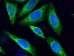
Immunocytochemistry/ Immunofluorescence: SSR2 Antibody (31G6) - BSA Free [NBP2-42647]
Immunocytochemistry/Immunofluorescence: SSR2 Antibody (31G6) - BSA Free [NBP2-42647] - Analysis of HepG2 cells using SSR2 antibody (31G6). Primary antibody dilution: 1:100. Image from verified customer review.Applications for SSR2 Antibody (31G6) - BSA Free
Application
Recommended Usage
Western Blot
1:500
Reviewed Applications
Read 1 review rated 5 using NBP2-42647 in the following applications:
Formulation, Preparation, and Storage
Purification
Protein A purified
Formulation
PBS (pH 7.4), 10% Glycerol
Format
BSA Free
Preservative
0.02% Sodium Azide
Concentration
1 mg/ml
Shipping
The product is shipped with polar packs. Upon receipt, store it immediately at the temperature recommended below.
Stability & Storage
Store at 4C short term. Aliquot and store at -20C long term. Avoid freeze-thaw cycles.
Background: SSR2
Alternate Names
DKFZp686F19123, Signal sequence receptor subunit beta, signal sequence receptor, beta (translocon-associated protein beta), SSR-beta, TLAP, translocon-associated protein beta, translocon-associated protein subunit beta, TRAP-beta, TRAPBTRAP-BETA
Gene Symbol
SSR2
UniProt
Additional SSR2 Products
Product Documents for SSR2 Antibody (31G6) - BSA Free
Certificate of Analysis
To download a Certificate of Analysis, please enter a lot or batch number in the search box below.
Product Specific Notices for SSR2 Antibody (31G6) - BSA Free
This product is for research use only and is not approved for use in humans or in clinical diagnosis. Primary Antibodies are guaranteed for 1 year from date of receipt.
Customer Reviews for SSR2 Antibody (31G6) - BSA Free (1)
5 out of 5
1 Customer Rating
Have you used SSR2 Antibody (31G6) - BSA Free?
Submit a review and receive an Amazon gift card!
$25/€18/£15/$25CAN/¥2500 Yen for a review with an image
$10/€7/£6/$10CAN/¥1110 Yen for a review without an image
Submit a review
Customer Images
Showing
1
-
1 of
1 review
Showing All
Filter By:
-
Application: ImmunocytochemistrySample Tested: HepG2 cellsSpecies: HumanVerified Customer | Posted 04/05/2022HepG2 cells. The dilution was 1:100.

There are no reviews that match your criteria.
Protocols
Find general support by application which include: protocols, troubleshooting, illustrated assays, videos and webinars.
- Appropriate Fixation of IHC/ICC Samples
- Cellular Response to Hypoxia Protocols
- ClariTSA™ Fluorophore Kits
- Detection & Visualization of Antibody Binding
- ELISA Sample Preparation & Collection Guide
- ELISA Troubleshooting Guide
- How to Run an R&D Systems DuoSet ELISA
- How to Run an R&D Systems Quantikine ELISA
- How to Run an R&D Systems Quantikine™ QuicKit™ ELISA
- ICC Cell Smear Protocol for Suspension Cells
- ICC Immunocytochemistry Protocol Videos
- ICC for Adherent Cells
- Immunocytochemistry (ICC) Protocol
- Immunocytochemistry Troubleshooting
- Immunofluorescence of Organoids Embedded in Cultrex Basement Membrane Extract
- Immunohistochemistry (IHC) and Immunocytochemistry (ICC) Protocols
- Preparing Samples for IHC/ICC Experiments
- Preventing Non-Specific Staining (Non-Specific Binding)
- Primary Antibody Selection & Optimization
- Protocol for VisUCyte™ HRP Polymer Detection Reagent
- Protocol for the Fluorescent ICC Staining of Cell Smears - Graphic
- Protocol for the Fluorescent ICC Staining of Cultured Cells on Coverslips - Graphic
- Protocol for the Preparation and Fluorescent ICC Staining of Cells on Coverslips
- Protocol for the Preparation and Fluorescent ICC Staining of Non-adherent Cells
- Protocol for the Preparation and Fluorescent ICC Staining of Stem Cells on Coverslips
- Protocol for the Preparation of a Cell Smear for Non-adherent Cell ICC - Graphic
- Quantikine HS ELISA Kit Assay Principle, Alkaline Phosphatase
- Quantikine HS ELISA Kit Principle, Streptavidin-HRP Polymer
- R&D Systems Quality Control Western Blot Protocol
- Sandwich ELISA (Colorimetric) – Biotin/Streptavidin Detection Protocol
- Sandwich ELISA (Colorimetric) – Direct Detection Protocol
- TUNEL and Active Caspase-3 Detection by IHC/ICC Protocol
- The Importance of IHC/ICC Controls
- Troubleshooting Guide: ELISA
- Troubleshooting Guide: Western Blot Figures
- Western Blot Conditions
- Western Blot Protocol
- Western Blot Protocol for Cell Lysates
- Western Blot Troubleshooting
- Western Blot Troubleshooting Guide
- View all Protocols, Troubleshooting, Illustrated assays and Webinars
Loading...