Survivin [p Ser20] Antibody - BSA Free
Novus Biologicals | Catalog # NB110-92717
Key Product Details
Species Reactivity
Validated:
Human, Mouse, Rat
Cited:
Human, Mouse, Rat
Applications
Validated:
Immunohistochemistry, Western Blot, Immunocytochemistry/ Immunofluorescence
Cited:
Western Blot, IF/IHC
Label
Unconjugated
Antibody Source
Polyclonal Rabbit IgG
Format
BSA Free
Loading...
Product Specifications
Immunogen
This Survivin [p Ser20] Antibody was developed against a synthetic peptide surrounding the phosphorylated serine 20 of the human Survivin protein. [Swiss-Prot #O15392]
Reactivity Notes
Mouse reactivity reported in scientific literature (PMID: 22814318). Rat reactivity reported in scientific literature (PMID: 17612487). Human reactivity reported in scientific literature (PMID: 24069188).
Modification
p Ser20
Localization
Cytoplasm and Mitochondria.
Clonality
Polyclonal
Host
Rabbit
Isotype
IgG
Theoretical MW
16 kDa.
Disclaimer note: The observed molecular weight of the protein may vary from the listed predicted molecular weight due to post translational modifications, post translation cleavages, relative charges, and other experimental factors.
Disclaimer note: The observed molecular weight of the protein may vary from the listed predicted molecular weight due to post translational modifications, post translation cleavages, relative charges, and other experimental factors.
Scientific Data Images for Survivin [p Ser20] Antibody - BSA Free
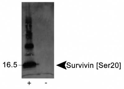
Western Blot: Survivin [p Ser20] Antibody [NB110-92717] - Detection of Survivin [Ser20] in phosphorylated recombinant protein (+), but not in unphosphorylated protein (-) using [NB110-92717]. Band detected at slightly higher than the predicted molecular weight of 16 kDa.
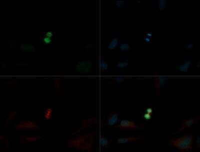
Immunocytochemistry/Immunofluorescence: Survivin [p Ser20] Antibody [NB110-92717] - Survivin [p Ser20] Antibody (NB110-92717) was tested in Hela cells with DyLight 488 (green). Nuclei and alpha-tubulin were counterstained with DAPI (blue) and DyLight 550 (red).
![Survivin [p Ser20] Antibody - BSA Free](https://resources.rndsystems.com/images/products/nb110-92717_rabbit-polyclonal-survivin-p-ser20-antibody-western-blot-132202613165617.jpg "Western Blot: Survivin [p Ser20] Antibody - BSA Free [NB110-92717] -")
Western Blot: Survivin [p Ser20] Antibody - BSA Free [NB110-92717] -
Silencing or inhibition of PLK1 and AURKB modulates survivin phosphorylation at S20 and T117 in AA TNBC cells.A, B Immunoblots showing the levels of PLK1, survivin, p-survivin (S20), and beta -actin after PLK1 silencing (A, B) or inhibition (C) in AA and EA TNBC cell lines. D–F Immunoblots showing the levels of AURKB, survivin, p-survivin (T117), and beta -actin after AURKB silencing (D, E), or inhibition (F) in AA and EA TNBC cell lines. Image collected and cropped by CiteAb from the following open publication (https://pubmed.ncbi.nlm.nih.gov/36627281), licensed under a CC-BY license. Not internally tested by Novus Biologicals.![Survivin [p Ser20] Antibody - BSA Free](https://resources.rndsystems.com/images/products/nb110-92717_rabbit-polyclonal-survivin-p-ser20-antibody-western-blot-13220261213181.jpg "Western Blot: Survivin [p Ser20] Antibody - BSA Free [NB110-92717] -")
Western Blot: Survivin [p Ser20] Antibody - BSA Free [NB110-92717] -
CPC complex formation is highest in S20-T117 double phospho-mimic survivin mutants.A Schematic representation of survivin-mutant plasmids. B–D Immunoblots (B, C) showing the levels of CPC proteins in input (B) and IP-bound (C) samples from cells expressing various survivin-WT and mutant plasmids, and their respective quantification (D). E Bar graphs showing the percentage of cell proliferation in control cells and in cells expressing survivin-WT and mutant plasmids. F Schematic illustration of YM155 treatment schedule in mice bearing tumors and surgically implanted with osmotic pumps. G–J Representative tumor images (G), changes in tumor volume (H), and changes in tumor size (I, J) in mice bearing AA (n = 12) and EA (n = 12) TNBC xenografts. Bars represent mean +/- SEM. Unpaired two-tailed Student’s t-test with Welch’s correction was used to determine statistical significance (*P < 0.05, **P < 0.005, ns = non-significant). Image collected and cropped by CiteAb from the following open publication (https://pubmed.ncbi.nlm.nih.gov/36627281), licensed under a CC-BY license. Not internally tested by Novus Biologicals.![Survivin [p Ser20] Antibody - BSA Free](https://resources.rndsystems.com/images/products/nb110-92717_rabbit-polyclonal-survivin-p-ser20-antibody-western-blot-132202613165616.jpg "Western Blot: Survivin [p Ser20] Antibody - BSA Free [NB110-92717] -")
Western Blot: Survivin [p Ser20] Antibody - BSA Free [NB110-92717] -
Silencing or inhibition of PLK1 and AURKB modulates survivin phosphorylation at S20 and T117 in AA TNBC cells.A, B Immunoblots showing the levels of PLK1, survivin, p-survivin (S20), and beta -actin after PLK1 silencing (A, B) or inhibition (C) in AA and EA TNBC cell lines. D–F Immunoblots showing the levels of AURKB, survivin, p-survivin (T117), and beta -actin after AURKB silencing (D, E), or inhibition (F) in AA and EA TNBC cell lines. Image collected and cropped by CiteAb from the following open publication (https://pubmed.ncbi.nlm.nih.gov/36627281), licensed under a CC-BY license. Not internally tested by Novus Biologicals.![Survivin [p Ser20] Antibody - BSA Free](https://resources.rndsystems.com/images/products/nb110-92717_rabbit-polyclonal-survivin-p-ser20-antibody-western-blot-13220261316563.jpg "Western Blot: Survivin [p Ser20] Antibody - BSA Free [NB110-92717] -")
Western Blot: Survivin [p Ser20] Antibody - BSA Free [NB110-92717] -
Silencing or inhibition of PLK1 and AURKB modulates survivin phosphorylation at S20 and T117 in AA TNBC cells.A, B Immunoblots showing the levels of PLK1, survivin, p-survivin (S20), and beta -actin after PLK1 silencing (A, B) or inhibition (C) in AA and EA TNBC cell lines. D–F Immunoblots showing the levels of AURKB, survivin, p-survivin (T117), and beta -actin after AURKB silencing (D, E), or inhibition (F) in AA and EA TNBC cell lines. Image collected and cropped by CiteAb from the following open publication (https://pubmed.ncbi.nlm.nih.gov/36627281), licensed under a CC-BY license. Not internally tested by Novus Biologicals.![Survivin [p Ser20] Antibody - BSA Free](https://resources.rndsystems.com/images/products/nb110-92717_rabbit-polyclonal-survivin-p-ser20-antibody-western-blot-132202613153818.jpg "Western Blot: Survivin [p Ser20] Antibody - BSA Free [NB110-92717] -")
Western Blot: Survivin [p Ser20] Antibody - BSA Free [NB110-92717] -
Silencing or inhibition of PLK1 and AURKB modulates survivin phosphorylation at S20 and T117 in AA TNBC cells.A, B Immunoblots showing the levels of PLK1, survivin, p-survivin (S20), and beta -actin after PLK1 silencing (A, B) or inhibition (C) in AA and EA TNBC cell lines. D–F Immunoblots showing the levels of AURKB, survivin, p-survivin (T117), and beta -actin after AURKB silencing (D, E), or inhibition (F) in AA and EA TNBC cell lines. Image collected and cropped by CiteAb from the following open publication (https://pubmed.ncbi.nlm.nih.gov/36627281), licensed under a CC-BY license. Not internally tested by Novus Biologicals.![Survivin [p Ser20] Antibody - BSA Free](https://resources.rndsystems.com/images/products/nb110-92717_rabbit-polyclonal-survivin-p-ser20-antibody-western-blot-132202612254924.jpg "Western Blot: Survivin [p Ser20] Antibody - BSA Free [NB110-92717] -")
Western Blot: Survivin [p Ser20] Antibody - BSA Free [NB110-92717] -
Silencing or inhibition of PLK1 and AURKB modulates survivin phosphorylation at S20 and T117 in AA TNBC cells.A, B Immunoblots showing the levels of PLK1, survivin, p-survivin (S20), and beta -actin after PLK1 silencing (A, B) or inhibition (C) in AA and EA TNBC cell lines. D–F Immunoblots showing the levels of AURKB, survivin, p-survivin (T117), and beta -actin after AURKB silencing (D, E), or inhibition (F) in AA and EA TNBC cell lines. Image collected and cropped by CiteAb from the following open publication (https://pubmed.ncbi.nlm.nih.gov/36627281), licensed under a CC-BY license. Not internally tested by Novus Biologicals.![Survivin [p Ser20] Antibody - BSA Free](https://resources.rndsystems.com/images/products/nb110-92717_rabbit-polyclonal-survivin-p-ser20-antibody-western-blot-1322026132588.jpg "Western Blot: Survivin [p Ser20] Antibody - BSA Free [NB110-92717] -")
Western Blot: Survivin [p Ser20] Antibody - BSA Free [NB110-92717] -
Silencing or inhibition of PLK1 and AURKB modulates survivin phosphorylation at S20 and T117 in AA TNBC cells.A, B Immunoblots showing the levels of PLK1, survivin, p-survivin (S20), and beta -actin after PLK1 silencing (A, B) or inhibition (C) in AA and EA TNBC cell lines. D–F Immunoblots showing the levels of AURKB, survivin, p-survivin (T117), and beta -actin after AURKB silencing (D, E), or inhibition (F) in AA and EA TNBC cell lines. Image collected and cropped by CiteAb from the following open publication (https://pubmed.ncbi.nlm.nih.gov/36627281), licensed under a CC-BY license. Not internally tested by Novus Biologicals.Applications for Survivin [p Ser20] Antibody - BSA Free
Application
Recommended Usage
Immunocytochemistry/ Immunofluorescence
1:100
Immunohistochemistry
1:10-1:500. Use reported in scientific literature (PMID 22814318)
Western Blot
reported in scientific literature (PMID 24069188)
Application Notes
The observed molecular weight of the protein may vary from the listed predicted molecular weight due to post translational modifications, post translation cleavages, relative charges, and other experimental factors.
Formulation, Preparation, and Storage
Purification
Immunogen affinity purified
Formulation
PBS, 30% Glycerol
Format
BSA Free
Preservative
0.1% Sodium Azide
Concentration
1.25 mg/ml
Shipping
The product is shipped with polar packs. Upon receipt, store it immediately at the temperature recommended below.
Stability & Storage
Store at 4C short term. Aliquot and store at -20C long term. Avoid freeze-thaw cycles.
Background: Survivin
Besides being highly abundant in fetal development and expressed in proliferating adult cells such as activated T lymphocytes, erythroblasts, and self-renewing stem cells, survivin is generally absent in adult tissues. However, it is elevated in common cancers such as lung, colon, pancreas, breast and prostate where it drives proliferation, metastasis, poor prognosis, and decreased patient survival (2).
Survivin has been shown to be involved in multiple cellular processes including cell cycle progression, mitotic spindle assembly, kinetochore attachment, angiogenesis, migration, and its anti-apoptotic activity has been linked to both its monomeric and homodimeric forms. Survivin impacts the function of other IAP members, c-IAP1 and c-IAP-2, or modulates the inhibitory activity of XIAP against caspases by forming a stable complex with XIAP and HBXIP. During the intrinsic apoptotic pathway, survivin may prevent the release of mitochondrial APAF1 into the cytoplasm or hinder the association of SMAC with other IAPS, which results in prolonged cell survival (3).
References
1. Sah NK, Seniya C. (2015) Survivin splice variants and their diagnostic significance. Tumour Biol. 36(9):6623-31. PMID: 26245993
2. Lladser A, Sanhueza C, Kiessling R, Quest AF. (2011) Is survivin the potential Achilles' heel of cancer? Adv Cancer Res. 111:1-37. PMID: 21704829
3. Wheatley SP, Altieri DC. (2019) Survivin at a glance. J Cell Sci. 132(7). PMID: 30948431
Alternate Names
API4, BIRC5
Gene Symbol
BIRC5
Additional Survivin Products
Product Documents for Survivin [p Ser20] Antibody - BSA Free
Certificate of Analysis
To download a Certificate of Analysis, please enter a lot or batch number in the search box below.
Product Specific Notices for Survivin [p Ser20] Antibody - BSA Free
This product is for research use only and is not approved for use in humans or in clinical diagnosis. Primary Antibodies are guaranteed for 1 year from date of receipt.
Related Research Areas
Citations for Survivin [p Ser20] Antibody - BSA Free
 Powered by Bioz
Customer Reviews for Survivin [p Ser20] Antibody - BSA Free
There are currently no reviews for this product. Be the first to review Survivin [p Ser20] Antibody - BSA Free and earn rewards!
Have you used Survivin [p Ser20] Antibody - BSA Free?
Submit a review and receive an Amazon gift card!
$25/€18/£15/$25CAN/¥2500 Yen for a review with an image
$10/€7/£6/$10CAN/¥1110 Yen for a review without an image
Submit a review
Protocols
View specific protocols for Survivin [p Ser20] Antibody - BSA Free (NB110-92717):
Survivin [p Ser20] Antibody:
Culture cells to appropriate density in 35 mm culture dishes or 6-well plates.
1. Remove culture medium and add 10% formalin to the dish. Fix at room temperature for 30 minutes.
2. Remove the formalin and add ice cold methanol. Incubate for 5-10 minutes.
3. Remove methanol and add washing solution (i.e. PBS). Be sure to not let the specimen dry out. Wash three times for 10 minutes.
4. To block nonspecific antibody binding incubate in 10% normal goat serum from 1 hour to overnight at room temperature.
5. Add primary antibody at appropriate dilution and incubate at room temperature from 2 hours to overnight at room temperature.
6. Remove primary antibody and replace with washing solution. Wash three times for 10 minutes.
7. Add secondary antibody at appropriate dilution. Incubate for 1 hour at room temperature.
8. Remove antibody and replace with wash solution, then wash for 10 minutes. Add Hoechst 33258 to wash solution at 1:25,0000 and incubate for 10 minutes. Wash a third time for 10 minutes.
9. Cells can be viewed directly after washing. The plates can also be stored in PBS containing Azide covered in Parafilm (TM). Cells can also be cover-slipped using Fluoromount, with appropriate sealing.
*The above information is only intended as a guide. The researcher should determine what protocol best meets their needs. Please follow safe laboratory procedures.
Find general support by application which include: protocols, troubleshooting, illustrated assays, videos and webinars.
- Antigen Retrieval Protocol (PIER)
- Antigen Retrieval for Frozen Sections Protocol
- Appropriate Fixation of IHC/ICC Samples
- Cellular Response to Hypoxia Protocols
- Chromogenic IHC Staining of Formalin-Fixed Paraffin-Embedded (FFPE) Tissue Protocol
- Chromogenic Immunohistochemistry Staining of Frozen Tissue
- ClariTSA™ Fluorophore Kits
- Detection & Visualization of Antibody Binding
- Fluorescent IHC Staining of Frozen Tissue Protocol
- Graphic Protocol for Heat-induced Epitope Retrieval
- Graphic Protocol for the Preparation and Fluorescent IHC Staining of Frozen Tissue Sections
- Graphic Protocol for the Preparation and Fluorescent IHC Staining of Paraffin-embedded Tissue Sections
- Graphic Protocol for the Preparation of Gelatin-coated Slides for Histological Tissue Sections
- ICC Cell Smear Protocol for Suspension Cells
- ICC Immunocytochemistry Protocol Videos
- ICC for Adherent Cells
- IHC Sample Preparation (Frozen sections vs Paraffin)
- Immunocytochemistry (ICC) Protocol
- Immunocytochemistry Troubleshooting
- Immunofluorescence of Organoids Embedded in Cultrex Basement Membrane Extract
- Immunofluorescent IHC Staining of Formalin-Fixed Paraffin-Embedded (FFPE) Tissue Protocol
- Immunohistochemistry (IHC) and Immunocytochemistry (ICC) Protocols
- Immunohistochemistry Frozen Troubleshooting
- Immunohistochemistry Paraffin Troubleshooting
- Preparing Samples for IHC/ICC Experiments
- Preventing Non-Specific Staining (Non-Specific Binding)
- Primary Antibody Selection & Optimization
- Protocol for Heat-Induced Epitope Retrieval (HIER)
- Protocol for Making a 4% Formaldehyde Solution in PBS
- Protocol for VisUCyte™ HRP Polymer Detection Reagent
- Protocol for the Fluorescent ICC Staining of Cell Smears - Graphic
- Protocol for the Fluorescent ICC Staining of Cultured Cells on Coverslips - Graphic
- Protocol for the Preparation & Fixation of Cells on Coverslips
- Protocol for the Preparation and Chromogenic IHC Staining of Frozen Tissue Sections
- Protocol for the Preparation and Chromogenic IHC Staining of Frozen Tissue Sections - Graphic
- Protocol for the Preparation and Chromogenic IHC Staining of Paraffin-embedded Tissue Sections
- Protocol for the Preparation and Chromogenic IHC Staining of Paraffin-embedded Tissue Sections - Graphic
- Protocol for the Preparation and Fluorescent ICC Staining of Cells on Coverslips
- Protocol for the Preparation and Fluorescent ICC Staining of Non-adherent Cells
- Protocol for the Preparation and Fluorescent ICC Staining of Stem Cells on Coverslips
- Protocol for the Preparation and Fluorescent IHC Staining of Frozen Tissue Sections
- Protocol for the Preparation and Fluorescent IHC Staining of Paraffin-embedded Tissue Sections
- Protocol for the Preparation of Gelatin-coated Slides for Histological Tissue Sections
- Protocol for the Preparation of a Cell Smear for Non-adherent Cell ICC - Graphic
- R&D Systems Quality Control Western Blot Protocol
- TUNEL and Active Caspase-3 Detection by IHC/ICC Protocol
- The Importance of IHC/ICC Controls
- Troubleshooting Guide: Immunohistochemistry
- Troubleshooting Guide: Western Blot Figures
- Western Blot Conditions
- Western Blot Protocol
- Western Blot Protocol for Cell Lysates
- Western Blot Troubleshooting
- Western Blot Troubleshooting Guide
- View all Protocols, Troubleshooting, Illustrated assays and Webinars
FAQs for Survivin [p Ser20] Antibody - BSA Free
Showing
1
-
2 of
2 FAQs
Showing All
-
Q: Can I use this antibody with species other than those listed?
A: The species we have listed are validated and therefore have a 100% guarantee to work with this antibody. We cannot guarantee that this will work with other species.
-
Q: What dilution is recommended for western blot and at what molecular weight should the band be detected for this antibody?
A: It is recommended to use a 3 ug/mL dilution for western blot and the theoretical molecular weight is 16 kDa so the band should be detected at this MW.
-
Q: Can I use this antibody with species other than those listed?
A: The species we have listed are validated and therefore have a 100% guarantee to work with this antibody. We cannot guarantee that this will work with other species.
-
Q: What dilution is recommended for western blot and at what molecular weight should the band be detected for this antibody?
A: It is recommended to use a 3 ug/mL dilution for western blot and the theoretical molecular weight is 16 kDa so the band should be detected at this MW.
Loading...
Associated Pathways
