TfR (Transferrin R) Antibody (DF1513)
Novus Biologicals | Catalog # NBP2-32945
Clone DF1513 was used by HLDA to establish CD designation.
Key Product Details
Species Reactivity
Human
Applications
Western Blot, Flow Cytometry, Immunocytochemistry/ Immunofluorescence, Simple Western
Label
Unconjugated
Antibody Source
Monoclonal Mouse IgG1 kappa Clone # DF1513
Loading...
Product Specifications
Immunogen
KG1 acute myeloid leukemia cell line
Localization
Cell surface
Specificity
It recognizes a ~90-95kDa protein which is identified as cell surface transferrin receptor (CD71), a disulfide-bonded homodimeric glycoprotein of 180-190kDa (Workshop IV). This monoclonal antibody is highly specific to CD71 and shows no cross-reaction with other related proteins. Ligand for transferrin receptor is the serum iron transport protein, transferrin. This receptor is broadly distributed in carcinomas, sarcomas, leukemias, and lymphomas. CD71/Transferrin receptor has been reported to be associated with cell proliferation in both normal and neoplastic tissues and useful in predicting clinical behavior or response to therapy in a number of malignancies including breast cancer.
Clonality
Monoclonal
Host
Mouse
Isotype
IgG1 kappa
Description
200ug/ml of antibody purified from Bioreactor Concentrate by Protein A or G. Prepared in 10 mM PBS with 0.05% BSA & 0.05% azide. Also available WITHOUT BSA & azide at 1.0 mg/ml. (NBP2-34602)
Antibody with azide - store at 2 to 8C. Antibody without azide - store at -20 to -80C.
Antibody with azide - store at 2 to 8C. Antibody without azide - store at -20 to -80C.
Scientific Data Images for TfR (Transferrin R) Antibody (DF1513)
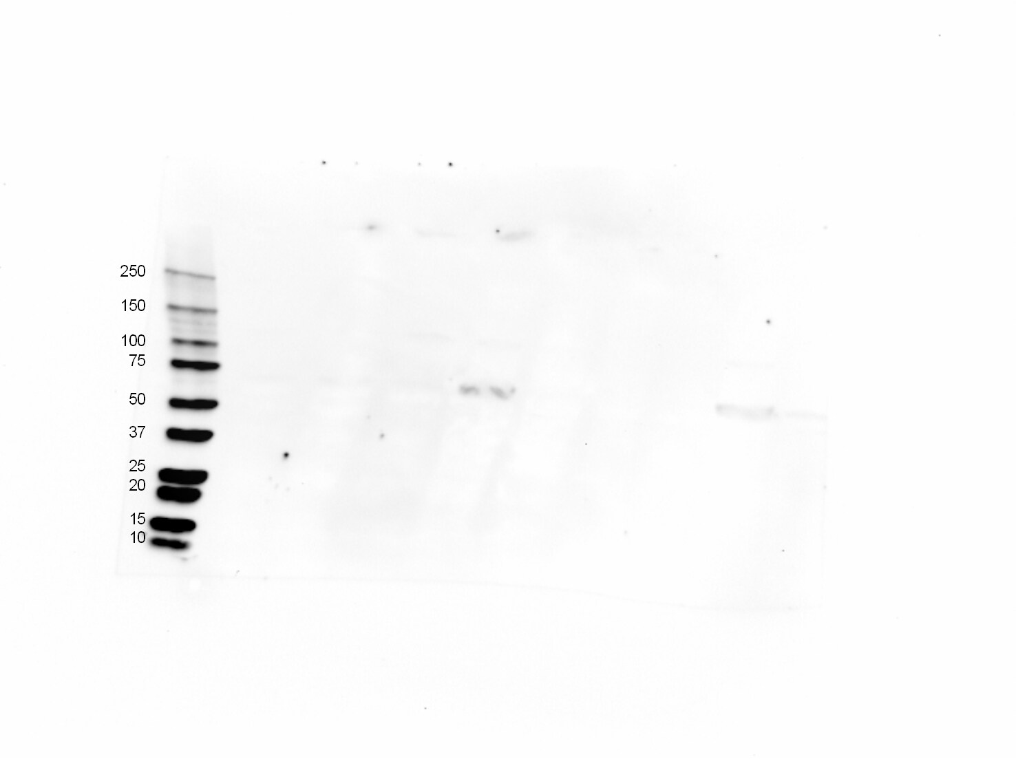
![Western Blot: TfR (Transferrin R) Antibody (DF1513) [NBP2-32945]](https://resources.rndsystems.com/images/products/TfR-Transferrin-R-Antibody-DF1513-Western-Blot-NBP2-32945-img0006.jpg "Western Blot: TfR (Transferrin R) Antibody (DF1513) [NBP2-32945]")
Western Blot: TfR (Transferrin R) Antibody (DF1513) [NBP2-32945]
Western Blot: TfR (Transferrin R) Antibody (DF1513) [NBP2-32945] - Western Blot Analysis of Jurkat, Raji, and THP-1 cell lysate using TfR (Transferrin R) Antibody (DF1513).![Immunocytochemistry/ Immunofluorescence: TfR (Transferrin R) Antibody (DF1513) [NBP2-32945]](https://resources.rndsystems.com/images/products/TfR-Transferrin-R-Antibody-DF1513-Immunocytochemistry-Immunofluorescence-NBP2-32945-img0005.jpg "Immunocytochemistry/ Immunofluorescence: TfR (Transferrin R) Antibody (DF1513) [NBP2-32945]")
Immunocytochemistry/ Immunofluorescence: TfR (Transferrin R) Antibody (DF1513) [NBP2-32945]
Immunocytochemistry/Immunofluorescence: TfR (Transferrin R) Antibody (DF1513) [NBP2-32945] - Immunofluorescence Analysis of Human Jurkat cells labeling CD71 with TfR (Transferrin R) Antibody (DF1513) followed by Goat anti-Mouse IgG-CF488 (Green). The nuclear counterstain is Red Dot (Red)![Flow Cytometry: TfR (Transferrin R) Antibody (DF1513) [NBP2-32945]](https://resources.rndsystems.com/images/products/TfR-Transferrin-R-Antibody-DF1513-Flow-Cytometry-NBP2-32945-img0007.jpg "Flow Cytometry: TfR (Transferrin R) Antibody (DF1513) [NBP2-32945]")
Flow Cytometry: TfR (Transferrin R) Antibody (DF1513) [NBP2-32945]
Flow Cytometry: TfR (Transferrin R) Antibody (DF1513) [NBP2-32945] - Flow Cytometric Analysis of human Jurkat cells using TfR (Transferrin R) antibody (DF1513) followed by Goat anti-Mouse IgG-CF488 (Orange); cells alone (Blue); Isotype Control (Red).![Simple Western: TfR (Transferrin R) Antibody (DF1513) [NBP2-32945]](https://resources.rndsystems.com/images/products/TfR-Transferrin-R-Antibody-DF1513-Simple-Western-NBP2-32945-img0001.jpg "Simple Western: TfR (Transferrin R) Antibody (DF1513) [NBP2-32945]")
Simple Western: TfR (Transferrin R) Antibody (DF1513) [NBP2-32945]
Simple Western: TfR (Transferrin R) Antibody (DF1513) [NBP2-32945] - Simple Western lane view shows a specific band for TfR (Transferrin R) in 0.2 mg/ml of Jurkat (left) and K562 (right) lysate(s). This experiment was performed under reducing conditions using the 12-230 kDa separation system.![Simple Western: TfR (Transferrin R) Antibody (DF1513) [NBP2-32945]](https://resources.rndsystems.com/images/products/TfR-Transferrin-R-Antibody-DF1513-Simple-Western-NBP2-32945-img0002.jpg "Simple Western: TfR (Transferrin R) Antibody (DF1513) [NBP2-32945]")
Simple Western: TfR (Transferrin R) Antibody (DF1513) [NBP2-32945]
Simple Western: TfR (Transferrin R) Antibody (DF1513) [NBP2-32945] - Electropherogram image of the corresponding Simple Western lane. TfR (Transferrin R) antibody was used at 10 ug/ml dilution of Jurkat and K562 lysates(s) respectively. Antibody (DF1513) [NBP2-32945] -")
Western Blot: TfR (Transferrin R) Antibody (DF1513) [NBP2-32945] -
The effect of NO on iron handling gene expression in human placental villous explants. (A–E) qPCR results, (F–J) protein analyses. Representative blots are shown in (K). HO-1 and TfR mRNA levels are significantly increased in the group treated with NO compared to the untreated controls. Protein abundance of HO-1 is significantly increased in the group treated with NO compared to the untreated controls. No significant change in expression is seen in the rest of the genes studied. HO-1: heme oxygenase 1, HAMP: hepcidin antimicrobial peptide, TfR: transferrin receptor, DMT1: divalent metal transporter 1, FPN: ferroportin 1. Data are reported from at least three independent experiments as mean +/- SD, *** p < 0.001, ns = not significant. Image collected and cropped by CiteAb from the following open publication (https://www.mdpi.com/1422-0067/24/6/5887), licensed under a CC-BY license. Not internally tested by Novus Biologicals. Antibody (DF1513) [NBP2-32945] -")
Western Blot: TfR (Transferrin R) Antibody (DF1513) [NBP2-32945] -
The effect of hypoxia on iron handling genes in response to NO in syncitialized BeWo cells (SCTs). (A–F) qPCR results and (G–K) protein analyses. Representative blots are shown in (K). Hypoxia alone did not significantly alter mRNA or protein levels of any of the studied iron homeostasis markers compared to normoxic controls. (A,B) The combination of hypoxia and NO resulted in a significant increase in HO-1 and HAMP mRNA levels, while NO increased HAMP mRNA levels significantly under hypoxic conditions. No significant effect of hypoxia or NO was seen in any of the other targets studied. HO-1: Heme Oxygenase 1, HAMP: Hepcidin antimicrobial peptide, TfR: Transferrin Receptor, TF: Transferrin, DMT1, Divalent metal transporter 1, FPN: Ferroportin 1. Data are reported from at least three independent experiments as mean +/- SD, * p < 0.05, ns = not significant. Image collected and cropped by CiteAb from the following open publication (https://www.mdpi.com/1422-0067/24/6/5887), licensed under a CC-BY license. Not internally tested by Novus Biologicals. Antibody (DF1513) [NBP2-32945] -")
Western Blot: TfR (Transferrin R) Antibody (DF1513) [NBP2-32945] -
The effect of NO on iron handling genes in syncytialized BeWo cells. (A–E), qPCR results, and (E–J) protein analyses. Representative blots are shown in (K). HO-1 and HAMP mRNA levels are significantly increased in the group treated with NO compared to the untreated controls. Protein abundance of HO-1 is significantly increased in the group treated with NO compared to the untreated controls. No significant change in expression is seen in the rest of the genes studied. HO-1: heme oxygenase 1, HAMP: hepcidin antimicrobial peptide, TfR: transferrin receptor, DMT1: divalent metal transporter 1, FPN: ferroportin 1. Data are reported from at least three independent experiments as mean +/- SD, ** p < 0.01, **** p < 0.0001, ns = not significant. Image collected and cropped by CiteAb from the following open publication (https://www.mdpi.com/1422-0067/24/6/5887), licensed under a CC-BY license. Not internally tested by Novus Biologicals.Applications for TfR (Transferrin R) Antibody (DF1513)
Application
Recommended Usage
Flow Cytometry
1-2 ug/million cells
Immunocytochemistry/ Immunofluorescence
1-2 ug/ml
Simple Western
10 ug/ml
Western Blot
1-2 ug/ml
Application Notes
Optimal dilution for a specific application should be determined.
See Simple Western Antibody Database for Simple Western validation: Tested in Jurkat and K562 lysates, separated by Size, antibody dilution of 10 ug/mL, apparent MW was 133 kDa
See Simple Western Antibody Database for Simple Western validation: Tested in Jurkat and K562 lysates, separated by Size, antibody dilution of 10 ug/mL, apparent MW was 133 kDa
Reviewed Applications
Read 1 review rated 4 using NBP2-32945 in the following applications:
Flow Cytometry Panel Builder
Bio-Techne Knows Flow Cytometry
Save time and reduce costly mistakes by quickly finding compatible reagents using the Panel Builder Tool.
Advanced Features
- Spectra Viewer - Custom analysis of spectra from multiple fluorochromes
- Spillover Popups - Visualize the spectra of individual fluorochromes
- Antigen Density Selector - Match fluorochrome brightness with antigen density

Formulation, Preparation, and Storage
Purification
Protein A or G purified
Formulation
10 mM PBS with 0.05% BSA
Preservative
0.05% Sodium Azide
Concentration
0.2 mg/ml
Shipping
The product is shipped with polar packs. Upon receipt, store it immediately at the temperature recommended below.
Stability & Storage
Store at 4C.
Background: TfR (Transferrin R)
Long Name
Transferrin Receptor
Alternate Names
CD71, TfR (TransferrinR), TFR1, TFRC, TRFR
Gene Symbol
TFRC
UniProt
Additional TfR (Transferrin R) Products
Product Documents for TfR (Transferrin R) Antibody (DF1513)
Certificate of Analysis
To download a Certificate of Analysis, please enter a lot or batch number in the search box below.
Product Specific Notices for TfR (Transferrin R) Antibody (DF1513)
This product is for research use only and is not approved for use in humans or in clinical diagnosis. Primary Antibodies are guaranteed for 1 year from date of receipt.
Citations for TfR (Transferrin R) Antibody (DF1513)
 Powered by Bioz
Customer Reviews for TfR (Transferrin R) Antibody (DF1513) (1)
4 out of 5
1 Customer Rating
Have you used TfR (Transferrin R) Antibody (DF1513)?
Submit a review and receive an Amazon gift card!
$25/€18/£15/$25CAN/¥2500 Yen for a review with an image
$10/€7/£6/$10CAN/¥1110 Yen for a review without an image
Submit a review
Customer Images
Showing
1
-
1 of
1 review
Showing All
Filter By:
-
Application: Western BlotSample Tested: Pancreas tissue, Stomach tissue, Testis tissue and Skin tissueSpecies: Canine and FelineVerified Customer | Posted 06/29/2017Lane 1: Canine Pancreas Lane 2: Canine Skin Lane 3: Canine Stomach Lane 4: Canine Testis Lane 5: Feline Pancreas Lane 6: Feline Skin Lane 7: Feline Stomach Lane 8: Feline TestisThis antibody will be used for IHC-P in both canine and feline tissue (results pending)

There are no reviews that match your criteria.
Protocols
Find general support by application which include: protocols, troubleshooting, illustrated assays, videos and webinars.
- 7-Amino Actinomycin D (7-AAD) Cell Viability Flow Cytometry Protocol
- Appropriate Fixation of IHC/ICC Samples
- Cellular Response to Hypoxia Protocols
- ClariTSA™ Fluorophore Kits
- Detection & Visualization of Antibody Binding
- Extracellular Membrane Flow Cytometry Protocol
- Flow Cytometry Protocol for Cell Surface Markers
- Flow Cytometry Protocol for Staining Membrane Associated Proteins
- Flow Cytometry Staining Protocols
- Flow Cytometry Troubleshooting Guide
- ICC Cell Smear Protocol for Suspension Cells
- ICC Immunocytochemistry Protocol Videos
- ICC for Adherent Cells
- Immunocytochemistry (ICC) Protocol
- Immunocytochemistry Troubleshooting
- Immunofluorescence of Organoids Embedded in Cultrex Basement Membrane Extract
- Immunohistochemistry (IHC) and Immunocytochemistry (ICC) Protocols
- Intracellular Flow Cytometry Protocol Using Alcohol (Methanol)
- Intracellular Flow Cytometry Protocol Using Detergents
- Intracellular Nuclear Staining Flow Cytometry Protocol Using Detergents
- Intracellular Staining Flow Cytometry Protocol Using Alcohol Permeabilization
- Intracellular Staining Flow Cytometry Protocol Using Detergents to Permeabilize Cells
- Preparing Samples for IHC/ICC Experiments
- Preventing Non-Specific Staining (Non-Specific Binding)
- Primary Antibody Selection & Optimization
- Propidium Iodide Cell Viability Flow Cytometry Protocol
- Protocol for Liperfluo
- Protocol for VisUCyte™ HRP Polymer Detection Reagent
- Protocol for the Characterization of Human Th22 Cells
- Protocol for the Characterization of Human Th9 Cells
- Protocol for the Fluorescent ICC Staining of Cell Smears - Graphic
- Protocol for the Fluorescent ICC Staining of Cultured Cells on Coverslips - Graphic
- Protocol for the Preparation and Fluorescent ICC Staining of Cells on Coverslips
- Protocol for the Preparation and Fluorescent ICC Staining of Non-adherent Cells
- Protocol for the Preparation and Fluorescent ICC Staining of Stem Cells on Coverslips
- Protocol for the Preparation of a Cell Smear for Non-adherent Cell ICC - Graphic
- Protocol: Annexin V and PI Staining by Flow Cytometry
- Protocol: Annexin V and PI Staining for Apoptosis by Flow Cytometry
- R&D Systems Quality Control Western Blot Protocol
- TUNEL and Active Caspase-3 Detection by IHC/ICC Protocol
- The Importance of IHC/ICC Controls
- Troubleshooting Guide: Fluorokine Flow Cytometry Kits
- Troubleshooting Guide: Western Blot Figures
- Western Blot Conditions
- Western Blot Protocol
- Western Blot Protocol for Cell Lysates
- Western Blot Troubleshooting
- Western Blot Troubleshooting Guide
- View all Protocols, Troubleshooting, Illustrated assays and Webinars
Loading...