TGF-beta 1 (Transforming Growth Factor beta 1) is a pleiotrophic cytokine that regulates immune function, proliferation, and epithelial-mesenchymal transition. TGF-beta 1 associates with LAP to form a latent complex. It can be is activated from latency by plasmin, matrix metalloproteases, thrombospondin 1, and a subset of integrins. TGF-beta 1 signals through complexes of TGF-beta RII with TGF-beta RI/ALK-5 or ALK-1. TGF-beta signaling is modulated by the accessory receptors TGF-beta RIII/Betaglycan and Endoglin/CD105.
TGF-beta Antibody (1D11.16.8) - BSA Free
Novus Biologicals | Catalog # NBP2-45137
Key Product Details
Species Reactivity
Validated:
Human, Mouse, Rat, Bovine, Canine, Goat, Hamster, Monkey
Cited:
Human, Mouse
Applications
Validated:
Immunohistochemistry, Immunohistochemistry-Paraffin, Sandwich ELISA, ELISA Capture (Matched Antibody Pair), Immunocytochemistry/ Immunofluorescence
Cited:
Immunohistochemistry-Paraffin, Immunohistochemistry-Frozen, IF/IHC
Label
Unconjugated
Antibody Source
Monoclonal Mouse IgG1 kappa Clone # 1D11.16.8
Format
BSA Free
Loading...
Product Specifications
Immunogen
Bovine bone-derived TGF-beta 1 and TGF-beta 2
Reactivity Notes
Goat reactivity reported from a verified customer review.
Localization
Cytoplasm and extracellular (secreted)
Specificity
This MAb recognizes TGF beta 1, 2 and 3. Three TGF betas have been identified in mammals. TGFbeta1, TGFbeta2 and TGFbeta3 are each synthesized as precursor proteins that are very similar in that each is cleaved to yield a 112 amino acid polypeptide that remains associated with the latent portion of the molecules. Biologically active TGFbeta requires dimerization of the monomers (usually homodimers) and release of the latent peptide portion. Overall, the mature region of the TGFbeta3 protein has approximately 80% identity to the mature region of both TGFbeta1 and TGFbeta2. However, the NH2 terminals or precursor regions of their molecules share only 27% sequence identity. TGFbeta's inhibit the growth of epithelial cells and stimulate the growth of mesenchymal cells.
Clonality
Monoclonal
Host
Mouse
Isotype
IgG1 kappa
Theoretical MW
13 kDa.
Disclaimer note: The observed molecular weight of the protein may vary from the listed predicted molecular weight due to post translational modifications, post translation cleavages, relative charges, and other experimental factors.
Disclaimer note: The observed molecular weight of the protein may vary from the listed predicted molecular weight due to post translational modifications, post translation cleavages, relative charges, and other experimental factors.
Scientific Data Images for TGF-beta Antibody (1D11.16.8) - BSA Free
![Immunocytochemistry/ Immunofluorescence: TGF-beta Antibody (1D11.16.8) - BSA Free [NBP2-45137]](https://resources.rndsystems.com/images/products/TGF-beta-Antibody-1D11-16-8-BSA-Free-Immunocytochemistry-Immunofluorescence-NBP2-45137-img0002.jpg "Immunocytochemistry/ Immunofluorescence: TGF-beta Antibody (1D11.16.8) - BSA Free [NBP2-45137]")
Immunocytochemistry/ Immunofluorescence: TGF-beta Antibody (1D11.16.8) - BSA Free [NBP2-45137]
Immunocytochemistry/Immunofluorescence: TGF-beta Antibody (1D11.16.8) - BSA Free [NBP2-45137] - U2OS cells were fixed in 4% paraformaldehyde for 10 minutes and permeabilized in 0.05% Triton X-100 in PBS for 5 minutes. The cells were incubated with TGF-beta Antibody [1D11.16.8] (NBP2-45137) at 1ug/ml overnight at 4C and detected with an anti-mouse DyLight 488 (Green) at a 1:1000 dilution for 60 minutes. Nuclei were counterstained with DAPI (Blue). Cells were imaged using a 40X objective.![Immunohistochemistry: TGF-beta Antibody (1D11.16.8) - BSA Free [NBP2-45137]](https://resources.rndsystems.com/images/products/TGF-beta-Antibody-1D11-16-8-Immunohistochemistry-NBP2-45137-img0001.jpg "Immunohistochemistry: TGF-beta Antibody (1D11.16.8) - BSA Free [NBP2-45137]")
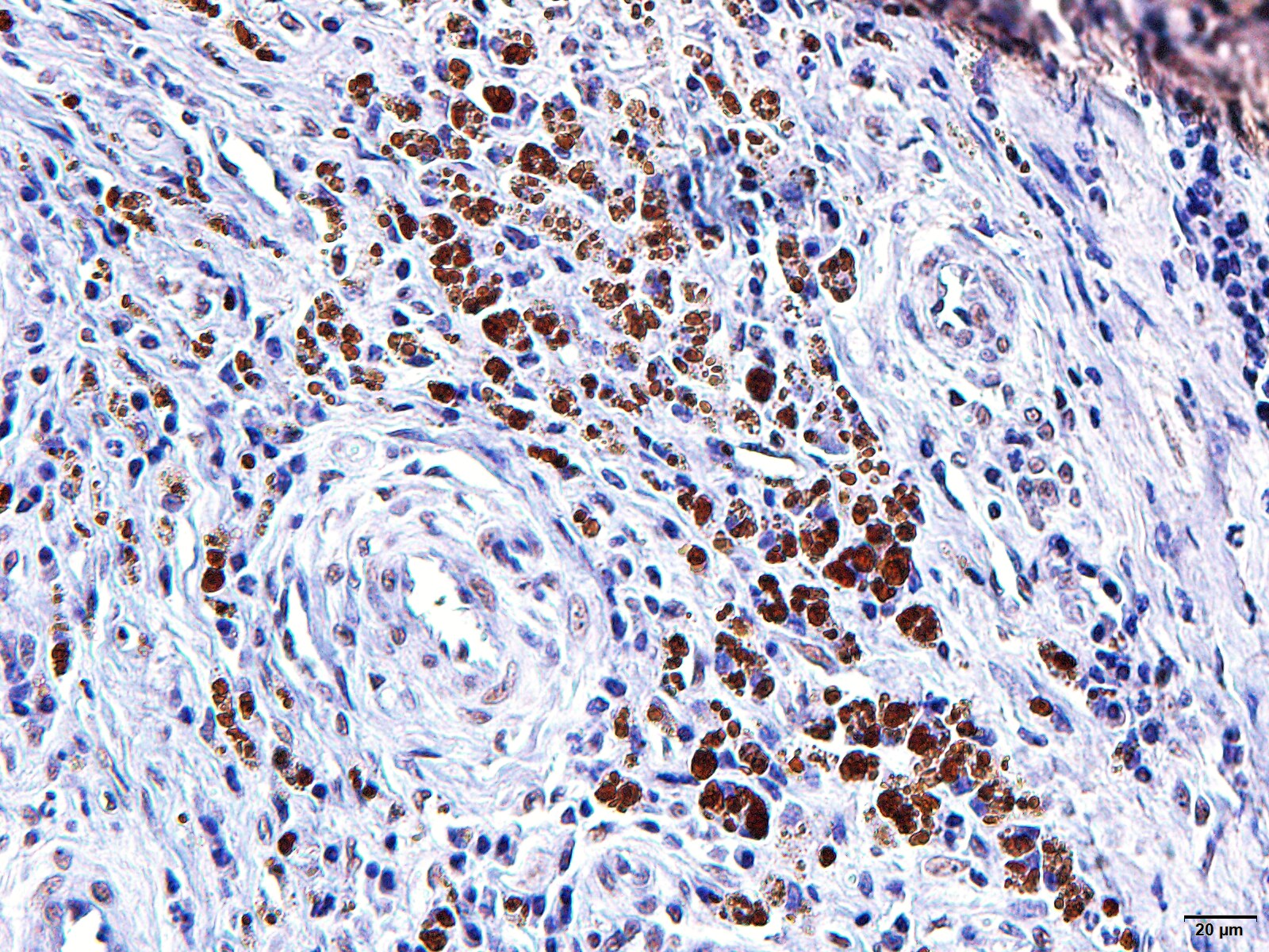
Immunohistochemistry: TGF-beta Antibody (1D11.16.8) - BSA Free [NBP2-45137]
Immunohistochemistry: TGF-beta Antibody (1D11.16.8) [NBP2-45137] - TGF-beta was detected in immersion fixed paraffin-embedded human skin using 25 ug/mL Mouse Anti-TGF-beta Monoclonal Antibody (Clone 1D11.16.8) overnight at 4 degrees C. Tissues was stained using the Anti-Mouse HRP-DAB Cell and Tissue Staining Kit (brown; Catalog # CTS002) and counterstained with Haematoxylin (blue). [NBP2-45137]")
Immunohistochemistry-Paraffin: Mouse Monoclonal TGF-beta Antibody (1D11.16.8) [NBP2-45137]
The sections of histological goat bone fragments were incubated with the primary antibody TGF-beta NBP2-45137 overnight. Subsequently, the secondary antibody NB7539 1:200; was applied and incubated for 1 hour. Image from a verified customer review.Applications for TGF-beta Antibody (1D11.16.8) - BSA Free
Application
Recommended Usage
Immunocytochemistry/ Immunofluorescence
8-25 ug/ml
Immunohistochemistry-Paraffin
0.5 - 1.0ug/ml
Application Notes
Hu-chromosome location: 19q13.1 (beta1); 1q41 (beta2); 14q24 (beta3)
For Cytokine Neutralization and Functional Assay, order Azide and BSA Free formulation (NBP2-47736)
For Cytokine Neutralization and Functional Assay, order Azide and BSA Free formulation (NBP2-47736)
Reviewed Applications
Read 1 review rated 5 using NBP2-45137 in the following applications:
Formulation, Preparation, and Storage
Purification
Protein A or G purified
Formulation
PBS
Format
BSA Free
Preservative
0.02% Sodium Azide
Concentration
1.0 mg/ml
Shipping
The product is shipped with polar packs. Upon receipt, store it immediately at the temperature recommended below.
Stability & Storage
Store at 4C short term. Store at -20C long term. Avoid freeze-thaw cycles.
Background: TGF-beta
Long Name
Transforming Growth Factor beta
Alternate Names
TGFbeta
Gene Symbol
TGFB1
Additional TGF-beta Products
Product Documents for TGF-beta Antibody (1D11.16.8) - BSA Free
Certificate of Analysis
To download a Certificate of Analysis, please enter a lot or batch number in the search box below.
Product Specific Notices for TGF-beta Antibody (1D11.16.8) - BSA Free
This product is for research use only and is not approved for use in humans or in clinical diagnosis. Primary Antibodies are guaranteed for 1 year from date of receipt.
Citations for TGF-beta Antibody (1D11.16.8) - BSA Free
 Powered by Bioz
Customer Reviews for TGF-beta Antibody (1D11.16.8) - BSA Free (1)
5 out of 5
1 Customer Rating
Have you used TGF-beta Antibody (1D11.16.8) - BSA Free?
Submit a review and receive an Amazon gift card!
$25/€18/£15/$25CAN/¥2500 Yen for a review with an image
$10/€7/£6/$10CAN/¥1110 Yen for a review without an image
Submit a review
Customer Images
Showing
1
-
1 of
1 review
Showing All
Filter By:
-
Application: Immunohistochemistry-ParaffinSample Tested: Bone Extracts and BoneSpecies: GoatVerified Customer | Posted 07/29/2025The sections of histological goat bone fragments were incubated with the primary antibody TGF-beta NBP2-45137 overnight. Subsequently, the secondary antibody NB7539 1:200; was applied and incubated for 1 hour.Goat NBP2-45137 antibody reactivity test
 Bio-Techne ResponseThis review reflects a new species or application tested on a primary antibody.
There are no reviews that match your criteria.
Protocols
Find general support by application which include: protocols, troubleshooting, illustrated assays, videos and webinars.
- Antigen Retrieval Protocol (PIER)
- Antigen Retrieval for Frozen Sections Protocol
- Appropriate Fixation of IHC/ICC Samples
- Cellular Response to Hypoxia Protocols
- Chromogenic IHC Staining of Formalin-Fixed Paraffin-Embedded (FFPE) Tissue Protocol
- Chromogenic Immunohistochemistry Staining of Frozen Tissue
- ClariTSA™ Fluorophore Kits
- Detection & Visualization of Antibody Binding
- ELISA Sample Preparation & Collection Guide
- ELISA Troubleshooting Guide
- Fluorescent IHC Staining of Frozen Tissue Protocol
- Graphic Protocol for Heat-induced Epitope Retrieval
- Graphic Protocol for the Preparation and Fluorescent IHC Staining of Frozen Tissue Sections
- Graphic Protocol for the Preparation and Fluorescent IHC Staining of Paraffin-embedded Tissue Sections
- Graphic Protocol for the Preparation of Gelatin-coated Slides for Histological Tissue Sections
- How to Run an R&D Systems DuoSet ELISA
- How to Run an R&D Systems Quantikine ELISA
- How to Run an R&D Systems Quantikine™ QuicKit™ ELISA
- ICC Cell Smear Protocol for Suspension Cells
- ICC Immunocytochemistry Protocol Videos
- ICC for Adherent Cells
- IHC Sample Preparation (Frozen sections vs Paraffin)
- Immunocytochemistry (ICC) Protocol
- Immunocytochemistry Troubleshooting
- Immunofluorescence of Organoids Embedded in Cultrex Basement Membrane Extract
- Immunofluorescent IHC Staining of Formalin-Fixed Paraffin-Embedded (FFPE) Tissue Protocol
- Immunohistochemistry (IHC) and Immunocytochemistry (ICC) Protocols
- Immunohistochemistry Frozen Troubleshooting
- Immunohistochemistry Paraffin Troubleshooting
- Preparing Samples for IHC/ICC Experiments
- Preventing Non-Specific Staining (Non-Specific Binding)
- Primary Antibody Selection & Optimization
- Protocol for Heat-Induced Epitope Retrieval (HIER)
- Protocol for Making a 4% Formaldehyde Solution in PBS
- Protocol for VisUCyte™ HRP Polymer Detection Reagent
- Protocol for the Fluorescent ICC Staining of Cell Smears - Graphic
- Protocol for the Fluorescent ICC Staining of Cultured Cells on Coverslips - Graphic
- Protocol for the Preparation & Fixation of Cells on Coverslips
- Protocol for the Preparation and Chromogenic IHC Staining of Frozen Tissue Sections
- Protocol for the Preparation and Chromogenic IHC Staining of Frozen Tissue Sections - Graphic
- Protocol for the Preparation and Chromogenic IHC Staining of Paraffin-embedded Tissue Sections
- Protocol for the Preparation and Chromogenic IHC Staining of Paraffin-embedded Tissue Sections - Graphic
- Protocol for the Preparation and Fluorescent ICC Staining of Cells on Coverslips
- Protocol for the Preparation and Fluorescent ICC Staining of Non-adherent Cells
- Protocol for the Preparation and Fluorescent ICC Staining of Stem Cells on Coverslips
- Protocol for the Preparation and Fluorescent IHC Staining of Frozen Tissue Sections
- Protocol for the Preparation and Fluorescent IHC Staining of Paraffin-embedded Tissue Sections
- Protocol for the Preparation of Gelatin-coated Slides for Histological Tissue Sections
- Protocol for the Preparation of a Cell Smear for Non-adherent Cell ICC - Graphic
- Quantikine HS ELISA Kit Assay Principle, Alkaline Phosphatase
- Quantikine HS ELISA Kit Principle, Streptavidin-HRP Polymer
- Sandwich ELISA (Colorimetric) – Biotin/Streptavidin Detection Protocol
- Sandwich ELISA (Colorimetric) – Direct Detection Protocol
- TUNEL and Active Caspase-3 Detection by IHC/ICC Protocol
- The Importance of IHC/ICC Controls
- Troubleshooting Guide: ELISA
- Troubleshooting Guide: Immunohistochemistry
- View all Protocols, Troubleshooting, Illustrated assays and Webinars
Loading...
Associated Pathways
