![Immunocytochemistry/ Immunofluorescence: TGN38 Antibody (2F7.1) [NB300-575]](https://resources.rndsystems.com/images/products/TGN38-Antibody-2F7-1-Immunocytochemistry-Immunofluorescence-NB300-575-img0016.jpg "Immunocytochemistry/ Immunofluorescence: TGN38 Antibody (2F7.1) [NB300-575]")
Loading...
Key Product Details
Species Reactivity
Validated:
Human, Mouse, Rat, Chinese Hamster, Monkey, Rabbit
Cited:
Human, Mouse, Rat
Applications
Validated:
Immunohistochemistry, Immunohistochemistry-Paraffin, Western Blot, Flow Cytometry, Immunocytochemistry/ Immunofluorescence
Cited:
Immunohistochemistry, Immunohistochemistry-Paraffin, Western Blot, Immunocytochemistry/ Immunofluorescence, IF/IHC
Label
Unconjugated
Antibody Source
Monoclonal Mouse IgG1 Clone # 2F7.1
Loading...
Product Specifications
Immunogen
Synthetic peptide corresponding to residues L(18) P S A S K P N N T S S E N N P P(34) C of rat TGN38.
Reactivity Notes
Chinese Hamster reactivity reported in scientific literature (PMID: 11408588). Rabbit reactivity reported in scientific literature (PMID: 10514494). Please note that this antibody is reactive to Mouse and derived from the same host, Mouse. Additional Mouse on Mouse blocking steps may be required for IHC and ICC experiments. Please contact Technical Support for more information. Use in Human reported in scientific literature (PMID:24576880).
Marker
TGN Marker
Specificity
Detects Trans Golgi Network (TGN) 38 from human and monkey samples and both recombinant and endogenous rat samples.
Clonality
Monoclonal
Host
Mouse
Isotype
IgG1
Scientific Data Images for TGN38 Antibody (2F7.1)
Immunocytochemistry/ Immunofluorescence: TGN38 Antibody (2F7.1) [NB300-575]
Immunocytochemistry/Immunofluorescence: TGN38 Antibody (2F7.1) [NB300-575] - HeLa and COS7 cells were rinsed with PBS and fixed with -20C methanol for 5 minutes. Cells were incubated with anti-TGN38 antibody diluted 1:100 in PBS containing 0.2% BSA for 1 hour. The cells were then rinsed in PBS and incubated with goat anti-mouse IgG conjugated to rhodamine for 30 minutes in PBS containing 0.2% BSA. Following washing, cells were imaged using a Zeiss Axioplan microscope. ICC/IF image submitted by a verified customer review.![Immunocytochemistry/ Immunofluorescence: TGN38 Antibody (2F7.1) [NB300-575]](https://resources.rndsystems.com/images/products/TGN38-Antibody-2F7-1-Immunocytochemistry-Immunofluorescence-NB300-575-img0013.jpg "Immunocytochemistry/ Immunofluorescence: TGN38 Antibody (2F7.1) [NB300-575]")
Immunocytochemistry/ Immunofluorescence: TGN38 Antibody (2F7.1) [NB300-575]
Immunocytochemistry/Immunofluorescence: TGN38 Antibody (2F7.1) [NB300-575] - Staining of TGN-38 in NRK cells.![Immunocytochemistry/ Immunofluorescence: TGN38 Antibody (2F7.1) [NB300-575]](https://resources.rndsystems.com/images/products/TGN38-Antibody-2F7-1-Immunocytochemistry-Immunofluorescence-NB300-575-img0014.jpg "Immunocytochemistry/ Immunofluorescence: TGN38 Antibody (2F7.1) [NB300-575]")
Immunocytochemistry/ Immunofluorescence: TGN38 Antibody (2F7.1) [NB300-575]
Immunocytochemistry/Immunofluorescence: TGN38 Antibody (2F7.1) [NB300-575] - Analysis of Phalloidin (green) and TGN38 (purple) in U2OS cells. Formalin fixed cells were permeabilized with 0.1% Triton X-100 in PBS for 10 minutes at room temperature and blocked with 2% BSA in PBS + 0.1% Triton X-100 for 30 minutes at room temperature. Cells were probed with a TGN38 monoclonal antibody at a dilution of 1:75 for at least 1 hour at room temperature, washed with PBS, and incubated with DyLight 680 goat anti-mouse IgG secondary antibody at a dilution of 1:250 for 30 minutes at room temperature. Actin was stained with DyLight 488 Phalloidin at a dilution of 1:300 (1 unit/mL final concentration) for 30 minutes. Images were taken on a Thermo Scientific ArrayScan VTI at 20X magnification.![Immunocytochemistry/ Immunofluorescence: TGN38 Antibody (2F7.1) [NB300-575]](https://resources.rndsystems.com/images/products/TGN38-Antibody-2F7-1-Immunocytochemistry-Immunofluorescence-NB300-575-img0015.jpg "Immunocytochemistry/ Immunofluorescence: TGN38 Antibody (2F7.1) [NB300-575]")
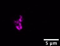
Immunocytochemistry/ Immunofluorescence: TGN38 Antibody (2F7.1) [NB300-575]
Immunocytochemistry/Immunofluorescence: TGN38 Antibody (2F7.1) [NB300-575] - Staining in NS-1 Cells.Applications for TGN38 Antibody (2F7.1)
Application
Recommended Usage
Flow Cytometry
1:20 - 1:50
Immunocytochemistry/ Immunofluorescence
1:1000
Immunohistochemistry
1:1000
Immunohistochemistry-Paraffin
1:1000
Western Blot
1:500
Application Notes
IP usage was reported in scientific literature (PMID: 10514494). WB: Detects an approx. 38 kDa protein representing deglycosylated recombinant rat TGN38. Under normal WB conditions, detects an 85-95 kDa band depending on cell type. IF: Staining of TGN38 in NRK cells results in staining predominantly in the TGN. IF: it has been shown that methanol works best as a fixative.
Reviewed Applications
Read 1 review rated 5 using NB300-575 in the following applications:
Flow Cytometry Panel Builder
Bio-Techne Knows Flow Cytometry
Save time and reduce costly mistakes by quickly finding compatible reagents using the Panel Builder Tool.
Advanced Features
- Spectra Viewer - Custom analysis of spectra from multiple fluorochromes
- Spillover Popups - Visualize the spectra of individual fluorochromes
- Antigen Density Selector - Match fluorochrome brightness with antigen density

Formulation, Preparation, and Storage
Purification
Protein A or G purified
Formulation
PBS, 1 mg/mL BSA
Preservative
0.05% Sodium Azide
Concentration
1 mg/ml
Shipping
The product is shipped with polar packs. Upon receipt, store it immediately at the temperature recommended below.
Stability & Storage
Store at -20C. Avoid freeze-thaw cycles.
Background: TGN38
Long Name
Trans-golgi Network Protein
Alternate Names
TGN46, TGN51, Tgoln1, Tgoln2, Ttgn1
Gene Symbol
TGOLN2
Additional TGN38 Products
Product Documents for TGN38 Antibody (2F7.1)
Certificate of Analysis
To download a Certificate of Analysis, please enter a lot or batch number in the search box below.
Product Specific Notices for TGN38 Antibody (2F7.1)
This product is for research use only and is not approved for use in humans or in clinical diagnosis. Primary Antibodies are guaranteed for 1 year from date of receipt.
Citations for TGN38 Antibody (2F7.1)
 Powered by Bioz
Customer Reviews for TGN38 Antibody (2F7.1) (1)
5 out of 5
1 Customer Rating
Have you used TGN38 Antibody (2F7.1)?
Submit a review and receive an Amazon gift card!
$25/€18/£15/$25CAN/¥2500 Yen for a review with an image
$10/€7/£6/$10CAN/¥1110 Yen for a review without an image
Submit a review
Customer Images
Showing
1
-
1 of
1 review
Showing All
Filter By:
-
Application: ImmunocytochemistrySample Tested: INS-1Species: RatVerified Customer | Posted 01/06/2020

There are no reviews that match your criteria.
Protocols
Find general support by application which include: protocols, troubleshooting, illustrated assays, videos and webinars.
- 7-Amino Actinomycin D (7-AAD) Cell Viability Flow Cytometry Protocol
- Antigen Retrieval Protocol (PIER)
- Antigen Retrieval for Frozen Sections Protocol
- Appropriate Fixation of IHC/ICC Samples
- Cellular Response to Hypoxia Protocols
- Chromogenic IHC Staining of Formalin-Fixed Paraffin-Embedded (FFPE) Tissue Protocol
- Chromogenic Immunohistochemistry Staining of Frozen Tissue
- ClariTSA™ Fluorophore Kits
- Detection & Visualization of Antibody Binding
- Extracellular Membrane Flow Cytometry Protocol
- Flow Cytometry Protocol for Cell Surface Markers
- Flow Cytometry Protocol for Staining Membrane Associated Proteins
- Flow Cytometry Staining Protocols
- Flow Cytometry Troubleshooting Guide
- Fluorescent IHC Staining of Frozen Tissue Protocol
- Graphic Protocol for Heat-induced Epitope Retrieval
- Graphic Protocol for the Preparation and Fluorescent IHC Staining of Frozen Tissue Sections
- Graphic Protocol for the Preparation and Fluorescent IHC Staining of Paraffin-embedded Tissue Sections
- Graphic Protocol for the Preparation of Gelatin-coated Slides for Histological Tissue Sections
- ICC Cell Smear Protocol for Suspension Cells
- ICC Immunocytochemistry Protocol Videos
- ICC for Adherent Cells
- IHC Sample Preparation (Frozen sections vs Paraffin)
- Immunocytochemistry (ICC) Protocol
- Immunocytochemistry Troubleshooting
- Immunofluorescence of Organoids Embedded in Cultrex Basement Membrane Extract
- Immunofluorescent IHC Staining of Formalin-Fixed Paraffin-Embedded (FFPE) Tissue Protocol
- Immunohistochemistry (IHC) and Immunocytochemistry (ICC) Protocols
- Immunohistochemistry Frozen Troubleshooting
- Immunohistochemistry Paraffin Troubleshooting
- Intracellular Flow Cytometry Protocol Using Alcohol (Methanol)
- Intracellular Flow Cytometry Protocol Using Detergents
- Intracellular Nuclear Staining Flow Cytometry Protocol Using Detergents
- Intracellular Staining Flow Cytometry Protocol Using Alcohol Permeabilization
- Intracellular Staining Flow Cytometry Protocol Using Detergents to Permeabilize Cells
- Preparing Samples for IHC/ICC Experiments
- Preventing Non-Specific Staining (Non-Specific Binding)
- Primary Antibody Selection & Optimization
- Propidium Iodide Cell Viability Flow Cytometry Protocol
- Protocol for Heat-Induced Epitope Retrieval (HIER)
- Protocol for Liperfluo
- Protocol for Making a 4% Formaldehyde Solution in PBS
- Protocol for VisUCyte™ HRP Polymer Detection Reagent
- Protocol for the Characterization of Human Th22 Cells
- Protocol for the Characterization of Human Th9 Cells
- Protocol for the Fluorescent ICC Staining of Cell Smears - Graphic
- Protocol for the Fluorescent ICC Staining of Cultured Cells on Coverslips - Graphic
- Protocol for the Preparation & Fixation of Cells on Coverslips
- Protocol for the Preparation and Chromogenic IHC Staining of Frozen Tissue Sections
- Protocol for the Preparation and Chromogenic IHC Staining of Frozen Tissue Sections - Graphic
- Protocol for the Preparation and Chromogenic IHC Staining of Paraffin-embedded Tissue Sections
- Protocol for the Preparation and Chromogenic IHC Staining of Paraffin-embedded Tissue Sections - Graphic
- Protocol for the Preparation and Fluorescent ICC Staining of Cells on Coverslips
- Protocol for the Preparation and Fluorescent ICC Staining of Non-adherent Cells
- Protocol for the Preparation and Fluorescent ICC Staining of Stem Cells on Coverslips
- Protocol for the Preparation and Fluorescent IHC Staining of Frozen Tissue Sections
- Protocol for the Preparation and Fluorescent IHC Staining of Paraffin-embedded Tissue Sections
- Protocol for the Preparation of Gelatin-coated Slides for Histological Tissue Sections
- Protocol for the Preparation of a Cell Smear for Non-adherent Cell ICC - Graphic
- Protocol: Annexin V and PI Staining by Flow Cytometry
- Protocol: Annexin V and PI Staining for Apoptosis by Flow Cytometry
- R&D Systems Quality Control Western Blot Protocol
- TUNEL and Active Caspase-3 Detection by IHC/ICC Protocol
- The Importance of IHC/ICC Controls
- Troubleshooting Guide: Fluorokine Flow Cytometry Kits
- Troubleshooting Guide: Immunohistochemistry
- Troubleshooting Guide: Western Blot Figures
- Western Blot Conditions
- Western Blot Protocol
- Western Blot Protocol for Cell Lysates
- Western Blot Troubleshooting
- Western Blot Troubleshooting Guide
- View all Protocols, Troubleshooting, Illustrated assays and Webinars
Loading...