TGN38 Antibody - Azide and BSA Free
Novus Biologicals | Catalog # NBP1-03495
![Immunocytochemistry/ Immunofluorescence: TGN38 Antibody [NBP1-03495]](https://resources.rndsystems.com/images/products/TGN38-Antibody-Immunocytochemistry-Immunofluorescence-NBP1-03495-img0007.jpg "Immunocytochemistry/ Immunofluorescence: TGN38 Antibody [NBP1-03495]")
Loading...
Key Product Details
Species Reactivity
Validated:
Human, Mouse, Rat, Monkey
Cited:
Human, Mouse
Applications
Validated:
Immunohistochemistry, Western Blot, Immunocytochemistry/ Immunofluorescence
Cited:
Western Blot, Immunocytochemistry/ Immunofluorescence
Label
Unconjugated
Antibody Source
Polyclonal Rabbit IgG
Format
Azide and BSA Free
Loading...
Product Specifications
Immunogen
A synthetic peptide from mouse TGN38 conjugated to blue carrier protein was used as the antigen.
Reactivity Notes
Marmoset (100%).
Marker
TGN Marker
Specificity
TGN38 (or TGN46, TGN48, TGN51 in human).
Clonality
Polyclonal
Host
Rabbit
Isotype
IgG
Description
PLEASE NOTE: 0.1mg sample size is provided in reconstituted format and the 0.5mg size is provided lyophilized. (Please see reconstitution instuctions).
Scientific Data Images for TGN38 Antibody - Azide and BSA Free
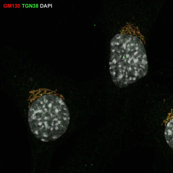
Immunocytochemistry/ Immunofluorescence: TGN38 Antibody [NBP1-03495]
Immunocytochemistry/Immunofluorescence: TGN38 Antibody [NBP1-03495] - Mouse embryonic fibroblasts, fixed with 4% PFA washed with PBS, permeabilized with 0.2% Triton X-100. Primary antibody at 1:100 in 3% BSA in PBS. Secondary antibodies incubation for 2 hours. Image from a verified customer review.![Immunocytochemistry/ Immunofluorescence: TGN38 Antibody [NBP1-03495]](https://resources.rndsystems.com/images/products/TGN38-Antibody-Immunocytochemistry-Immunofluorescence-NBP1-03495-img0001.jpg "Immunocytochemistry/ Immunofluorescence: TGN38 Antibody [NBP1-03495]")
Immunocytochemistry/ Immunofluorescence: TGN38 Antibody [NBP1-03495]
Immunocytochemistry/Immunofluorescence: TGN38 Antibody [NBP1-03495] - Human Melanoma cell line C 32.![Immunocytochemistry/ Immunofluorescence: TGN38 Antibody [NBP1-03495]](https://resources.rndsystems.com/images/products/TGN38-Antibody-Immunocytochemistry-Immunofluorescence-NBP1-03495-img0003.jpg "Immunocytochemistry/ Immunofluorescence: TGN38 Antibody [NBP1-03495]")
Immunocytochemistry/ Immunofluorescence: TGN38 Antibody [NBP1-03495]
Immunocytochemistry/Immunofluorescence: TGN38 Antibody [NBP1-03495] - Human Melanoma cell line C 32![Immunocytochemistry/ Immunofluorescence: TGN38 Antibody [NBP1-03495]](https://resources.rndsystems.com/images/products/TGN38-Antibody-Immunocytochemistry-Immunofluorescence-NBP1-03495-img0005.jpg "Immunocytochemistry/ Immunofluorescence: TGN38 Antibody [NBP1-03495]")
Immunocytochemistry/ Immunofluorescence: TGN38 Antibody [NBP1-03495]
Immunocytochemistry/Immunofluorescence: TGN38 Antibody [NBP1-03495] - Human Melanoma cell line C 32 was cultured overnight on round cover slides placed in a 24 well tissue culture plate. Culture media removed and washed twice with PBS before fixing with 2% formalin for 10 minutes. Cells were then washed three times with PBS and incubated with Tris 0.01M containing Triton X 0.005% for 15 minutes. Cells were washed and incubated with 100 ul of Rabbit antibody to TGN diluted 1:100 in the blocking buffer for 30 minutes. Welles were then washed 7 times with PBS and incubated with 100 ul of anti Rb-FITC conjugate diluted 1:100 in the blocking buffer for further 30 minutes. Cells were washed as before and nuclear counter stained with Hoechst and mounted on to slides.![Immunocytochemistry/ Immunofluorescence: TGN38 Antibody [NBP1-03495]](https://resources.rndsystems.com/images/products/TGN38-Antibody-Immunocytochemistry-Immunofluorescence-NBP1-03495-img0006.jpg "Immunocytochemistry/ Immunofluorescence: TGN38 Antibody [NBP1-03495]")
Immunocytochemistry/ Immunofluorescence: TGN38 Antibody [NBP1-03495]
Immunocytochemistry/Immunofluorescence: TGN38 Antibody [NBP1-03495] - Human Melanoma cell line C 32 was cultured overnight on round cover slides placed in a 24 well tissue culture plate. Culture media removed and washed twice with PBS before fixing with 2% formalin for 10 minutes. Cells were then washed three times with PBS and incubated with Tris 0.01M containing Triton X 0.005% for 15 minutes. Cells were washed and incubated with 100 ul of Rabbit antibody to TGN diluted 1:100 in the blocking buffer for 30 minutes. Welles were then washed 7 times with PBS and incubated with 100 ul of anti Rb-FITC conjugate diluted 1:100 in the blocking buffer for further 30 minutes. Cells were washed as before and nuclear counter stained with Hoechst and mounted on to slides.
Immunocytochemistry/ Immunofluorescence: TGN38 Antibody - Azide and BSA Free [NBP1-03495] -
Localization of vesicles with accumulated ceramides in ASC specks by live-cell fluorescence confocal microscopy and cryo-ET.a Distribution of BODIPY TR ceramide (BCer) during ASC speck formation in iBMDMs expressing ASC-mCerulean, or WT iBMDMs stained with FAM-FLICA. Areas of speck formation are boxed. See Supplementary Movie 3 for full time courses. Right, percentage of whole-cell fluorescence that mapped to the area of speck formation, before and after speck formation, t0 and tS (1–3 min and 20–50 min after nigericin addition), respectively. 12 specks from ASC-mCerulean iBMDMs and 11 specks from WT iBMDMs were analyzed. See Source Data File for source data. Scale bars, 10 um. b Immunofluorescence microscopy of LPS-primed WT iBMDMs with or without nigericin stimulation. Anti-NLRP3 partially colocalized with anti-TGN38. The TGN was dispersed following stimulation. Scale bar, 10 um. See Supplementary Movie 4 for a Z-stack series. The images are representative of three independent experiments, with at least 20 cells imaged per experiment. c Immunofluorescence microscopy of LPS-primed ASC-mCerulean iBMDMs with or without nigericin stimulation, and with nigericin stimulation and caspase inhibitor Z-VAD-FMK. There is little overlap between ASC-mCerulean and anti-TGN38 fluorescence. Scale bars, 10 um. The images are representative of three independent experiments, with at least 20 cells imaged per experiment. d 1.36-nm thick virtual tomographic slice within an ASC speck. Scale bar, 200 nm. Lower panel, 3-D segmentation model. e Vesicles from cryo-ET reconstructions of ASC-mCerulean iBMDMs. Scale bar, 100 nm. The histogram shows the vesicle size distribution. Image collected and cropped by CiteAb from the following open publication (https://pubmed.ncbi.nlm.nih.gov/37945612), licensed under a CC-BY license. Not internally tested by Novus Biologicals.
Immunocytochemistry/ Immunofluorescence: TGN38 Antibody - Azide and BSA Free [NBP1-03495] -
Localization of vesicles with accumulated ceramides in ASC specks by live-cell fluorescence confocal microscopy and cryo-ET.a Distribution of BODIPY TR ceramide (BCer) during ASC speck formation in iBMDMs expressing ASC-mCerulean, or WT iBMDMs stained with FAM-FLICA. Areas of speck formation are boxed. See Supplementary Movie 3 for full time courses. Right, percentage of whole-cell fluorescence that mapped to the area of speck formation, before and after speck formation, t0 and tS (1–3 min and 20–50 min after nigericin addition), respectively. 12 specks from ASC-mCerulean iBMDMs and 11 specks from WT iBMDMs were analyzed. See Source Data File for source data. Scale bars, 10 um. b Immunofluorescence microscopy of LPS-primed WT iBMDMs with or without nigericin stimulation. Anti-NLRP3 partially colocalized with anti-TGN38. The TGN was dispersed following stimulation. Scale bar, 10 um. See Supplementary Movie 4 for a Z-stack series. The images are representative of three independent experiments, with at least 20 cells imaged per experiment. c Immunofluorescence microscopy of LPS-primed ASC-mCerulean iBMDMs with or without nigericin stimulation, and with nigericin stimulation and caspase inhibitor Z-VAD-FMK. There is little overlap between ASC-mCerulean and anti-TGN38 fluorescence. Scale bars, 10 um. The images are representative of three independent experiments, with at least 20 cells imaged per experiment. d 1.36-nm thick virtual tomographic slice within an ASC speck. Scale bar, 200 nm. Lower panel, 3-D segmentation model. e Vesicles from cryo-ET reconstructions of ASC-mCerulean iBMDMs. Scale bar, 100 nm. The histogram shows the vesicle size distribution. Image collected and cropped by CiteAb from the following open publication (https://pubmed.ncbi.nlm.nih.gov/37945612), licensed under a CC-BY license. Not internally tested by Novus Biologicals.Applications for TGN38 Antibody - Azide and BSA Free
Application
Recommended Usage
Immunocytochemistry/ Immunofluorescence
1:50-1:250
Immunohistochemistry
10-50 ug/ml
Western Blot
10-50 ug/ml
Application Notes
The 0.1 mg size ships reconstituted and the 0.5 mg size ships lyophilized. ICC/IF reactivity reported in (PMID: 27505890) and a verified customer review.
Reviewed Applications
Read 1 review rated 5 using NBP1-03495 in the following applications:
Formulation, Preparation, and Storage
Purification
Ammonium sulfate precipitation
Reconstitution
Reconstitute 0.5 mg size in 0.5 ml of sterile water. Centrifuge to remove any insoluble material. Glycerol may be added (1:1) for additional stability. Please note the 0.1 mg size is provided in reconstituted format.
Formulation
Contains 0.02% benzalkonium chloride.
Format
Azide and BSA Free
Preservative
No Preservative
Concentration
Please see the vial label for concentration. If unlisted please contact technical services.
Shipping
The product is shipped with polar packs. Upon receipt, store it immediately at the temperature recommended below.
Stability & Storage
Store at 4C short term. Aliquot and store at -20C long term. Avoid freeze-thaw cycles.
Calculators
Background: TGN38
Long Name
Trans-golgi Network Protein
Alternate Names
TGN46, TGN51, Tgoln1, Tgoln2, Ttgn1
Gene Symbol
TGOLN2
UniProt
Additional TGN38 Products
Product Documents for TGN38 Antibody - Azide and BSA Free
Certificate of Analysis
To download a Certificate of Analysis, please enter a lot or batch number in the search box below.
Product Specific Notices for TGN38 Antibody - Azide and BSA Free
This product is for research use only and is not approved for use in humans or in clinical diagnosis. Primary Antibodies are guaranteed for 1 year from date of receipt.
Citations for TGN38 Antibody - Azide and BSA Free
 Powered by Bioz
Customer Reviews for TGN38 Antibody - Azide and BSA Free (1)
5 out of 5
1 Customer Rating
Have you used TGN38 Antibody - Azide and BSA Free?
Submit a review and receive an Amazon gift card!
$25/€18/£15/$25CAN/¥2500 Yen for a review with an image
$10/€7/£6/$10CAN/¥1110 Yen for a review without an image
Submit a review
Customer Images
Showing
1
-
1 of
1 review
Showing All
Filter By:
-
Application: immune-fluorescenceSample Tested: MEFSpecies: MouseVerified Customer | Posted 08/05/2019MEF cells were fixed by 4% PFA in PBS, for 15 min at room temperature. The cells were washed with PBS, and permeabilized with 0.2% Triton X100 in PBS, for 5 min at room temperature. The cells were washed with PBS, and then incubated with the indicated antibodies diluted as 1:100 in 3% BSA in PBS, at 4 degree overnight. On the following day, the cells were washed with PBS, and incubated with corresponding secondary antibodies, for 2 h at room temperature. Then the cells were washed with PBS, and mounted with moviol.

There are no reviews that match your criteria.
Protocols
Find general support by application which include: protocols, troubleshooting, illustrated assays, videos and webinars.
- Antigen Retrieval Protocol (PIER)
- Antigen Retrieval for Frozen Sections Protocol
- Appropriate Fixation of IHC/ICC Samples
- Cellular Response to Hypoxia Protocols
- Chromogenic IHC Staining of Formalin-Fixed Paraffin-Embedded (FFPE) Tissue Protocol
- Chromogenic Immunohistochemistry Staining of Frozen Tissue
- ClariTSA™ Fluorophore Kits
- Detection & Visualization of Antibody Binding
- Fluorescent IHC Staining of Frozen Tissue Protocol
- Graphic Protocol for Heat-induced Epitope Retrieval
- Graphic Protocol for the Preparation and Fluorescent IHC Staining of Frozen Tissue Sections
- Graphic Protocol for the Preparation and Fluorescent IHC Staining of Paraffin-embedded Tissue Sections
- Graphic Protocol for the Preparation of Gelatin-coated Slides for Histological Tissue Sections
- ICC Cell Smear Protocol for Suspension Cells
- ICC Immunocytochemistry Protocol Videos
- ICC for Adherent Cells
- IHC Sample Preparation (Frozen sections vs Paraffin)
- Immunocytochemistry (ICC) Protocol
- Immunocytochemistry Troubleshooting
- Immunofluorescence of Organoids Embedded in Cultrex Basement Membrane Extract
- Immunofluorescent IHC Staining of Formalin-Fixed Paraffin-Embedded (FFPE) Tissue Protocol
- Immunohistochemistry (IHC) and Immunocytochemistry (ICC) Protocols
- Immunohistochemistry Frozen Troubleshooting
- Immunohistochemistry Paraffin Troubleshooting
- Preparing Samples for IHC/ICC Experiments
- Preventing Non-Specific Staining (Non-Specific Binding)
- Primary Antibody Selection & Optimization
- Protocol for Heat-Induced Epitope Retrieval (HIER)
- Protocol for Making a 4% Formaldehyde Solution in PBS
- Protocol for VisUCyte™ HRP Polymer Detection Reagent
- Protocol for the Fluorescent ICC Staining of Cell Smears - Graphic
- Protocol for the Fluorescent ICC Staining of Cultured Cells on Coverslips - Graphic
- Protocol for the Preparation & Fixation of Cells on Coverslips
- Protocol for the Preparation and Chromogenic IHC Staining of Frozen Tissue Sections
- Protocol for the Preparation and Chromogenic IHC Staining of Frozen Tissue Sections - Graphic
- Protocol for the Preparation and Chromogenic IHC Staining of Paraffin-embedded Tissue Sections
- Protocol for the Preparation and Chromogenic IHC Staining of Paraffin-embedded Tissue Sections - Graphic
- Protocol for the Preparation and Fluorescent ICC Staining of Cells on Coverslips
- Protocol for the Preparation and Fluorescent ICC Staining of Non-adherent Cells
- Protocol for the Preparation and Fluorescent ICC Staining of Stem Cells on Coverslips
- Protocol for the Preparation and Fluorescent IHC Staining of Frozen Tissue Sections
- Protocol for the Preparation and Fluorescent IHC Staining of Paraffin-embedded Tissue Sections
- Protocol for the Preparation of Gelatin-coated Slides for Histological Tissue Sections
- Protocol for the Preparation of a Cell Smear for Non-adherent Cell ICC - Graphic
- R&D Systems Quality Control Western Blot Protocol
- TUNEL and Active Caspase-3 Detection by IHC/ICC Protocol
- The Importance of IHC/ICC Controls
- Troubleshooting Guide: Immunohistochemistry
- Troubleshooting Guide: Western Blot Figures
- Western Blot Conditions
- Western Blot Protocol
- Western Blot Protocol for Cell Lysates
- Western Blot Troubleshooting
- Western Blot Troubleshooting Guide
- View all Protocols, Troubleshooting, Illustrated assays and Webinars
Loading...