TIF1 alpha Antibody
Novus Biologicals | Catalog # NB100-2596
![Western Blot: TIF1 alpha Antibody [NB100-2596]](https://resources.rndsystems.com/images/products/TIF1-alpha-Antibody-Western-Blot-NB100-2596-img0011.jpg "Western Blot: TIF1 alpha Antibody [NB100-2596]")
Loading...
Key Product Details
Validated by
Independent Antibodies, Biological Validation
Species Reactivity
Human, Mouse
Applications
Immunohistochemistry, Immunohistochemistry-Paraffin, Western Blot, Immunoprecipitation, Chromatin Immunoprecipitation Sequencing
Label
Unconjugated
Antibody Source
Polyclonal Rabbit IgG
Loading...
Product Specifications
Immunogen
The immunogen recognized by this antibody maps to a region between residue 700 and 750 of human Transcriptional Intermediary Factor 1 Alpha (Tripartite Motif-Containing 24 ) using the numbering given in entry NP_056989.2 (GeneID 8805).
Clonality
Polyclonal
Host
Rabbit
Isotype
IgG
Scientific Data Images for TIF1 alpha Antibody
Western Blot: TIF1 alpha Antibody [NB100-2596]
Western Blot: TIF1 alpha Antibody [NB100-2596] - Detection of Human and Mouse TIF1 Alpha/TRIM24 by Western Blot. Samples: Whole cell lysate (50 ug) from HeLa, 293T, Jurkat, mouse TCMK-1, and mouse NIH3T3 cells prepared using NETN lysis buffer. Antibody: Affinity purified rabbit anti-TIF1 Alpha/TRIM24 antibody NB100-2596 used for WB at 0.1 ug/ml. Detection: Chemiluminescence with an exposure time of 30 seconds.![Immunohistochemistry-Paraffin: TIF1 alpha Antibody [NB100-2596]](https://resources.rndsystems.com/images/products/TIF1-alpha-Antibody-Immunohistochemistry-Paraffin-NB100-2596-img0013.jpg "Immunohistochemistry-Paraffin: TIF1 alpha Antibody [NB100-2596]")
Immunohistochemistry-Paraffin: TIF1 alpha Antibody [NB100-2596]
Immunohistochemistry-Paraffin: TIF1 alpha Antibody [NB100-2596] - Section of mouse teratoma. Antibody: Affinity purified rabbit anti-TIF1alpha/TRIM24 used at a dilution of 1:200 (1ug/ml). Detection: DAB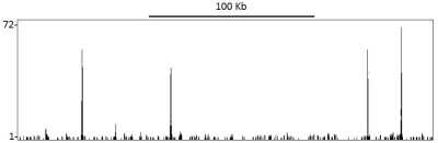
Chromatin Immunoprecipitation Sequencing: TIF1 alpha Antibody [NB100-2596] - Localization of TIF1 Alpha/TRIM24 Binding Sites by ChIP-sequencing.Chromatin from Breast cancer cell line T47D was immunoprecipitated with anti-TIF1 Alpha/TRIM24 antibody NB100-2596 and analyzed by DNA sequencing. The figure illustrates the peak distribution of TIF1 Alpha/TRIM24 binding within a 250 Kb region of chromosome 19 as detected using anti-TIF1 Alpha/TRIM24 antibody NB100-2596. ChIP-seq validation performed by Active Motif, Carlsbad, CA.
![Western Blot: TIF1 alpha Antibody [NB100-2596]](https://resources.rndsystems.com/images/products/TIF1-alpha-Antibody-Western-Blot-NB100-2596-img0003.jpg "Western Blot: TIF1 alpha Antibody [NB100-2596]")
![Immunohistochemistry-Paraffin: TIF1 alpha Antibody [NB100-2596]](https://resources.rndsystems.com/images/products/TIF1-alpha-Antibody-Immunohistochemistry-Paraffin-NB100-2596-img0012.jpg "Immunohistochemistry-Paraffin: TIF1 alpha Antibody [NB100-2596]")
Immunohistochemistry-Paraffin: TIF1 alpha Antibody [NB100-2596]
Immunohistochemistry-Paraffin: TIF1 alpha Antibody [NB100-2596] - Section of human breast carcinoma. Antibody: Affinity purified rabbit anti-TIF1 alpha/TRIM24 used at a dilution of 1:200 (1ug/ml). Detection: DABApplications for TIF1 alpha Antibody
Application
Recommended Usage
Immunohistochemistry
1:200 - 1:1000
Immunohistochemistry-Paraffin
1:200 - 1:1000
Immunoprecipitation
2 - 10 ug/mg lysate
Western Blot
1:2000-1:10000
Application Notes
Epitope retrieval with citrate buffer pH6.0 is recommended for FFPE tissue sections. ChIP-Seq ChIP-Seq 4 ug/30 ug chromatin.
Formulation, Preparation, and Storage
Purification
Immunogen affinity purified
Formulation
TBS and 0.1% BSA
Preservative
0.09% Sodium Azide
Concentration
0.2 mg/ml
Shipping
The product is shipped with polar packs. Upon receipt, store it immediately at the temperature recommended below.
Stability & Storage
Store at 4C. Do not freeze.
Background: TIF1 alpha
Long Name
Transcription intermediary factor 1-alpha
Alternate Names
RNF82, TIF1, TIF1-alpha, TIF1A, TRIM24
Gene Symbol
TRIM24
UniProt
Additional TIF1 alpha Products
Product Documents for TIF1 alpha Antibody
Certificate of Analysis
To download a Certificate of Analysis, please enter a lot or batch number in the search box below.
Product Specific Notices for TIF1 alpha Antibody
This product is for research use only and is not approved for use in humans or in clinical diagnosis. Primary Antibodies are guaranteed for 1 year from date of receipt.
Citations for TIF1 alpha Antibody
 Powered by Bioz
Customer Reviews for TIF1 alpha Antibody
There are currently no reviews for this product. Be the first to review TIF1 alpha Antibody and earn rewards!
Have you used TIF1 alpha Antibody?
Submit a review and receive an Amazon gift card!
$25/€18/£15/$25CAN/¥2500 Yen for a review with an image
$10/€7/£6/$10CAN/¥1110 Yen for a review without an image
Submit a review
Protocols
Find general support by application which include: protocols, troubleshooting, illustrated assays, videos and webinars.
- Antigen Retrieval Protocol (PIER)
- Antigen Retrieval for Frozen Sections Protocol
- Appropriate Fixation of IHC/ICC Samples
- Cellular Response to Hypoxia Protocols
- Chromogenic IHC Staining of Formalin-Fixed Paraffin-Embedded (FFPE) Tissue Protocol
- Chromogenic Immunohistochemistry Staining of Frozen Tissue
- ClariTSA™ Fluorophore Kits
- Detection & Visualization of Antibody Binding
- Fluorescent IHC Staining of Frozen Tissue Protocol
- Graphic Protocol for Heat-induced Epitope Retrieval
- Graphic Protocol for the Preparation and Fluorescent IHC Staining of Frozen Tissue Sections
- Graphic Protocol for the Preparation and Fluorescent IHC Staining of Paraffin-embedded Tissue Sections
- Graphic Protocol for the Preparation of Gelatin-coated Slides for Histological Tissue Sections
- IHC Sample Preparation (Frozen sections vs Paraffin)
- Immunofluorescent IHC Staining of Formalin-Fixed Paraffin-Embedded (FFPE) Tissue Protocol
- Immunohistochemistry (IHC) and Immunocytochemistry (ICC) Protocols
- Immunohistochemistry Frozen Troubleshooting
- Immunohistochemistry Paraffin Troubleshooting
- Immunoprecipitation Protocol
- Preparing Samples for IHC/ICC Experiments
- Preventing Non-Specific Staining (Non-Specific Binding)
- Primary Antibody Selection & Optimization
- Protocol for Heat-Induced Epitope Retrieval (HIER)
- Protocol for Making a 4% Formaldehyde Solution in PBS
- Protocol for VisUCyte™ HRP Polymer Detection Reagent
- Protocol for the Preparation & Fixation of Cells on Coverslips
- Protocol for the Preparation and Chromogenic IHC Staining of Frozen Tissue Sections
- Protocol for the Preparation and Chromogenic IHC Staining of Frozen Tissue Sections - Graphic
- Protocol for the Preparation and Chromogenic IHC Staining of Paraffin-embedded Tissue Sections
- Protocol for the Preparation and Chromogenic IHC Staining of Paraffin-embedded Tissue Sections - Graphic
- Protocol for the Preparation and Fluorescent IHC Staining of Frozen Tissue Sections
- Protocol for the Preparation and Fluorescent IHC Staining of Paraffin-embedded Tissue Sections
- Protocol for the Preparation of Gelatin-coated Slides for Histological Tissue Sections
- R&D Systems Quality Control Western Blot Protocol
- TUNEL and Active Caspase-3 Detection by IHC/ICC Protocol
- The Importance of IHC/ICC Controls
- Troubleshooting Guide: Immunohistochemistry
- Troubleshooting Guide: Western Blot Figures
- Western Blot Conditions
- Western Blot Protocol
- Western Blot Protocol for Cell Lysates
- Western Blot Troubleshooting
- Western Blot Troubleshooting Guide
- View all Protocols, Troubleshooting, Illustrated assays and Webinars
Loading...