Tight Junction Protein 1 Antibody - BSA Free
Novus Biologicals | Catalog # NBP1-85046
![Immunohistochemistry-Paraffin: Tight Junction Protein 1 Antibody [NBP1-85046]](https://resources.rndsystems.com/images/products/Tight-Junction-Protein-1-Antibody-Immunohistochemistry-Paraffin-NBP1-85046-img0013.jpg "Immunohistochemistry-Paraffin: Tight Junction Protein 1 Antibody [NBP1-85046]")
Loading...
Key Product Details
Validated by
Orthogonal Validation
Species Reactivity
Validated:
Human
Cited:
Human, Mouse, Rat
Applications
Validated:
Immunohistochemistry, Immunohistochemistry-Paraffin, Immunohistochemistry-Frozen, Immunocytochemistry/ Immunofluorescence
Cited:
Immunohistochemistry-Paraffin, Western Blot, IF/IHC
Label
Unconjugated
Antibody Source
Polyclonal Rabbit IgG
Format
BSA Free
Loading...
Product Specifications
Immunogen
This antibody was developed against Recombinant Protein corresponding to amino acids: ASSQPAKPTKVTLVKSRKNEEYGLRLASHIFVKEISQDSLAARDGNIQEGDVVLKINGTVTENMSLTDAKTLIERSKGKLKMVVQRDERATLLNVPDLSDSIHSANASERDDISEIQSLASDHSGR
Reactivity Notes
Rat reactivity reported in scientific literature (PMID: 31174067). Analysis of Tight Junction Protein 1 in RIPA whole cell lysates of Mouse primary choroid plexus epithelial cells using anti-Tight Junction Protein 1 antibody verified customer review.
Marker
Intercellular Junctions/Tight Junction Marker
Clonality
Polyclonal
Host
Rabbit
Isotype
IgG
Scientific Data Images for Tight Junction Protein 1 Antibody - BSA Free
Immunohistochemistry-Paraffin: Tight Junction Protein 1 Antibody [NBP1-85046]
Immunohistochemistry-Paraffin: Tight Junction Protein 1 Antibody [NBP1-85046] - Staining in human cerebral cortex and skeletal muscle tissues using anti-TJP1 antibody. Corresponding TJP1 RNA-seq data are presented for the same tissues.![Western Blot: Tight Junction Protein 1 Antibody [NBP1-85046]](https://resources.rndsystems.com/images/products/Tight-Junction-Protein-1-Antibody-Western-Blot-NBP1-85046-img0006.jpg "Western Blot: Tight Junction Protein 1 Antibody [NBP1-85046]")
Western Blot: Tight Junction Protein 1 Antibody [NBP1-85046]
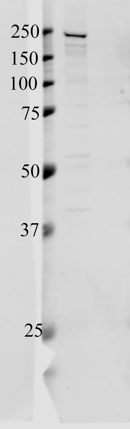
Western Blot: Tight Junction Protein 1 Antibody [NBP1-85046] - Analysis of Tight Junction Protein 1 in RIPA whole cell lysates of mouse primary choroid plexus epithelial cells using anti-Tight Junction Protein 1 antibody. Image from verified customer review.![Immunocytochemistry/ Immunofluorescence: Tight Junction Protein 1 Antibody [NBP1-85046]](https://resources.rndsystems.com/images/products/Tight-Junction-Protein-1-Antibody-Immunocytochemistry-Immunofluorescence-NBP1-85046-img0007.jpg "Immunocytochemistry/ Immunofluorescence: Tight Junction Protein 1 Antibody [NBP1-85046]")
Immunocytochemistry/ Immunofluorescence: Tight Junction Protein 1 Antibody [NBP1-85046]
Immunocytochemistry/Immunofluorescence: Tight Junction Protein 1 Antibody [NBP1-85046] - Staining of human cell line U-2 OS shows localization to cytosol & cell junctions. Antibody staining is shown in green.![Immunohistochemistry-Paraffin: Tight Junction Protein 1 Antibody [NBP1-85046]](https://resources.rndsystems.com/images/products/Tight-Junction-Protein-1-Antibody-Immunohistochemistry-Paraffin-NBP1-85046-img0011.jpg "Immunohistochemistry-Paraffin: Tight Junction Protein 1 Antibody [NBP1-85046]")
Immunohistochemistry-Paraffin: Tight Junction Protein 1 Antibody [NBP1-85046]
Immunohistochemistry-Paraffin: Tight Junction Protein 1 Antibody [NBP1-85046] - Staining of human cerebral cortex shows high expression.![Immunohistochemistry-Paraffin: Tight Junction Protein 1 Antibody [NBP1-85046]](https://resources.rndsystems.com/images/products/Tight-Junction-Protein-1-Antibody-Immunohistochemistry-Paraffin-NBP1-85046-img0012.jpg "Immunohistochemistry-Paraffin: Tight Junction Protein 1 Antibody [NBP1-85046]")
Immunohistochemistry-Paraffin: Tight Junction Protein 1 Antibody [NBP1-85046]
Immunohistochemistry-Paraffin: Tight Junction Protein 1 Antibody [NBP1-85046] - Staining of human skeletal muscle shows low expression as expected.
Western Blot: Tight Junction Protein 1 Antibody [NBP1-85046] -
Western Blot: Tight Junction Protein 1 Antibody [NBP1-85046] - Expression of tight junction proteins in cerebellum of rats with mild liver damage. Expression of (A,B) occludin & (C,D) ZO-1 at 2 & 4 weeks was analyzed by Western blot. Values are mean ± SEM of 10–12 rats per group. One-way ANOVA with Tukey’s test was performed for occludin at 2 weeks (F(3,27) = 4.99, p < 0.01) & 4 weeks (F(3,47) = 0.611, p > 0.05) & for ZO-1 at 2 weeks (W(3,23) = 0.715, p > 0.05) & 4 weeks (F(3,43) = 3.698, p < 0.05). Values significantly different from control rats are indicated by asterisks & from CCl4 rats by a. * p < 0.05; a p < 0.05. Image collected & cropped by CiteAb from the following publication (https://pubmed.ncbi.nlm.nih.gov/34440206), licensed under a CC-BY license. Not internally tested by Novus Biologicals.
Western Blot: Tight Junction Protein 1 Antibody [NBP1-85046] -
Western Blot: Tight Junction Protein 1 Antibody [NBP1-85046] - Expression of tight junction proteins in cerebellum of rats with mild liver damage. Expression of (A,B) occludin & (C,D) ZO-1 at 2 & 4 weeks was analyzed by Western blot. Values are mean ± SEM of 10–12 rats per group. One-way ANOVA with Tukey’s test was performed for occludin at 2 weeks (F(3,27) = 4.99, p < 0.01) & 4 weeks (F(3,47) = 0.611, p > 0.05) & for ZO-1 at 2 weeks (W(3,23) = 0.715, p > 0.05) & 4 weeks (F(3,43) = 3.698, p < 0.05). Values significantly different from control rats are indicated by asterisks & from CCl4 rats by a. * p < 0.05; a p < 0.05. Image collected & cropped by CiteAb from the following publication (https://pubmed.ncbi.nlm.nih.gov/34440206), licensed under a CC-BY license. Not internally tested by Novus Biologicals.Applications for Tight Junction Protein 1 Antibody - BSA Free
Application
Recommended Usage
Immunocytochemistry/ Immunofluorescence
0.25 - 2 ug/mL
Immunohistochemistry
1:50 - 1:200
Immunohistochemistry-Frozen
validated from a verified customer review.
Immunohistochemistry-Paraffin
1:50 - 1:200
Application Notes
IHC-Paraffin, HIER pH 6 retrieval is recommended. ICC/IF, Fixation Permeabilization: Use PFA/Triton X-100.
Reviewed Applications
Read 2 reviews rated 4.5 using NBP1-85046 in the following applications:
Formulation, Preparation, and Storage
Purification
Affinity purified
Formulation
PBS (pH 7.2) and 40% Glycerol
Format
BSA Free
Preservative
0.02% Sodium Azide
Concentration
Concentrations vary lot to lot. See vial label for concentration. If unlisted please contact technical services.
Shipping
The product is shipped with polar packs. Upon receipt, store it immediately at the temperature recommended below.
Stability & Storage
Store at 4C short term. Aliquot and store at -20C long term. Avoid freeze-thaw cycles.
Background: Tight Junction Protein 1
Alternate Names
DKFZp686M05161, MGC133289, Tight junction protein 1, tight junction protein 1 (zona occludens 1), tight junction protein ZO-1, TJP1, ZO1, ZO-1, zona occludens 1, Zona occludens protein 1, zonula occludens 1 protein, Zonula occludens protein 1
Gene Symbol
TJP1
Additional Tight Junction Protein 1 Products
Product Documents for Tight Junction Protein 1 Antibody - BSA Free
Certificate of Analysis
To download a Certificate of Analysis, please enter a lot or batch number in the search box below.
Product Specific Notices for Tight Junction Protein 1 Antibody - BSA Free
This product is for research use only and is not approved for use in humans or in clinical diagnosis. Primary Antibodies are guaranteed for 1 year from date of receipt.
Citations for Tight Junction Protein 1 Antibody - BSA Free
 Powered by Bioz
Customer Reviews for Tight Junction Protein 1 Antibody - BSA Free (2)
4.5 out of 5
2 Customer Ratings
Have you used Tight Junction Protein 1 Antibody - BSA Free?
Submit a review and receive an Amazon gift card!
$25/€18/£15/$25CAN/¥2500 Yen for a review with an image
$10/€7/£6/$10CAN/¥1110 Yen for a review without an image
Submit a review
Customer Images
Showing
1
-
2 of
2 reviews
Showing All
Filter By:
-
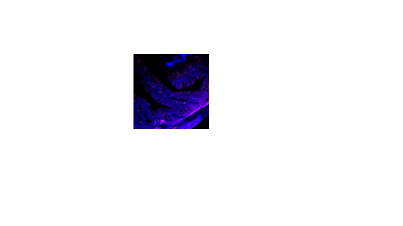
Application: Immunohistochemistry-FrozenSample Tested: Rat intestineSpecies: RatVerified Customer | Posted 05/21/2021Tight Junction expression (red color) in rat intestine. Blue color shows DAPI staining.

-
Application: Western BlotSample Tested: RIPA whole cell lysates of mouse primary choroid plexus epithelial cellsSpecies: MouseVerified Customer | Posted 08/26/2015Murine choroid plexus primary cell lysate in RIPA
There are no reviews that match your criteria.
Protocols
Find general support by application which include: protocols, troubleshooting, illustrated assays, videos and webinars.
- Antigen Retrieval Protocol (PIER)
- Antigen Retrieval for Frozen Sections Protocol
- Appropriate Fixation of IHC/ICC Samples
- Cellular Response to Hypoxia Protocols
- Chromogenic IHC Staining of Formalin-Fixed Paraffin-Embedded (FFPE) Tissue Protocol
- Chromogenic Immunohistochemistry Staining of Frozen Tissue
- ClariTSA™ Fluorophore Kits
- Detection & Visualization of Antibody Binding
- Fluorescent IHC Staining of Frozen Tissue Protocol
- Graphic Protocol for Heat-induced Epitope Retrieval
- Graphic Protocol for the Preparation and Fluorescent IHC Staining of Frozen Tissue Sections
- Graphic Protocol for the Preparation and Fluorescent IHC Staining of Paraffin-embedded Tissue Sections
- Graphic Protocol for the Preparation of Gelatin-coated Slides for Histological Tissue Sections
- ICC Cell Smear Protocol for Suspension Cells
- ICC Immunocytochemistry Protocol Videos
- ICC for Adherent Cells
- IHC Sample Preparation (Frozen sections vs Paraffin)
- Immunocytochemistry (ICC) Protocol
- Immunocytochemistry Troubleshooting
- Immunofluorescence of Organoids Embedded in Cultrex Basement Membrane Extract
- Immunofluorescent IHC Staining of Formalin-Fixed Paraffin-Embedded (FFPE) Tissue Protocol
- Immunohistochemistry (IHC) and Immunocytochemistry (ICC) Protocols
- Immunohistochemistry Frozen Troubleshooting
- Immunohistochemistry Paraffin Troubleshooting
- Preparing Samples for IHC/ICC Experiments
- Preventing Non-Specific Staining (Non-Specific Binding)
- Primary Antibody Selection & Optimization
- Protocol for Heat-Induced Epitope Retrieval (HIER)
- Protocol for Making a 4% Formaldehyde Solution in PBS
- Protocol for VisUCyte™ HRP Polymer Detection Reagent
- Protocol for the Fluorescent ICC Staining of Cell Smears - Graphic
- Protocol for the Fluorescent ICC Staining of Cultured Cells on Coverslips - Graphic
- Protocol for the Preparation & Fixation of Cells on Coverslips
- Protocol for the Preparation and Chromogenic IHC Staining of Frozen Tissue Sections
- Protocol for the Preparation and Chromogenic IHC Staining of Frozen Tissue Sections - Graphic
- Protocol for the Preparation and Chromogenic IHC Staining of Paraffin-embedded Tissue Sections
- Protocol for the Preparation and Chromogenic IHC Staining of Paraffin-embedded Tissue Sections - Graphic
- Protocol for the Preparation and Fluorescent ICC Staining of Cells on Coverslips
- Protocol for the Preparation and Fluorescent ICC Staining of Non-adherent Cells
- Protocol for the Preparation and Fluorescent ICC Staining of Stem Cells on Coverslips
- Protocol for the Preparation and Fluorescent IHC Staining of Frozen Tissue Sections
- Protocol for the Preparation and Fluorescent IHC Staining of Paraffin-embedded Tissue Sections
- Protocol for the Preparation of Gelatin-coated Slides for Histological Tissue Sections
- Protocol for the Preparation of a Cell Smear for Non-adherent Cell ICC - Graphic
- TUNEL and Active Caspase-3 Detection by IHC/ICC Protocol
- The Importance of IHC/ICC Controls
- Troubleshooting Guide: Immunohistochemistry
- View all Protocols, Troubleshooting, Illustrated assays and Webinars
Loading...