TLR7 Antibody - BSA Free
Novus Biologicals | Catalog # NBP2-24906
Key Product Details
Species Reactivity
Validated:
Human, Mouse, Rat
Cited:
Human, Mouse, Rat
Applications
Validated:
Immunohistochemistry, Immunohistochemistry-Paraffin, Immunohistochemistry-Frozen, Western Blot, Flow Cytometry, Flow (Intracellular), Immunocytochemistry/ Immunofluorescence, Simple Western, Immunoprecipitation, Bioactivity, Dot Blot, Proximity Ligation Assay
Cited:
Immunohistochemistry, Immunohistochemistry-Paraffin, Immunohistochemistry-Frozen, Western Blot, Block/Neutralize, Flow Cytometry, Immunocytochemistry/ Immunofluorescence, Immunoprecipitation, Bioassay, Cytometric Bead Assay Standard, Flow Cytometry Control, Proximity Ligation Assay, IF/IHC
Label
Unconjugated
Antibody Source
Polyclonal Rabbit IgG
Format
BSA Free
Loading...
Product Specifications
Immunogen
Partial synthetic peptide made to an internal portion of the human TLR7 protein (between amino acids 700-750) [UniProt 9NYK1]
Reactivity Notes
Use in Human reported in scientific literature (PMID:33806288).
Clonality
Polyclonal
Host
Rabbit
Isotype
IgG
Scientific Data Images for TLR7 Antibody - BSA Free
![Immunohistochemistry: TLR7 Antibody - BSA Free [NBP2-24906]](https://resources.rndsystems.com/images/products/TLR7-Antibody-Immunohistochemistry-NBP2-24906-img0022.jpg "Immunohistochemistry: TLR7 Antibody - BSA Free [NBP2-24906]")
Immunohistochemistry: TLR7 Antibody - BSA Free [NBP2-24906]
TLR7-Antibody-Immunohistochemistry-NBP2-24906-img0022.jpg![Flow Cytometry: TLR7 Antibody - BSA Free [NBP2-24906]](https://resources.rndsystems.com/images/products/TLR7-Antibody-Flow-Cytometry-NBP2-24906-img0025.jpg "Flow Cytometry: TLR7 Antibody - BSA Free [NBP2-24906]")
Flow Cytometry: TLR7 Antibody - BSA Free [NBP2-24906]
Flow Cytometry: TLR7 Antibody [NBP2-24906] - An intracellular stain was performed on THP-1 cells with NBP2-24906 (blue) and a matched isotype control NBP2-24891 (orange). Cells were fixed with 4% PFA and then permeabilized with 0.1% saponin. Cells were incubated in an antibody dilution of 1 ug/mL for 30 minutes at room temperature, followed by Rabbit IgG (H+L) Cross-Adsorbed Secondary Antibody, Dylight 550 (SA5-10033, Thermo Fisher).![Immunocytochemistry/ Immunofluorescence: TLR7 Antibody - BSA Free [NBP2-24906]](https://resources.rndsystems.com/images/products/TLR7-Antibody-Immunocytochemistry-Immunofluorescence-NBP2-24906-img0002.jpg "Immunocytochemistry/ Immunofluorescence: TLR7 Antibody - BSA Free [NBP2-24906]")
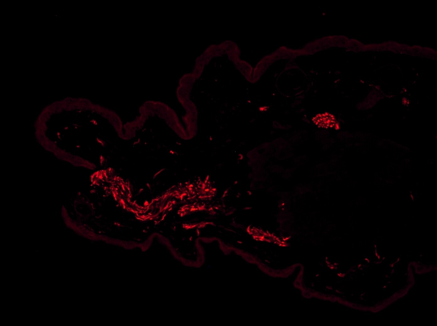
Immunocytochemistry/ Immunofluorescence: TLR7 Antibody - BSA Free [NBP2-24906]
Immunocytochemistry/Immunofluorescence: TLR7 Antibody [NBP2-24906] - Immunolocalization of TLR7 (A) and glial fibrillary acidic protein (GFAP) (B) in murine myenteric plexus. Most of the GFAP positive cells were also positive for TLR7 in the merged image (C, yellow staining). (Courtesy of Barajon I, et al., Journal of Histochemistry and Cytochemistry, 57(11): 1013-;1023, 2009.)![Immunohistochemistry: TLR7 Antibody - BSA Free [NBP2-24906]](https://resources.rndsystems.com/images/products/TLR7-Antibody-Immunohistochemistry-NBP2-24906-img0023.jpg "Immunohistochemistry: TLR7 Antibody - BSA Free [NBP2-24906]")
Immunohistochemistry: TLR7 Antibody - BSA Free [NBP2-24906]
TLR7-Antibody-Immunohistochemistry-NBP2-24906-img0023.jpg![Immunohistochemistry-Frozen: TLR7 Antibody - BSA Free [NBP2-24906]](https://resources.rndsystems.com/images/products/TLR7-Antibody-Immunohistochemistry-Frozen-NBP2-24906-img0006.jpg "Immunohistochemistry-Frozen: TLR7 Antibody - BSA Free [NBP2-24906]")
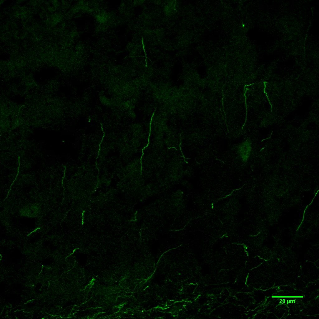
Immunohistochemistry-Frozen: TLR7 Antibody - BSA Free [NBP2-24906]
Immunohistochemistry-Frozen: TLR7 Antibody [NBP2-24906] - analysis of frozen mouse ear skin tissue using anti-TLR7 antibody. Image from verified customer review.![Immunohistochemistry-Frozen: TLR7 Antibody - BSA Free [NBP2-24906]](https://resources.rndsystems.com/images/products/TLR7-Antibody-Immunohistochemistry-Frozen-NBP2-24906-img0005.jpg "Immunohistochemistry-Frozen: TLR7 Antibody - BSA Free [NBP2-24906]")
Immunohistochemistry-Frozen: TLR7 Antibody - BSA Free [NBP2-24906]
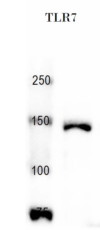
Immunohistochemistry-Frozen: TLR7 Antibody [NBP2-24906] - analysis of TLR7 in acetone-fixed, frozen mouse ear skin section using anti-TLR7 antibody. Image from verified customer review.![Western Blot: TLR7 AntibodyBSA Free [NBP2-24906]](https://resources.rndsystems.com/images/products/TLR7-Antibody-Western-Blot-NBP2-24906-img0018.jpg "Western Blot: TLR7 AntibodyBSA Free [NBP2-24906]")
Western Blot: TLR7 AntibodyBSA Free [NBP2-24906]
Western Blot: TLR7 Antibody [NBP2-24906] - Analysis using the Azide Free version of NBP2-24906. Detection of TLR7 in RAW cell lysate using this antibody.![Immunocytochemistry/ Immunofluorescence: TLR7 Antibody - BSA Free [NBP2-24906]](https://resources.rndsystems.com/images/products/TLR7-Antibody-Immunocytochemistry-Immunofluorescence-NBP2-24906-img0017.jpg "Immunocytochemistry/ Immunofluorescence: TLR7 Antibody - BSA Free [NBP2-24906]")
Immunocytochemistry/ Immunofluorescence: TLR7 Antibody - BSA Free [NBP2-24906]
Immunocytochemistry/Immunofluorescence: TLR7 Antibody [NBP2-24906] - Analysis using the Azide Free version of NBP2-24906. Staining of Raw 246.7 cells with Dylight 488 (green). Nuclei and alpha-tubulin were counterstained with DAPI (blue) and Dylight 550 (red). Image objective 40x. An antibody dilution of 1:10 was used.![Immunohistochemistry-Paraffin: TLR7 Antibody - BSA Free [NBP2-24906]](https://resources.rndsystems.com/images/products/TLR7-Antibody---BSA-Free-Immunohistochemistry-Paraffin-NBP2-24906-img0026.jpg "Immunohistochemistry-Paraffin: TLR7 Antibody - BSA Free [NBP2-24906]")
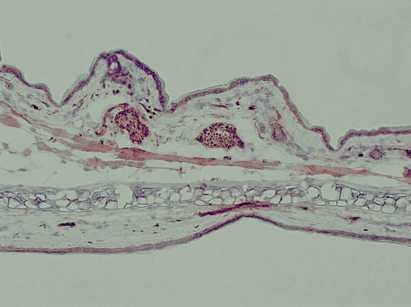
Immunohistochemistry-Paraffin: TLR7 Antibody - BSA Free [NBP2-24906]
Immunohistochemistry-Paraffin: TLR7 Antibody - BSA Free [NBP2-24906] - Analysis of a FFPE tissue section of human skin using 1:200 dilution of TLR7 antibody (NBP2-24906). The staining was developed using HRP labeled anti-rabbit secondary antibody and DAB reagent, and nuclei of cells were counter-stained with hematoxylin.
Immunohistochemistry-Paraffin: TLR7 Antibody - BSA Free [NBP2-24906] -
Immunohistochemistry-Paraffin: TLR7 Antibody - BSA Free [NBP2-24906] - Analysis of human colon tissue using NBP2-24905 (top) and an isotype control (bottom) at 5 ug/ml.
Immunohistochemistry: TLR7 Antibody - BSA Free [NBP2-24906] -
Immunohistochemistry: TLR7 Antibody - BSA Free [NBP2-24906] - Expression of TLR7 in lung tissues of non-smokers, smokers, & COPD patients. Total protein & mRNA were obtained from lung tissues of non-smokers (n = 15), smokers (n = 12), & COPD patients (n = 15). TLR7 protein & mRNA expression were determined by western blot (A) & real time PCR (B) in lung parenchyma. (A) Representative images of western blot for TLR7 & corresponding densitometry expressed as ratio to beta -actin. (B) TLR7 mRNA expression given as the ratio to GAPDH. (C, D, E) Lung sections were immunostained for TLR7 & quantified by means of immunohistochemical score of TLR7 in alveolar macrophages (C) & bronchial epithelial cells (D). (E) Representative immunohistochemistry images are shown. The control IgG isotype showed negative staining. Data are presented as individual values & mean ± standard deviation. Exact P values were obtained using Kruskal-Wallis & Dunn’s post-hoc tests. Image collected & cropped by CiteAb from the following publication (https://respiratory-research.biomedcentral.com/articles/10.1186/s12931-…), licensed under a CC-BY license. Not internally tested by Novus Biologicals.
Western Blot: TLR7 Antibody - BSA Free [NBP2-24906] -
Expression of TLR7 in brain tissues at different time points after induction of status epilepticus.Quantitative real-time PCR analysis of the expression of TLR7 mRNA in the hippocampus (a) and cortex (b) at different time points after induction of SE (n = 3). ns not significant; *P < 0.05; ****P < 0.0001; one-way ANOVA with Tukey’s post hoc test. Bars represent the mean +/- SEM. The expression of FL-TLR7 and C-terminal TLR7 in the hippocampus (c, d) and temporal cortex (e, f) on the third day after induction of SE by intrahippocampal injection with KA were both significantly higher than that of the control group (n = 9 mice per group). **P < 0.01; unpaired two-tailed Student’s t-test. Bars represent the mean +/- SEM. Image collected and cropped by CiteAb from the following open publication (https://pubmed.ncbi.nlm.nih.gov/37258573), licensed under a CC-BY license. Not internally tested by Novus Biologicals.
Western Blot: TLR7 Antibody - BSA Free [NBP2-24906] -
Expression of TLR7 in brain tissues at different time points after induction of status epilepticus.Quantitative real-time PCR analysis of the expression of TLR7 mRNA in the hippocampus (a) and cortex (b) at different time points after induction of SE (n = 3). ns not significant; *P < 0.05; ****P < 0.0001; one-way ANOVA with Tukey’s post hoc test. Bars represent the mean +/- SEM. The expression of FL-TLR7 and C-terminal TLR7 in the hippocampus (c, d) and temporal cortex (e, f) on the third day after induction of SE by intrahippocampal injection with KA were both significantly higher than that of the control group (n = 9 mice per group). **P < 0.01; unpaired two-tailed Student’s t-test. Bars represent the mean +/- SEM. Image collected and cropped by CiteAb from the following open publication (https://pubmed.ncbi.nlm.nih.gov/37258573), licensed under a CC-BY license. Not internally tested by Novus Biologicals.Applications for TLR7 Antibody - BSA Free
Application
Recommended Usage
Bioactivity
reported in scientific literature (PMID 28894449)
Dot Blot
reported in scientific literature (PMID 27248820)
Flow Cytometry
2-5 ug/ 1x10^6 cells
Immunocytochemistry/ Immunofluorescence
1:10-1:2000
Immunohistochemistry
1:200. Use reported in scientific literature (PMID 28219705)
Immunohistochemistry-Frozen
1:10-1:2000. Use reported by customer review
Immunohistochemistry-Paraffin
1:200. Use reported in scientific literature (PMID 25264223)
Immunoprecipitation
reported in scientific literature (PMID 17452530)
Proximity Ligation Assay
reported in scientific literature (PMID 30579042)
Simple Western
1:20
Western Blot
1-3 ug/ml
Reviewed Applications
Read 4 reviews rated 5 using NBP2-24906 in the following applications:
Flow Cytometry Panel Builder
Bio-Techne Knows Flow Cytometry
Save time and reduce costly mistakes by quickly finding compatible reagents using the Panel Builder Tool.
Advanced Features
- Spectra Viewer - Custom analysis of spectra from multiple fluorochromes
- Spillover Popups - Visualize the spectra of individual fluorochromes
- Antigen Density Selector - Match fluorochrome brightness with antigen density

Formulation, Preparation, and Storage
Purification
Immunogen affinity purified
Formulation
PBS
Format
BSA Free
Preservative
0.02% Sodium Azide
Concentration
1.0 mg/ml
Shipping
The product is shipped with polar packs. Upon receipt, store it immediately at the temperature recommended below.
Stability & Storage
Store at 4C short term. Aliquot and store at -20C long term. Avoid freeze-thaw cycles.
Background: TLR7
While TLRs play an important role in innate immune response, dysfunction in the TLR-MyD88 signaling cascade has also been reported in various autoimmune disorders (5,6). Elevated expression of TLR7 is associated with increased risk of system lupus erythematosus (SLE), an autoimmune disease involving B cell hyperactivity (6,7). Studies involving mouse models has also found that increased TLR7 expression predisposes mice to a lupus-like disease (7). Therapeutics targeting TLR7 have been developed to either enhance or inhibit its activity depending on the circumstance. For example, TLR7 agonists such as imiquimod, resiquimod, and 852A are used to increase TLR7 activity for treatment of cancers and to fight viral infections (7,8). On the other hand, TLR7 antagonists inhibit its activation and have been developed to combat chronic immune stimulation as seen in inflammatory and autoimmune diseases (8).
References
1. Petes C, Odoardi N, Gee K. The Toll for Trafficking: Toll-Like Receptor 7 Delivery to the Endosome. Front Immunol. 2017;8:1075. https://doi.org/10.3389/fimmu.2017.01075
2. Maeda K, Akira S. TLR7 Structure: Cut in Z-Loop. Immunity. 2016;45(4):705-707. https://doi.org/10.1016/j.immuni.2016.10.003
3. Krieg AM, Vollmer J. Toll-like receptors 7, 8, and 9: linking innate immunity to autoimmunity. Immunol Rev. 2007;220:251-269. https://doi.org/10.1111/j.1600-065X.2007.00572.x
4. Uniprot (Q9NYK1)
5. Zheng C, Chen J, Chu F, Zhu J, Jin T. Inflammatory Role of TLR-MyD88 Signaling in Multiple Sclerosis. Front Mol Neurosci. 2020;12:314. https://doi.org/10.3389/fnmol.2019.00314
6. Chi H, Li C, Zhao FS, et al. Anti-tumor Activity of Toll-Like Receptor 7 Agonists. Front Pharmacol. 2017;8:304. https://doi.org/10.3389/fphar.2017.00304
7. Fillatreau S, Manfroi B, Dorner T. Toll-like receptor signalling in B cells during systemic lupus erythematosus. Nat Rev Rheumatol. 2021;17(2):98-108. https://doi.org/10.1038/s41584-020-00544-4
8. Patinote C, Karroum NB, Moarbess G, et al. Agonist and antagonist ligands of toll-like receptors 7 and 8: Ingenious tools for therapeutic purposes. Eur J Med Chem. 2020;193:112238. https://doi.org/10.1016/j.ejmech.2020.112238
Long Name
Toll-like Receptor 7
Alternate Names
toll-like receptor 7
Gene Symbol
TLR7
Additional TLR7 Products
Product Documents for TLR7 Antibody - BSA Free
Certificate of Analysis
To download a Certificate of Analysis, please enter a lot or batch number in the search box below.
Product Specific Notices for TLR7 Antibody - BSA Free
This product is for research use only and is not approved for use in humans or in clinical diagnosis. Primary Antibodies are guaranteed for 1 year from date of receipt.
Citations for TLR7 Antibody - BSA Free
 Powered by Bioz
Customer Reviews for TLR7 Antibody - BSA Free (4)
5 out of 5
4 Customer Ratings
Have you used TLR7 Antibody - BSA Free?
Submit a review and receive an Amazon gift card!
$25/€18/£15/$25CAN/¥2500 Yen for a review with an image
$10/€7/£6/$10CAN/¥1110 Yen for a review without an image
Submit a review
Customer Images
Showing
1
-
4 of
4 reviews
Showing All
Filter By:
-
Application: Immunohistochemistry-FrozenSample Tested: Fixe mouse brain tissueSpecies: MouseVerified Customer | Posted 08/02/2019Immunostaing of 4% PFA fixed mouse brain tissue, stained with Rabbi anti-TLR7 (dilution 1:500; Alexa 488 green).

-
Application: Western BlotSample Tested: Dorsal root ganglion neuronsSpecies: RatVerified Customer | Posted 12/02/2017
-
Application: Immunohistochemistry-FrozenSample Tested: Mouse Ear Skin (frozen section)Species: MouseVerified Customer | Posted 08/29/2015Mouse Ear Skin (frozen section)
-
Application: ImmunofluorescenceSample Tested: Mouse Ear Skin (frozen section)Species: MouseVerified Customer | Posted 08/28/2015Mouse Ear Skin
There are no reviews that match your criteria.
Protocols
View specific protocols for TLR7 Antibody - BSA Free (NBP2-24906):
Protocol for Flow Cytometry Intracellular Staining
Sample Preparation.
1. Grow cells to 60-85% confluency. Flow cytometry requires between 2 x 10⁵ and 1 x 10⁶ cells for optimal performance.
2. If cells are adherent, harvest gently by washing once with staining buffer and then scraping. Avoid using trypsin as this can disrupt certain epitopes of interest. If enzymatic harvest is required, use Accutase, Collagenase, or TrypLE Express for a less damaging option.
3. Reserve 100 uL for counting, then transfer cell volume into a 50 mL conical tube and centrifuge for 8 minutes at 400 RCF.
a. Count cells using a hemocytometer and a 1:1 trypan blue exclusion stain to determine cell viability before starting the flow protocol. If cells appear blue, do not proceed.
4. Re-suspend cells to a concentration of 1 x 10⁶ cells/mL in staining buffer (NBP2-26247).
5. Aliquot out 100 uL samples in accordance with your experimental samples.
Tip: When cell surface and intracellular staining are required in the same sample, it is advisable that the cell surface staining be performed first since the fixation and permeabilization steps might reduce the availability of surface antigens.
Intracellular Staining.
Tip: When performing intracellular staining, it is important to use appropriate fixation and permeabilization reagents based upon the target and its subcellular location. Generally, our Intracellular Flow Assay Kit (NBP2-29450) is a good place to start as it contains an optimized combination of reagents for intracellular staining as well as an inhibitor of intracellular protein transport (necessary if staining secreted proteins). Certain targets may require more gentle or transient permeabilization protocols such as the commonly employed methanol or saponin-based methods.
Protocol for Cytoplasmic Targets:
1. Fix the cells by adding 100 uL fixation solution (such as 4% PFA) to each sample for 10-15 minutes.
2. Permeabilize cells by adding 100 uL of a permeabilization buffer to every 1 x 10⁶ cells present in the sample. Mix well and incubate at room temperature for 15 minutes.
a. For cytoplasmic targets, use a gentle permeabilization solution such as 1X PBS + 0.5% Saponin or 1X PBS + 0.5% Tween-20.
b. To maintain the permeabilized state throughout your experiment, use staining buffer + 0.1% of the permeabilization reagent (i.e. 0.1% Tween-20 or 0.1% Saponin).
3. Following the 15 minute incubation, add 2 mL of the staining buffer + 0.1% permeabilizer to each sample.
4. Centrifuge for 1 minute at 400 RCF.
5. Discard supernatant and re-suspend in 100 uL of staining buffer + 0.1% permeabilizer.
6. Add appropriate amount of each antibody (eg. 1 test or 1 ug per sample, as experimentally determined).
7. Mix well and incubate at room temperature for 30 minutes- 1 hour. Gently mix samples every 10-15 minutes.
8. Following the primary/conjugate incubation, add 1-2 mL/sample of staining buffer +0.1% permeabilizer and centrifuge for 1 minute at 400 RCF.
9. Wash twice by re-suspending cells in staining buffer (2 mL for tubes or 200 uL for wells) and centrifuging at 400 RCF for 5 minutes. Discard supernatant.
10. Add appropriate amount of secondary antibody (as experimentally determined) to each sample.
11. Incubate at room temperature in dark for 20 minutes.
12. Add 1-2 mL of staining buffer and centrifuge at 400 RCF for 1 minute and discard supernatant.
13. Wash twice by re-suspending cells in staining buffer (2 mL for tubes or 200 uL for wells) and centrifuging at 400 RCF for 5 minutes. Discard supernatant.
14. Resuspend in an appropriate volume of staining buffer (usually 500 uL per sample) and proceed with analysis on your flow cytometer.
Immunocytochemistry Protocol
Culture cells to appropriate density in 35 mm culture dishes or 6-well plates.
1. Remove culture medium and wash the cells briefly in PBS. Add 4% paraformaldehyde to the dish and fix at room temperature for 10 minutes.
2. Remove the paraformaldehyde and wash the cells in PBS.
3. Permeabilize the cells with 0.1% Triton X100 or other suitable detergent for 2 min.
4. Remove the permeabilization buffer and wash three times for 5 minutes each in PBS. Be sure to not let the specimen dry out.
5. To block nonspecific antibody binding, incubate in 10% normal goat serum from 1 hour to overnight at room temperature.
6. Add primary antibody at appropriate dilution and incubate overnight at 4C.
7. Remove primary antibody and replace with PBS. Wash three times for 5 minutes each.
8. Add secondary antibody at appropriate dilution. Incubate for 1 hour at room temperature.
9. Remove secondary antibody and replace with PBS. Wash three times for 5 minutes each.
10. Counter stain DNA with DAPI if required.
Immunohistochemistry-Paraffin Embedded Sections
Antigen Unmasking:
Bring slides to a boil in 10 mM sodium citrate buffer (pH 6.0) then maintain at a sub-boiling temperature for 10 minutes. Cool slides on bench-top for 30 minutes (keep slides in the sodium citrate buffer all the time).
Staining:
1. Wash sections in deionized water three times for 5 minutes each.
2. Wash sections in PBS for 5 minutes.
3. Block each section with 100-400 ul blocking solution (1% BSA in PBS) for 1 hour at room temperature.
4. Remove blocking solution and add 100-400 ul diluted primary antibody. Incubate overnight at 4 C.
5. Remove antibody solution and wash sections in wash buffer three times for 5 minutes each.
6. Add 100-400 ul HRP polymer conjugated secondary antibody. Incubate 30 minutes at room temperature.
7. Wash sections three times in wash buffer for 5 minutes each.
8. Add 100-400 ul DAB substrate to each section and monitor staining closely.
9. As soon as the sections develop, immerse slides in deionized water.
10. Counterstain sections in hematoxylin.
11. Wash sections in deionized water two times for 5 minutes each.
12. Dehydrate sections.
13. Mount coverslips.
Western Blot Protocol
1. Perform SDS-PAGE on samples to be analyzed, loading 10-25 ug of total protein per lane.
2. Transfer proteins to PVDF membrane according to the instructions provided by the manufacturer of the membrane and transfer apparatus.
3. Stain the membrane with Ponceau S (or similar product) to assess transfer success, and mark molecular weight standards where appropriate.
4. Rinse the blot TBS -0.05% Tween 20 (TBST).
5. Block the membrane in 5% Non-fat milk in TBST (blocking buffer) for at least 1 hour.
6. Wash the membrane in TBST three times for 10 minutes each.
7. Dilute primary antibody in blocking buffer and incubate overnight at 4C with gentle rocking.
8. Wash the membrane in TBST three times for 10 minutes each.
9. Incubate the membrane in diluted HRP conjugated secondary antibody in blocking buffer (as per manufacturer's instructions) for 1 hour at room temperature.
10. Wash the blot in TBST three times for 10 minutes each (this step can be repeated as required to reduce background).
11. Apply the detection reagent of choice in accordance with the manufacturer's instructions.
Find general support by application which include: protocols, troubleshooting, illustrated assays, videos and webinars.
- 7-Amino Actinomycin D (7-AAD) Cell Viability Flow Cytometry Protocol
- Antigen Retrieval Protocol (PIER)
- Antigen Retrieval for Frozen Sections Protocol
- Appropriate Fixation of IHC/ICC Samples
- Cellular Response to Hypoxia Protocols
- Chromogenic IHC Staining of Formalin-Fixed Paraffin-Embedded (FFPE) Tissue Protocol
- Chromogenic Immunohistochemistry Staining of Frozen Tissue
- ClariTSA™ Fluorophore Kits
- Detection & Visualization of Antibody Binding
- Extracellular Membrane Flow Cytometry Protocol
- Flow Cytometry Protocol for Cell Surface Markers
- Flow Cytometry Protocol for Staining Membrane Associated Proteins
- Flow Cytometry Staining Protocols
- Flow Cytometry Troubleshooting Guide
- Fluorescent IHC Staining of Frozen Tissue Protocol
- Graphic Protocol for Heat-induced Epitope Retrieval
- Graphic Protocol for the Preparation and Fluorescent IHC Staining of Frozen Tissue Sections
- Graphic Protocol for the Preparation and Fluorescent IHC Staining of Paraffin-embedded Tissue Sections
- Graphic Protocol for the Preparation of Gelatin-coated Slides for Histological Tissue Sections
- ICC Cell Smear Protocol for Suspension Cells
- ICC Immunocytochemistry Protocol Videos
- ICC for Adherent Cells
- IHC Sample Preparation (Frozen sections vs Paraffin)
- Immunocytochemistry (ICC) Protocol
- Immunocytochemistry Troubleshooting
- Immunofluorescence of Organoids Embedded in Cultrex Basement Membrane Extract
- Immunofluorescent IHC Staining of Formalin-Fixed Paraffin-Embedded (FFPE) Tissue Protocol
- Immunohistochemistry (IHC) and Immunocytochemistry (ICC) Protocols
- Immunohistochemistry Frozen Troubleshooting
- Immunohistochemistry Paraffin Troubleshooting
- Immunoprecipitation Protocol
- Intracellular Flow Cytometry Protocol Using Alcohol (Methanol)
- Intracellular Flow Cytometry Protocol Using Detergents
- Intracellular Nuclear Staining Flow Cytometry Protocol Using Detergents
- Intracellular Staining Flow Cytometry Protocol Using Alcohol Permeabilization
- Intracellular Staining Flow Cytometry Protocol Using Detergents to Permeabilize Cells
- Preparing Samples for IHC/ICC Experiments
- Preventing Non-Specific Staining (Non-Specific Binding)
- Primary Antibody Selection & Optimization
- Propidium Iodide Cell Viability Flow Cytometry Protocol
- Protocol for Heat-Induced Epitope Retrieval (HIER)
- Protocol for Liperfluo
- Protocol for Making a 4% Formaldehyde Solution in PBS
- Protocol for VisUCyte™ HRP Polymer Detection Reagent
- Protocol for the Characterization of Human Th22 Cells
- Protocol for the Characterization of Human Th9 Cells
- Protocol for the Fluorescent ICC Staining of Cell Smears - Graphic
- Protocol for the Fluorescent ICC Staining of Cultured Cells on Coverslips - Graphic
- Protocol for the Preparation & Fixation of Cells on Coverslips
- Protocol for the Preparation and Chromogenic IHC Staining of Frozen Tissue Sections
- Protocol for the Preparation and Chromogenic IHC Staining of Frozen Tissue Sections - Graphic
- Protocol for the Preparation and Chromogenic IHC Staining of Paraffin-embedded Tissue Sections
- Protocol for the Preparation and Chromogenic IHC Staining of Paraffin-embedded Tissue Sections - Graphic
- Protocol for the Preparation and Fluorescent ICC Staining of Cells on Coverslips
- Protocol for the Preparation and Fluorescent ICC Staining of Non-adherent Cells
- Protocol for the Preparation and Fluorescent ICC Staining of Stem Cells on Coverslips
- Protocol for the Preparation and Fluorescent IHC Staining of Frozen Tissue Sections
- Protocol for the Preparation and Fluorescent IHC Staining of Paraffin-embedded Tissue Sections
- Protocol for the Preparation of Gelatin-coated Slides for Histological Tissue Sections
- Protocol for the Preparation of a Cell Smear for Non-adherent Cell ICC - Graphic
- Protocol: Annexin V and PI Staining by Flow Cytometry
- Protocol: Annexin V and PI Staining for Apoptosis by Flow Cytometry
- R&D Systems Quality Control Western Blot Protocol
- TUNEL and Active Caspase-3 Detection by IHC/ICC Protocol
- The Importance of IHC/ICC Controls
- Troubleshooting Guide: Fluorokine Flow Cytometry Kits
- Troubleshooting Guide: Immunohistochemistry
- Troubleshooting Guide: Western Blot Figures
- Western Blot Conditions
- Western Blot Protocol
- Western Blot Protocol for Cell Lysates
- Western Blot Troubleshooting
- Western Blot Troubleshooting Guide
- View all Protocols, Troubleshooting, Illustrated assays and Webinars
FAQs for TLR7 Antibody - BSA Free
Showing
1
-
1 of
1 FAQ
Showing All
-
Q: Regarding antibody NBP2-24906 is the western blot of RAW264.7 cells detected with colorimetric staining? or is it luminescence (ECL)?
A: Image 5 of 13 is ECL


