![Western Blot: TOP2B Antibody [NB100-40842]](https://resources.rndsystems.com/images/products/TOP2B-Antibody-Western-Blot-NB100-40842-img0010.jpg "Western Blot: TOP2B Antibody [NB100-40842]")
Loading...
Key Product Details
Validated by
Independent Antibodies, Biological Validation
Species Reactivity
Validated:
Human, Mouse
Cited:
Human, Mouse
Applications
Validated:
Immunohistochemistry, Immunohistochemistry-Paraffin, Western Blot, Immunoprecipitation
Cited:
Western Blot, Immunocytochemistry/ Immunofluorescence, Immunoprecipitation
Label
Unconjugated
Antibody Source
Polyclonal Rabbit IgG
Loading...
Product Specifications
Immunogen
The immunogen recognized by this antibody maps to a region between residue 1570 and the C-terminus (residue 1621) of human DNA Topoisomerase II Beta using the numbering given in entry NP_001059.2 (GeneID 7155).
Clonality
Polyclonal
Host
Rabbit
Isotype
IgG
Theoretical MW
183 kDa.
Disclaimer note: The observed molecular weight of the protein may vary from the listed predicted molecular weight due to post translational modifications, post translation cleavages, relative charges, and other experimental factors.
Disclaimer note: The observed molecular weight of the protein may vary from the listed predicted molecular weight due to post translational modifications, post translation cleavages, relative charges, and other experimental factors.
Scientific Data Images for TOP2B Antibody
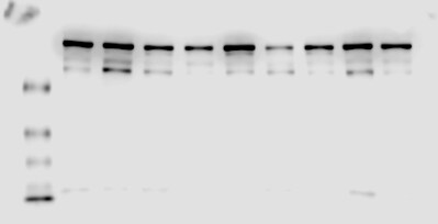
Western Blot: TOP2B Antibody [NB100-40842]
Western Blot: TOP2B Antibody [NB100-40842] - Whole cell lysate (50 ug) from HEK293T, HeLa, Jurkat, TCMK-1, and NIH 3T3 cells prepared using NETN lysis buffer. Antibody: Affinity purified rabbit anti-Topo II Beta antibody used for WB at 0.04 ug/ml. Detection: Chemiluminescence withan exposure time of 30 seconds![Immunohistochemistry-Paraffin: TOP2B Antibody [NB100-40842]](https://resources.rndsystems.com/images/products/TOP2B-Antibody-Immunohistochemistry-Paraffin-NB100-40842-img0009.jpg "Immunohistochemistry-Paraffin: TOP2B Antibody [NB100-40842]")
Immunohistochemistry-Paraffin: TOP2B Antibody [NB100-40842]
Immunohistochemistry-Paraffin: TOP2B Antibody [NB100-40842] - FFPE section of human small cell lung cancer. Antibody: Affinity purified rabbitanti-Topo II Beta used at a dilution of 1:1,000 (0.2ug/ml). Detection: DAB![Western Blot: TOP2B Antibody [NB100-40842]](https://resources.rndsystems.com/images/products/TOP2B-Antibody-Western-Blot-NB100-40842-img0007.jpg "Western Blot: TOP2B Antibody [NB100-40842]")
![Immunohistochemistry-Paraffin: TOP2B Antibody [NB100-40842]](https://resources.rndsystems.com/images/products/TOP2B-Antibody-Immunohistochemistry-Paraffin-NB100-40842-img0008.jpg "Immunohistochemistry-Paraffin: TOP2B Antibody [NB100-40842]")
Immunohistochemistry-Paraffin: TOP2B Antibody [NB100-40842]
Immunohistochemistry-Paraffin: TOP2B Antibody [NB100-40842] - Sample: FFPE section of human ovarian cancer. Antibody: Affinity purified rabbit anti-Topo II Beta used at a dilution of 1:1,000 (0.2ug/ml). Detection: DABApplications for TOP2B Antibody
Application
Recommended Usage
Immunohistochemistry
1:200 - 1:1000
Immunohistochemistry-Paraffin
1:200 - 1:1000
Immunoprecipitation
2-10 ug/mg lysate
Western Blot
1:2000-1:10000
Application Notes
Epitope retrieval with citrate buffer pH 6.0 is recommended for FFPE tissue sections.
Reviewed Applications
Read 1 review rated 4 using NB100-40842 in the following applications:
Formulation, Preparation, and Storage
Purification
Immunogen affinity purified
Formulation
TBS and 0.1% BSA
Preservative
0.09% Sodium Azide
Concentration
0.2 mg/ml
Shipping
The product is shipped with polar packs. Upon receipt, store it immediately at the temperature recommended below.
Stability & Storage
Store at 4C. Do not freeze.
Background: TOP2B
Long Name
DNA Topoisomerase II beta
Alternate Names
antigen MLAA-44, DNA topoisomerase 2-beta, DNA topoisomerase II beta, DNA topoisomerase II, 180 kD, DNA topoisomerase II, beta isozyme, EC 5.99.1.3, top2beta, TOPIIB, topo II beta, topoisomerase (DNA) II beta (180kD), topoisomerase (DNA) II beta 180kDa, topoisomerase II beta, topoisomerase IIb, U937 associated antigen
Entrez Gene IDs
7155 (Human)
Gene Symbol
TOP2B
UniProt
Additional TOP2B Products
Product Documents for TOP2B Antibody
Certificate of Analysis
To download a Certificate of Analysis, please enter a lot or batch number in the search box below.
Product Specific Notices for TOP2B Antibody
This product is for research use only and is not approved for use in humans or in clinical diagnosis. Primary Antibodies are guaranteed for 1 year from date of receipt.
Citations for TOP2B Antibody
 Powered by Bioz
Customer Reviews for TOP2B Antibody (1)
4 out of 5
1 Customer Rating
Have you used TOP2B Antibody?
Submit a review and receive an Amazon gift card!
$25/€18/£15/$25CAN/¥2500 Yen for a review with an image
$10/€7/£6/$10CAN/¥1110 Yen for a review without an image
Submit a review
Customer Images
Showing
1
-
1 of
1 review
Showing All
Filter By:
-
Application: Western BlotSample Tested:Species: HumanVerified Customer | Posted 08/24/2015NB100-40842

There are no reviews that match your criteria.
Protocols
Find general support by application which include: protocols, troubleshooting, illustrated assays, videos and webinars.
- Antigen Retrieval Protocol (PIER)
- Antigen Retrieval for Frozen Sections Protocol
- Appropriate Fixation of IHC/ICC Samples
- Cellular Response to Hypoxia Protocols
- Chromogenic IHC Staining of Formalin-Fixed Paraffin-Embedded (FFPE) Tissue Protocol
- Chromogenic Immunohistochemistry Staining of Frozen Tissue
- ClariTSA™ Fluorophore Kits
- Detection & Visualization of Antibody Binding
- Fluorescent IHC Staining of Frozen Tissue Protocol
- Graphic Protocol for Heat-induced Epitope Retrieval
- Graphic Protocol for the Preparation and Fluorescent IHC Staining of Frozen Tissue Sections
- Graphic Protocol for the Preparation and Fluorescent IHC Staining of Paraffin-embedded Tissue Sections
- Graphic Protocol for the Preparation of Gelatin-coated Slides for Histological Tissue Sections
- IHC Sample Preparation (Frozen sections vs Paraffin)
- Immunofluorescent IHC Staining of Formalin-Fixed Paraffin-Embedded (FFPE) Tissue Protocol
- Immunohistochemistry (IHC) and Immunocytochemistry (ICC) Protocols
- Immunohistochemistry Frozen Troubleshooting
- Immunohistochemistry Paraffin Troubleshooting
- Immunoprecipitation Protocol
- Preparing Samples for IHC/ICC Experiments
- Preventing Non-Specific Staining (Non-Specific Binding)
- Primary Antibody Selection & Optimization
- Protocol for Heat-Induced Epitope Retrieval (HIER)
- Protocol for Making a 4% Formaldehyde Solution in PBS
- Protocol for VisUCyte™ HRP Polymer Detection Reagent
- Protocol for the Preparation & Fixation of Cells on Coverslips
- Protocol for the Preparation and Chromogenic IHC Staining of Frozen Tissue Sections
- Protocol for the Preparation and Chromogenic IHC Staining of Frozen Tissue Sections - Graphic
- Protocol for the Preparation and Chromogenic IHC Staining of Paraffin-embedded Tissue Sections
- Protocol for the Preparation and Chromogenic IHC Staining of Paraffin-embedded Tissue Sections - Graphic
- Protocol for the Preparation and Fluorescent IHC Staining of Frozen Tissue Sections
- Protocol for the Preparation and Fluorescent IHC Staining of Paraffin-embedded Tissue Sections
- Protocol for the Preparation of Gelatin-coated Slides for Histological Tissue Sections
- R&D Systems Quality Control Western Blot Protocol
- TUNEL and Active Caspase-3 Detection by IHC/ICC Protocol
- The Importance of IHC/ICC Controls
- Troubleshooting Guide: Immunohistochemistry
- Troubleshooting Guide: Western Blot Figures
- Western Blot Conditions
- Western Blot Protocol
- Western Blot Protocol for Cell Lysates
- Western Blot Troubleshooting
- Western Blot Troubleshooting Guide
- View all Protocols, Troubleshooting, Illustrated assays and Webinars
Loading...