Key Product Details
Species Reactivity
Human, Canine, Monkey, Primate
Applications
Immunohistochemistry, Immunohistochemistry-Paraffin, Western Blot
Label
Unconjugated
Antibody Source
Monoclonal Mouse IgG1 Clone # OTI10H5
Loading...
Product Specifications
Immunogen
Full length human recombinant protein of human TRPM4 (NP_060106) produced in HEK293T cell.
Specificity
This antibody is specific for Homo sapiens transient receptor potential cation channel, subfamily M, member 4 (TRPM4).
Clonality
Monoclonal
Host
Mouse
Isotype
IgG1
Theoretical MW
134.3 kDa.
Disclaimer note: The observed molecular weight of the protein may vary from the listed predicted molecular weight due to post translational modifications, post translation cleavages, relative charges, and other experimental factors.
Disclaimer note: The observed molecular weight of the protein may vary from the listed predicted molecular weight due to post translational modifications, post translation cleavages, relative charges, and other experimental factors.
Scientific Data Images for TRPM4 Antibody (OTI10H5)
![Western Blot: TRPM4 Antibody (OTI10H5) [NBP1-48036]](https://resources.rndsystems.com/images/products/TRPM4-Antibody-10H5-Western-Blot-NBP1-48036-img0007.jpg "Western Blot: TRPM4 Antibody (OTI10H5) [NBP1-48036]")
Western Blot: TRPM4 Antibody (OTI10H5) [NBP1-48036]
Western Blot: TRPM4 Antibody (OTI10H5) [NBP1-48036] - Analysis of extracts (35ug) from 9 different cell lines by using anti-TRPM4 monoclonal antibody.![Immunohistochemistry-Paraffin: TRPM4 Antibody (OTI10H5) [NBP1-48036]](https://resources.rndsystems.com/images/products/TRPM4-Antibody-OTI10H5-Immunohistochemistry-Paraffin-NBP1-48036-img0009.jpg "Immunohistochemistry-Paraffin: TRPM4 Antibody (OTI10H5) [NBP1-48036]")
Immunohistochemistry-Paraffin: TRPM4 Antibody (OTI10H5) [NBP1-48036]
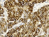
Immunohistochemistry-Paraffin: TRPM4 Antibody (OTI10H5) [NBP1-48036] - Human endometrial carcinoma tissue section. Fixed paraffin embedded sections were deparaffinized. EDTA was used for antigen retrieval. Samples were incubated O/N in 4 C with the primary antibody (1:200 dilution). IHC-P image submitted by a verified review.![Western Blot: TRPM4 Antibody (OTI10H5) [NBP1-48036]](https://resources.rndsystems.com/images/products/TRPM4-Antibody-10H5-Western-Blot-NBP1-48036-img0001.jpg "Western Blot: TRPM4 Antibody (OTI10H5) [NBP1-48036]")
Western Blot: TRPM4 Antibody (OTI10H5) [NBP1-48036]
Western Blot: TRPM4 Antibody (10H5) [NBP1-48036] - HEK293T cells were transfected with the pCMV6-ENTRY control (Left lane) or pCMV6-ENTRY TRPM4 (Right lane) cDNA for 48 hrs and lysed. Equivalent amounts of cell lysates (5 ug per lane) were separated by SDS-PAGE and immunoblotted with anti-TRPM4.![Immunohistochemistry-Paraffin: TRPM4 Antibody (OTI10H5) [NBP1-48036]](https://resources.rndsystems.com/images/products/TRPM4-Antibody-10H5-Immunohistochemistry-Paraffin-NBP1-48036-img0002.jpg "Immunohistochemistry-Paraffin: TRPM4 Antibody (OTI10H5) [NBP1-48036]")
Immunohistochemistry-Paraffin: TRPM4 Antibody (OTI10H5) [NBP1-48036]
Immunohistochemistry-Paraffin: TRPM4 Antibody (10H5) [NBP1-48036] - Staining of paraffin-embedded Carcinoma of Human liver tissue using anti-TRPM4 mouse monoclonal antibody.![Immunohistochemistry-Paraffin: TRPM4 Antibody (OTI10H5) [NBP1-48036]](https://resources.rndsystems.com/images/products/TRPM4-Antibody-10H5-Immunohistochemistry-Paraffin-NBP1-48036-img0003.jpg "Immunohistochemistry-Paraffin: TRPM4 Antibody (OTI10H5) [NBP1-48036]")
Immunohistochemistry-Paraffin: TRPM4 Antibody (OTI10H5) [NBP1-48036]
Immunohistochemistry-Paraffin: TRPM4 Antibody (10H5) [NBP1-48036] - Staining of paraffin-embedded Human Kidney tissue using anti-TRPM4 mouse monoclonal antibody.![Immunohistochemistry-Paraffin: TRPM4 Antibody (OTI10H5) [NBP1-48036]](https://resources.rndsystems.com/images/products/TRPM4-Antibody-10H5-Immunohistochemistry-Paraffin-NBP1-48036-img0004.jpg "Immunohistochemistry-Paraffin: TRPM4 Antibody (OTI10H5) [NBP1-48036]")
Immunohistochemistry-Paraffin: TRPM4 Antibody (OTI10H5) [NBP1-48036]
Immunohistochemistry-Paraffin: TRPM4 Antibody (10H5) [NBP1-48036] - Staining of paraffin-embedded Human liver tissue using anti-TRPM4 mouse monoclonal antibody.![Immunohistochemistry-Paraffin: TRPM4 Antibody (OTI10H5) [NBP1-48036]](https://resources.rndsystems.com/images/products/TRPM4-Antibody-10H5-Immunohistochemistry-Paraffin-NBP1-48036-img0005.jpg "Immunohistochemistry-Paraffin: TRPM4 Antibody (OTI10H5) [NBP1-48036]")
Immunohistochemistry-Paraffin: TRPM4 Antibody (OTI10H5) [NBP1-48036]
Immunohistochemistry-Paraffin: TRPM4 Antibody (10H5) [NBP1-48036] - Staining of paraffin-embedded Human Ovary tissue using anti-TRPM4 mouse monoclonal antibody.![Immunohistochemistry-Paraffin: TRPM4 Antibody (OTI10H5) [NBP1-48036]](https://resources.rndsystems.com/images/products/TRPM4-Antibody-10H5-Immunohistochemistry-Paraffin-NBP1-48036-img0006.jpg "Immunohistochemistry-Paraffin: TRPM4 Antibody (OTI10H5) [NBP1-48036]")
Immunohistochemistry-Paraffin: TRPM4 Antibody (OTI10H5) [NBP1-48036]
Immunohistochemistry-Paraffin: TRPM4 Antibody (10H5) [NBP1-48036] - Staining of paraffin-embedded Human pancreas tissue using anti-TRPM4 mouse monoclonal antibody.![Immunohistochemistry: TRPM4 Antibody (OTI10H5) [NBP1-48036]](https://resources.rndsystems.com/images/products/TRPM4-Antibody-OTI10H5-Immunohistochemistry-NBP1-48036-img0008.jpg "Immunohistochemistry: TRPM4 Antibody (OTI10H5) [NBP1-48036]")
Immunohistochemistry: TRPM4 Antibody (OTI10H5) [NBP1-48036]
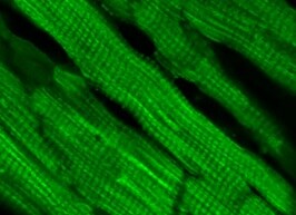
Immunohistochemistry: TRPM4 Antibody (OTI10H5) [NBP1-48036] - Human heart tissue was blocked with 10% serum and stained with the primary antibody at 1:100 dilution overnight at 4 degrees. A secondary FITC-conjugated anti-mouse antibody was used in 1:400 dilution for 2 hours at room temperature. IHC image submitted by a verified customer review.[NBP1-48036]")
TRPM4 Antibody (OTI10H5)[NBP1-48036]
TRPM4 Antibody (OTI10H5)[NBP1-48036]Simple Western� analysis of endogenous protein ACTN1 from HELA lysates (0.2 mg/mL) using ACTN1 Mouse Monoclonal Antibody. The virtual lane view (left) shows the target (as indicated) at 1:50 dilution of primary antibody. The corresponding electropherogram view (right) plots chemiluminescence by molecular weight along the capillary at a 1:50 dilution of primary antibody. This experiment was performed under reducing conditions on the Jess� Simple Western instrument from ProteinSimple, a Bio-Techne brand, using the 12�230 kDa Separation Module.Applications for TRPM4 Antibody (OTI10H5)
Application
Recommended Usage
Immunohistochemistry
1:50
Immunohistochemistry-Paraffin
1:50
Western Blot
1:5000-10000
Reviewed Applications
Read 2 reviews rated 5 using NBP1-48036 in the following applications:
Formulation, Preparation, and Storage
Purification
Immunogen affinity purified
Formulation
PBS (pH 7.3), 1.0% BSA and 50% Glycerol
Preservative
0.02% Sodium Azide
Concentration
1 mg/ml
Shipping
The product is shipped with polar packs. Upon receipt, store it immediately at the temperature recommended below.
Stability & Storage
Store at -20C. Avoid freeze-thaw cycles.
Background: TRPM4
Long Name
Transient receptor potential cation channel subfamily M member 4
Alternate Names
hTRPM4, LTrpC-4, LTrpC4, Melastatin-4
Entrez Gene IDs
54795 (Human)
Gene Symbol
TRPM4
Additional TRPM4 Products
Product Documents for TRPM4 Antibody (OTI10H5)
Certificate of Analysis
To download a Certificate of Analysis, please enter a lot or batch number in the search box below.
Product Specific Notices for TRPM4 Antibody (OTI10H5)
This product is for research use only and is not approved for use in humans or in clinical diagnosis. Primary Antibodies are guaranteed for 1 year from date of receipt.
Customer Reviews for TRPM4 Antibody (OTI10H5) (2)
5 out of 5
2 Customer Ratings
Have you used TRPM4 Antibody (OTI10H5)?
Submit a review and receive an Amazon gift card!
$25/€18/£15/$25CAN/¥2500 Yen for a review with an image
$10/€7/£6/$10CAN/¥1110 Yen for a review without an image
Submit a review
Customer Images
Showing
1
-
2 of
2 reviews
Showing All
Filter By:
-
Application: Immunohistochemistry-ParaffinSample Tested: Endometrial carcinomaSpecies: HumanVerified Customer | Posted 12/13/2021Human endometrial carcinoma tissueFixed paraffin embedded sections were deparaffinized. EDTA was used for antigen retrieval. Samples were incubated O/N in 4 C with the primary antibody (1:200 dilution).

-
Application: IHC/ImmunofluorescenceSample Tested: Heart tissueSpecies: HumanVerified Customer | Posted 08/27/2021The tissue was blocked with 10% serum and stained with the primary antibody at 1:100 dilution overnight at 4 degrees. A secondary FITC-conjugated anti-mouse antibody was used in 1:400 dilution for 2 hours at room temperature.
There are no reviews that match your criteria.
Protocols
Find general support by application which include: protocols, troubleshooting, illustrated assays, videos and webinars.
- Antigen Retrieval Protocol (PIER)
- Antigen Retrieval for Frozen Sections Protocol
- Appropriate Fixation of IHC/ICC Samples
- Cellular Response to Hypoxia Protocols
- Chromogenic IHC Staining of Formalin-Fixed Paraffin-Embedded (FFPE) Tissue Protocol
- Chromogenic Immunohistochemistry Staining of Frozen Tissue
- ClariTSA™ Fluorophore Kits
- Detection & Visualization of Antibody Binding
- Fluorescent IHC Staining of Frozen Tissue Protocol
- Graphic Protocol for Heat-induced Epitope Retrieval
- Graphic Protocol for the Preparation and Fluorescent IHC Staining of Frozen Tissue Sections
- Graphic Protocol for the Preparation and Fluorescent IHC Staining of Paraffin-embedded Tissue Sections
- Graphic Protocol for the Preparation of Gelatin-coated Slides for Histological Tissue Sections
- IHC Sample Preparation (Frozen sections vs Paraffin)
- Immunofluorescent IHC Staining of Formalin-Fixed Paraffin-Embedded (FFPE) Tissue Protocol
- Immunohistochemistry (IHC) and Immunocytochemistry (ICC) Protocols
- Immunohistochemistry Frozen Troubleshooting
- Immunohistochemistry Paraffin Troubleshooting
- Preparing Samples for IHC/ICC Experiments
- Preventing Non-Specific Staining (Non-Specific Binding)
- Primary Antibody Selection & Optimization
- Protocol for Heat-Induced Epitope Retrieval (HIER)
- Protocol for Making a 4% Formaldehyde Solution in PBS
- Protocol for VisUCyte™ HRP Polymer Detection Reagent
- Protocol for the Preparation & Fixation of Cells on Coverslips
- Protocol for the Preparation and Chromogenic IHC Staining of Frozen Tissue Sections
- Protocol for the Preparation and Chromogenic IHC Staining of Frozen Tissue Sections - Graphic
- Protocol for the Preparation and Chromogenic IHC Staining of Paraffin-embedded Tissue Sections
- Protocol for the Preparation and Chromogenic IHC Staining of Paraffin-embedded Tissue Sections - Graphic
- Protocol for the Preparation and Fluorescent IHC Staining of Frozen Tissue Sections
- Protocol for the Preparation and Fluorescent IHC Staining of Paraffin-embedded Tissue Sections
- Protocol for the Preparation of Gelatin-coated Slides for Histological Tissue Sections
- R&D Systems Quality Control Western Blot Protocol
- TUNEL and Active Caspase-3 Detection by IHC/ICC Protocol
- The Importance of IHC/ICC Controls
- Troubleshooting Guide: Immunohistochemistry
- Troubleshooting Guide: Western Blot Figures
- Western Blot Conditions
- Western Blot Protocol
- Western Blot Protocol for Cell Lysates
- Western Blot Troubleshooting
- Western Blot Troubleshooting Guide
- View all Protocols, Troubleshooting, Illustrated assays and Webinars
Loading...