Tubulin alpha-1B [ac Lys40] Antibody (RM318)
Novus Biologicals | Catalog # NBP2-61591
Recombinant Monoclonal Antibody
Loading...
Key Product Details
Validated by
Biological Validation
Species Reactivity
Validated:
Vertebrate
Cited:
Mouse
Applications
Validated:
Western Blot, Immunocytochemistry/ Immunofluorescence
Cited:
Western Blot
Label
Unconjugated
Antibody Source
Recombinant Monoclonal Rabbit IgG Clone # RM318 expressed in HEK293
Loading...
Product Specifications
Immunogen
An acetyl-peptide corresponding to Acetyl-Tubulin alpha-1B [ac Lys40]
Modification
ac Lys40
Specificity
This antibody reacts to Tubulin alpha-1B acetylated at Lysine 40. No cross reactivity to non-acetylated alpha-Tubulin at Lysine 40
Clonality
Monoclonal
Host
Rabbit
Isotype
IgG
Scientific Data Images for Tubulin alpha-1B [ac Lys40] Antibody (RM318)
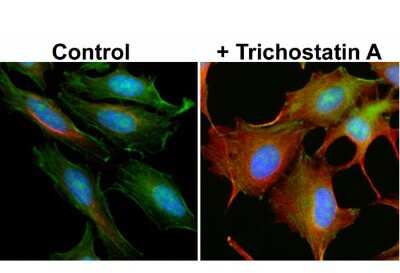
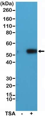
Immunocytochemistry/Immunofluorescence: Tubulin alpha-1B [ac Lys40] Antibody (RM318) [NBP2-61591] - Immunocytochemistry of HeLa cells non-treated or treated with Trichostatin A (TSA), using NBP2-61591, red). Actin filaments have been labeled with fluorescein phalloidin green), and nuclei with DAPI (blue).
Applications for Tubulin alpha-1B [ac Lys40] Antibody (RM318)
Application
Recommended Usage
Immunocytochemistry/ Immunofluorescence
1:1000-1:5000
Western Blot
1:1000 - 1:2000
Formulation, Preparation, and Storage
Purification
Protein A purified
Formulation
50% Glycerol/PBS, 1% BSA
Preservative
0.09% Sodium Azide
Concentration
Please see the vial label for concentration. If unlisted please contact technical services.
Shipping
The product is shipped with polar packs. Upon receipt, store it immediately at the temperature recommended below.
Stability & Storage
Store at -20C. Avoid freeze-thaw cycles.
Background: Tubulin alpha-1B
Long Name
Tubulin alpha-1B chain
Alternate Names
TUBA1B, Tubulin K-alpha-1
Gene Symbol
TUBA1B
Additional Tubulin alpha-1B Products
Product Documents for Tubulin alpha-1B [ac Lys40] Antibody (RM318)
Certificate of Analysis
To download a Certificate of Analysis, please enter a lot or batch number in the search box below.
Product Specific Notices for Tubulin alpha-1B [ac Lys40] Antibody (RM318)
This product is for research use only and is not approved for use in humans or in clinical diagnosis. Primary Antibodies are guaranteed for 1 year from date of receipt.
Citations for Tubulin alpha-1B [ac Lys40] Antibody (RM318)
 Powered by Bioz
Customer Reviews for Tubulin alpha-1B [ac Lys40] Antibody (RM318)
There are currently no reviews for this product. Be the first to review Tubulin alpha-1B [ac Lys40] Antibody (RM318) and earn rewards!
Have you used Tubulin alpha-1B [ac Lys40] Antibody (RM318)?
Submit a review and receive an Amazon gift card!
$25/€18/£15/$25CAN/¥2500 Yen for a review with an image
$10/€7/£6/$10CAN/¥1110 Yen for a review without an image
Submit a review
Protocols
Find general support by application which include: protocols, troubleshooting, illustrated assays, videos and webinars.
- Appropriate Fixation of IHC/ICC Samples
- Cellular Response to Hypoxia Protocols
- ClariTSA™ Fluorophore Kits
- Detection & Visualization of Antibody Binding
- ICC Cell Smear Protocol for Suspension Cells
- ICC Immunocytochemistry Protocol Videos
- ICC for Adherent Cells
- Immunocytochemistry (ICC) Protocol
- Immunocytochemistry Troubleshooting
- Immunofluorescence of Organoids Embedded in Cultrex Basement Membrane Extract
- Immunohistochemistry (IHC) and Immunocytochemistry (ICC) Protocols
- Preparing Samples for IHC/ICC Experiments
- Preventing Non-Specific Staining (Non-Specific Binding)
- Primary Antibody Selection & Optimization
- Protocol for VisUCyte™ HRP Polymer Detection Reagent
- Protocol for the Fluorescent ICC Staining of Cell Smears - Graphic
- Protocol for the Fluorescent ICC Staining of Cultured Cells on Coverslips - Graphic
- Protocol for the Preparation and Fluorescent ICC Staining of Cells on Coverslips
- Protocol for the Preparation and Fluorescent ICC Staining of Non-adherent Cells
- Protocol for the Preparation and Fluorescent ICC Staining of Stem Cells on Coverslips
- Protocol for the Preparation of a Cell Smear for Non-adherent Cell ICC - Graphic
- R&D Systems Quality Control Western Blot Protocol
- TUNEL and Active Caspase-3 Detection by IHC/ICC Protocol
- The Importance of IHC/ICC Controls
- Troubleshooting Guide: Western Blot Figures
- Western Blot Conditions
- Western Blot Protocol
- Western Blot Protocol for Cell Lysates
- Western Blot Troubleshooting
- Western Blot Troubleshooting Guide
- View all Protocols, Troubleshooting, Illustrated assays and Webinars
Loading...