Tyrosine Hydroxylase Antibody - Azide Free
Novus Biologicals | Catalog # NB300-110
![Western Blot: Tyrosine Hydroxylase Antibody [NB300-110]](https://resources.rndsystems.com/images/products/Tyrosine-Hydroxylase-Antibody-Western-Blot-NB300-110-img0006.jpg "Western Blot: Tyrosine Hydroxylase Antibody [NB300-110]")
Loading...
Key Product Details
Species Reactivity
Validated:
Human, Mouse, Rat, Amphibian, Avian, Mammal, Rabbit, Reptile
Cited:
Human, Mouse, Rat, Amphibian, Avian, Finch, Rabbit, Reptile
Predicted:
Chicken (100%). Backed by our 100% Guarantee.
Applications
Validated:
Immunohistochemistry, Immunohistochemistry-Paraffin, Immunohistochemistry-Frozen, Immunohistochemistry Free-Floating, Western Blot, Immunocytochemistry/ Immunofluorescence, Single Cell Western
Cited:
Immunohistochemistry, Immunohistochemistry-Paraffin, Immunohistochemistry-Frozen, Immunohistochemistry Free-Floating, Western Blot, Immunocytochemistry/ Immunofluorescence, IF/IHC
Label
Unconjugated
Antibody Source
Polyclonal Sheep IgG
Format
Azide Free
Loading...
Product Specifications
Immunogen
SDS-denatured, native rat tyrosine hydroxylase purified from pheochromocytoma.
Reactivity Notes
The antibody recognizes all mammalian and at least some non-mammalian forms of the enzyme in Western blot and in IHC/IF. Amphibian reactivity reported in scientific literature (PMID: 28867550).
Localization
NB300-110 can be used as a marker for dopaminergic and noradrenergic neurons.
Marker
Neuronal Marker
Specificity
Specific for the ~60 kDa tyrosine hydroxylase protein.
Clonality
Polyclonal
Host
Sheep
Isotype
IgG
Theoretical MW
60 kDa.
Disclaimer note: The observed molecular weight of the protein may vary from the listed predicted molecular weight due to post translational modifications, post translation cleavages, relative charges, and other experimental factors.
Disclaimer note: The observed molecular weight of the protein may vary from the listed predicted molecular weight due to post translational modifications, post translation cleavages, relative charges, and other experimental factors.
Scientific Data Images for Tyrosine Hydroxylase Antibody - Azide Free
Western Blot: Tyrosine Hydroxylase Antibody [NB300-110]
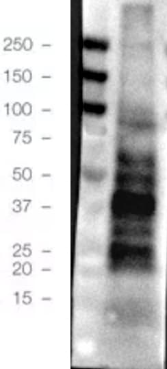
Western Blot: Tyrosine Hydroxylase Antibody [NB300-110] - Analysis of rat caudate lysate showing specific immunolabeling of the ~60 kDa tyrosine hydroxylase protein.![Western Blot: Tyrosine Hydroxylase Antibody [NB300-110]](https://resources.rndsystems.com/images/products/Tyrosine-Hydroxylase-Antibody-Western-Blot-NB300-110-img0009.jpg "Western Blot: Tyrosine Hydroxylase Antibody [NB300-110]")
Western Blot: Tyrosine Hydroxylase Antibody [NB300-110]
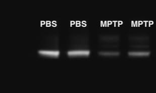
Western Blot: Tyrosine Hydroxylase Antibody [NB300-110] - Mice were injected with PBS (control group) or the dopaminergic neurotoxin MPTP. There is low concentration of TH in the striatum of mouse injected with MPTP. 30 ug total protein per lane. Antibody at 1:3000. Western blot image submitted by a verified customer review.![Immunocytochemistry/ Immunofluorescence: Tyrosine Hydroxylase Antibody [NB300-110]](https://resources.rndsystems.com/images/products/Tyrosine%20Hydroxylase%20Antibody-Immunocytochemistry-Immunofluorescence-NB300-110-img0013.jpg "Immunocytochemistry/ Immunofluorescence: Tyrosine Hydroxylase Antibody [NB300-110]")
Immunocytochemistry/ Immunofluorescence: Tyrosine Hydroxylase Antibody [NB300-110]
Tyrosine Hydroxylase Antibody-Immunocytochemistry-Immunofluorescence-NB300-110-img0013.jpg![Immunohistochemistry: Tyrosine Hydroxylase Antibody [NB300-110]](https://resources.rndsystems.com/images/products/Tyrosine-Hydroxylase-Antibody-Immunohistochemistry-NB300-110-img0008.jpg "Immunohistochemistry: Tyrosine Hydroxylase Antibody [NB300-110]")
Immunohistochemistry: Tyrosine Hydroxylase Antibody [NB300-110]
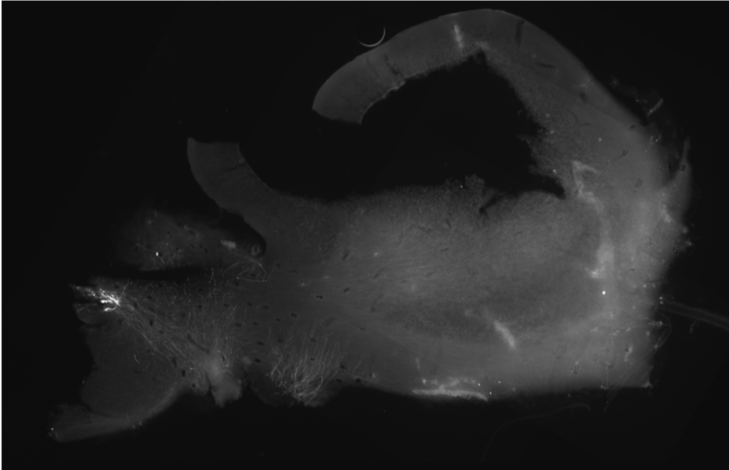
Immunohistochemistry: Tyrosine Hydroxylase Antibody [NB300-110] - Mouse cryo-sections. Antibody at 1:500. Secondary antibody: dk-anti-sp Alexa Fluor488 at 1:500; 1 h. IHC image submitted by a verified customer review.![Immunocytochemistry/ Immunofluorescence: Tyrosine Hydroxylase Antibody [NB300-110]](https://resources.rndsystems.com/images/products/Tyrosine-Hydroxylase-Antibody-Immunocytochemistry-Immunofluorescence-NB300-110-img0012.jpg "Immunocytochemistry/ Immunofluorescence: Tyrosine Hydroxylase Antibody [NB300-110]")
Immunocytochemistry/ Immunofluorescence: Tyrosine Hydroxylase Antibody [NB300-110]
Tyrosine-Hydroxylase-Antibody-Immunocytochemistry-Immunofluorescence-NB300-110-img0012.jpg![Immunohistochemistry-Paraffin: Tyrosine Hydroxylase Antibody [NB300-110]](https://resources.rndsystems.com/images/products/Tyrosine-Hydroxylase-Antibody-Immunohistochemistry-Paraffin-NB300-110-img0011.jpg "Immunohistochemistry-Paraffin: Tyrosine Hydroxylase Antibody [NB300-110]")
Immunohistochemistry-Paraffin: Tyrosine Hydroxylase Antibody [NB300-110]
Immunohistochemistry-Paraffin: Tyrosine Hydroxylase Antibody [NB300-110] - Sagittal section of Pleurodeles waltl (amphibian) brain. The cell bodies of dopaminergic neurons in ventral tegmental area and dopaminergic fibers in striatum are stained with the TH antibody. IHC image submitted by a verified customer review.![Immunohistochemistry: Tyrosine Hydroxylase Antibody [NB300-110]](https://resources.rndsystems.com/images/products/Tyrosine-Hydroxylase-Antibody-Immunohistochemistry-NB300-110-img0003.jpg "Immunohistochemistry: Tyrosine Hydroxylase Antibody [NB300-110]")
Immunohistochemistry: Tyrosine Hydroxylase Antibody [NB300-110]
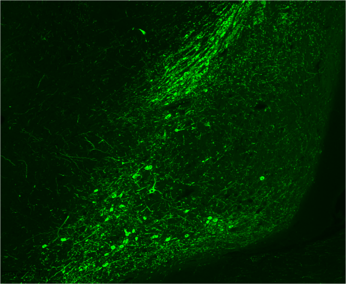
Immunohistochemistry: Tyrosine Hydroxylase Antibody [NB300-110] - Immunohistochemistry analysis of gelatin section of mouse brain (substantia nigra pars compacta) using Tyrosine Hydroxylase antibody. IHC image submitted by a verified customer review.
Immunocytochemistry/ Immunofluorescence: Tyrosine Hydroxylase Antibody [NB300-110] -
Immunocytochemistry/ Immunofluorescence: Tyrosine Hydroxylase Antibody [NB300-110] - Activation of the DDR in mice transduced with AAV2/6 h-syn.a h-syn expression increases 53BP1 & gamma H2AX foci, & ATM phosphorylation in nigral dopaminergic neurons. b The DDR is not activated by viral delivery of GFP. Scale bar: 50 μm. (**p < 0.01; ***p < 0.001; Student’s t test). All bar graphs show mean + /− s.e.m Image collected & cropped by CiteAb from the following publication (https://pubmed.ncbi.nlm.nih.gov/30050065), licensed under a CC-BY license. Not internally tested by Novus Biologicals.
Immunocytochemistry/ Immunofluorescence: Tyrosine Hydroxylase Antibody [NB300-110] -
Immunocytochemistry/ Immunofluorescence: Tyrosine Hydroxylase Antibody [NB300-110] - Activation of the DDR in mice injected with alpha -syn PFF in the striatum.Augmented levels 53BP1 & gamma H2AX foci, & ATM phosphorylation in dopamine neurons of the substantia nigra. Scale bar: 50 μm. (**p < 0.01; ***p < 0.001; Student’s t test). All bar graphs show mean + /− s.e.m Image collected & cropped by CiteAb from the following publication (https://pubmed.ncbi.nlm.nih.gov/30050065), licensed under a CC-BY license. Not internally tested by Novus Biologicals.
Immunocytochemistry/ Immunofluorescence: Tyrosine Hydroxylase Antibody [NB300-110] -
Immunocytochemistry/ Immunofluorescence: Tyrosine Hydroxylase Antibody [NB300-110] - Cytoplasmic Wlds protects dopaminergic neurons from MPP+ toxicity. (A) Dissociated dopaminergic cultures from both WT & cyto Wlds mice were co-stained with TH & Wlds antibodies to confirm the subcellular localization of Wlds. (B) Cultures were treated with 2 μm MPP+ for 48 hours prior to fixing & staining. (C) Quantification of TH+ cell bodies & (D) TH+ neurites shows that cytoplasmic WldS protected both cell bodies & neurites against MPP+. Data are normalized to control cultures & denote the mean ± SEM of representative determinations made in three separate cultures. *p < 0.05. Image collected & cropped by CiteAb from the following publication (https://pubmed.ncbi.nlm.nih.gov/22315973), licensed under a CC-BY license. Not internally tested by Novus Biologicals.
Immunocytochemistry/ Immunofluorescence: Tyrosine Hydroxylase Antibody [NB300-110] -
Immunocytochemistry/ Immunofluorescence: Tyrosine Hydroxylase Antibody [NB300-110] - Activation of the DDR in mice transduced with AAV2/6 h-syn.a h-syn expression increases 53BP1 & gamma H2AX foci, & ATM phosphorylation in nigral dopaminergic neurons. b The DDR is not activated by viral delivery of GFP. Scale bar: 50 μm. (**p < 0.01; ***p < 0.001; Student’s t test). All bar graphs show mean + /− s.e.m Image collected & cropped by CiteAb from the following publication (https://pubmed.ncbi.nlm.nih.gov/30050065), licensed under a CC-BY license. Not internally tested by Novus Biologicals.
Western Blot: Tyrosine Hydroxylase Antibody [NB300-110] -
Western Blot: Tyrosine Hydroxylase Antibody [NB300-110] - Rab10 is a mediator of GCase activity by LRRK2 in fibroblasts & DA neurons.Western blot analysis of fibroblasts, from healthy controls or from patients with the LRRK2 G2019S mutation treated with lentivirus encoding Rab8 & Rab10 shRNA, probed for Rab8, Rab10, & GAPDH (loading control) a. Examination of relative lysosomal GCase activity in fibroblasts upon Rab8 & Rab10 knock-down b–e. Western blot analysis of fibroblasts from 3 patients with LRRK2 G2019S & from 3 healthy controls were probed for phospho-Rab10 (p-Rab10), Rab10, and GAPDH (loading control). The data is presented as the average p-Rab10 signal for the 3 G2019S samples relative to the 3 controls f. Western blot analysis of fibroblasts from 3 controls treated with MLi-2 were probed for phospho-Rab10 (p-Rab10), Rab10, GCase, & tubulin (loading control). Data is presented as the ratio of p-Rab10 to total Rab10 for the treated relative to untreated cells g. Representative western blots of lysates from LRRK2 G2019S & R1441C DA neurons relative to the corresponding isogenic controls h or relative to MLi-2 treated neurons i were probed for p-Rab10, Rab10 & beta -3-tubulin (loading control). The data are presented as the mean ± SEM, n = 3; *p < 0.05, **p < 0.01, using one-way ANOVA followed by Tukey’s multiple comparison post hoc testb–e, or paired two-tailed t-test f, g. Source data are provided as a Source Data file. Image collected & cropped by CiteAb from the following publication (https://pubmed.ncbi.nlm.nih.gov/31804465), licensed under a CC-BY license. Not internally tested by Novus Biologicals.
Western Blot: Tyrosine Hydroxylase Antibody [NB300-110] -
Western Blot: Tyrosine Hydroxylase Antibody [NB300-110] - LRRK2 kinase inhibitors rescue PD-related pathophysiologic phenotypes in LRRK2 & GBA1 mutant neurons. Measurement of insoluble oxidized dopamine by near-IR fluorescence from LRRK2 G2019S.a & R1441C c mutant DA neurons treated with 6166 or MLi-2. Treated cultures were also subjected to western blot analysis of phospho-S129 aSyn (P-S129), total aSyn, & tyrosine hydrolase (TH) with beta -3-tubulin used as a loading control b, d. Measurement of relative levels of insoluble oxidized dopamine by near-IR fluorescence from DA neurons containing GBA1 E326K e or N370S h mutations. Treated cultures were also subjected to western blot analysis of P-S129, total aSyn, & TH with beta -3-tubulin used as a loading control f, i. Representative images from additional DA neurons containing GBA1 E326K g or N370S j mutations treated with MLi-2 & stained with antibodies targeted to P-S129 & beta -3-tubulin, scale bars, 50 µm. The data are presented as the mean ± SEM, n = 3; *p < 0.05, **p < 0.01 relative to untreated, one-way ANOVA followed by Tukey’s multiple comparison post hoc test. Source data are provided as a Source Data file. Image collected & cropped by CiteAb from the following publication (https://pubmed.ncbi.nlm.nih.gov/31804465), licensed under a CC-BY license. Not internally tested by Novus Biologicals.
Immunocytochemistry/ Immunofluorescence: Tyrosine Hydroxylase Antibody [NB300-110] -
Immunocytochemistry/ Immunofluorescence: Tyrosine Hydroxylase Antibody [NB300-110] - Comparison of Mouse & Human Dopaminergic Neuronal Development(A) WNT1 compartments marking lateral population of the floor plate in human & mouse tissue (scale bar, 100 μm).(B) Bar plot of cell types of the human & mouse dopaminergic lineage, showing the expression of key genes. Bars show average mRNA expression, scaled to the absolute molecule counts indicated on the right axis. Error bars show SEM.(C) Validation of mNbM by in situ hybridization for Igfbpl1 & Nhlh1 (scale bar, 50 μm).(D) Neuroblasts in human & mouse ventral midbrain (scale bar, 100 μm; magnification, 20 μm).(E) Selected genes showing similar (left) or distinct (right) expression in mouse & human ventral midbrain. Blue, expressed above baseline in mouse (>99.8% posterior probability); green, expressed above baseline in human (>99.8% posterior probability); gray, not expressed above baseline.(F) Validation of LMO3 expression by immunohistochemistry in a subset of TH+ neurons in the E18.5 mouse ventral midbrain (scale bar left, 100 μm; right, 20 μm).(G) Validation of BNC2 expression by immunohistochemistry in TH+ neurons in the E18.5 mouse ventral midbrain (scale bar left, 100 μm; right, 20 μm). Image collected & cropped by CiteAb from the following publication (https://linkinghub.elsevier.com/retrieve/pii/S0092867416313095), licensed under a CC-BY license. Not internally tested by Novus Biologicals.
Immunocytochemistry/ Immunofluorescence: Tyrosine Hydroxylase Antibody [NB300-110] -
Immunocytochemistry/ Immunofluorescence: Tyrosine Hydroxylase Antibody [NB300-110] - Developmental song exposure & social context affect cFOS expression in TH neurons of the caudal VTA. Sagittal drawings (A,C) & photomicrographs of tyrosine hydroxylase (TH; green label) expression (B,D) in the ventral tegmental area (VTA) & periaqueductal gray (PAG; panels A,B), & the substantia nigra pars compacta (SNc) & locus coeruleus (LC; panels C,D). (E) Examples of co-localized expression of TH (green) & cFOS (red) in the caudal VTA of a normally-reared bird that heard silence (left panel), UD song (middle panel; UD song) & FD song (FD; right panel). White arrows indicate examples of colocalized expression. (F) Percent of TH neurons co-localized with cFOS in the caudal VTA (cVTA), rostral VTA (rVTA) & SNc. Box-and-whisker plots for normally-reared (yellow colors) & song-naïve (green colors) hearing UD (UD; lighter colors) or female-directed (FD; darker colors) songs. Each box spans the interquartile range, horizontal white lines indicate the median & whiskers show the minima & maxima. Levels of cFOS expression in TH neurons for silence controls are plotted as the mean (gray dashed line) +/− standard error (gray boxes). *Indicates a significant difference at p < 0.05 for all comparisons within a brain area. Image collected & cropped by CiteAb from the following publication (https://pubmed.ncbi.nlm.nih.gov/30082796), licensed under a CC-BY license. Not internally tested by Novus Biologicals.
Immunocytochemistry/ Immunofluorescence: Tyrosine Hydroxylase Antibody [NB300-110] -
Immunocytochemistry/ Immunofluorescence: Tyrosine Hydroxylase Antibody [NB300-110] - Comparison of Mouse & Human Dopaminergic Neuronal Development(A) WNT1 compartments marking lateral population of the floor plate in human & mouse tissue (scale bar, 100 μm).(B) Bar plot of cell types of the human & mouse dopaminergic lineage, showing the expression of key genes. Bars show average mRNA expression, scaled to the absolute molecule counts indicated on the right axis. Error bars show SEM.(C) Validation of mNbM by in situ hybridization for Igfbpl1 & Nhlh1 (scale bar, 50 μm).(D) Neuroblasts in human & mouse ventral midbrain (scale bar, 100 μm; magnification, 20 μm).(E) Selected genes showing similar (left) or distinct (right) expression in mouse & human ventral midbrain. Blue, expressed above baseline in mouse (>99.8% posterior probability); green, expressed above baseline in human (>99.8% posterior probability); gray, not expressed above baseline.(F) Validation of LMO3 expression by immunohistochemistry in a subset of TH+ neurons in the E18.5 mouse ventral midbrain (scale bar left, 100 μm; right, 20 μm).(G) Validation of BNC2 expression by immunohistochemistry in TH+ neurons in the E18.5 mouse ventral midbrain (scale bar left, 100 μm; right, 20 μm). Image collected & cropped by CiteAb from the following publication (https://linkinghub.elsevier.com/retrieve/pii/S0092867416313095), licensed under a CC-BY license. Not internally tested by Novus Biologicals.
Immunocytochemistry/ Immunofluorescence: Tyrosine Hydroxylase Antibody [NB300-110] -
Immunocytochemistry/ Immunofluorescence: Tyrosine Hydroxylase Antibody [NB300-110] - Conversion of hNES cells into hPRogFPM & their differentiation into midbrain dopaminergic neurons. a Schematic representation of the conversion & differentiation protocols. b, cRT-qPCR analysis at day 8, showing the expression of midbrain-hindbrain TFs, such as OTX2, GBX2, LMX1A, & FOXA2 (b), as well as the dopaminergic neuron markers, NR4A2, TH, SLC18A22, & SLC6A3 (c). d Immunocytochemistry analysis of the presence of OTX2 & TH in control unconverted NES cultures, compared with NES cells converted with SAF+Dkk1 & differentiated until day 8. e, f Percentage of OTX2+ & TH+ cells in the conditions in d. P = 0.02673 (e), P = 0.03233 (f), n = 3. g–i Expression of the key midbrain TFs, LMX1A, NR4A2, & PBX1, in TH+cells derived from SAI2-NES cells after conversion & differentiation. j, k TH+ cells express the mature neuronal marker, MAP2 (j), & some acquire mature neuronal morphologies, with long processes & varicosities at day 8 (k). Scale 50μm. Box plots (b, c, e, f): Center line, median; hinges, 25% & 75% quartiles; whiskers, 1.5 interquartile range. Statistics: (b, c) ANOVA, followed by pair-wise t-test with Bonferroni correction for multiple testing. (e, f). Two sample t-test; *P ≤ 0,05; **P ≤ 0,01; ***P ≤ 0,001. N = 3 (GBX2, FOXA2, TH, SLC6A3), n = 4 (LMX1A, OTX2, NR4A2, SLC18A2) Image collected & cropped by CiteAb from the following publication (https://pubmed.ncbi.nlm.nih.gov/29968757), licensed under a CC-BY license. Not internally tested by Novus Biologicals.
Immunocytochemistry/ Immunofluorescence: Tyrosine Hydroxylase Antibody [NB300-110] -
Immunocytochemistry/ Immunofluorescence: Tyrosine Hydroxylase Antibody [NB300-110] - Comparison of Mouse & Human Dopaminergic Neuronal Development(A) WNT1 compartments marking lateral population of the floor plate in human & mouse tissue (scale bar, 100 μm).(B) Bar plot of cell types of the human & mouse dopaminergic lineage, showing the expression of key genes. Bars show average mRNA expression, scaled to the absolute molecule counts indicated on the right axis. Error bars show SEM.(C) Validation of mNbM by in situ hybridization for Igfbpl1 & Nhlh1 (scale bar, 50 μm).(D) Neuroblasts in human & mouse ventral midbrain (scale bar, 100 μm; magnification, 20 μm).(E) Selected genes showing similar (left) or distinct (right) expression in mouse & human ventral midbrain. Blue, expressed above baseline in mouse (>99.8% posterior probability); green, expressed above baseline in human (>99.8% posterior probability); gray, not expressed above baseline.(F) Validation of LMO3 expression by immunohistochemistry in a subset of TH+ neurons in the E18.5 mouse ventral midbrain (scale bar left, 100 μm; right, 20 μm).(G) Validation of BNC2 expression by immunohistochemistry in TH+ neurons in the E18.5 mouse ventral midbrain (scale bar left, 100 μm; right, 20 μm). Image collected & cropped by CiteAb from the following publication (https://linkinghub.elsevier.com/retrieve/pii/S0092867416313095), licensed under a CC-BY license. Not internally tested by Novus Biologicals.
Immunocytochemistry/ Immunofluorescence: Tyrosine Hydroxylase Antibody [NB300-110] -
Immunocytochemistry/ Immunofluorescence: Tyrosine Hydroxylase Antibody [NB300-110] - alpha -Synuclein overexpression or intracerebral seeding impacts the dopaminergic system.a–c Intranigral injection of AAV2/6 serotype expressing human alpha -syn (h-syn) results in increased protein expression paralleled by reduction in tyrosine hydroxylase (TH) levels. (d) h-syn is also expressed in dopaminergic cell bodies (arrows). e Unbiased stereological counts demonstrate a reduction in nigral dopaminergic cell bodies. f, g Intracranial injection of alpha -syn pre-formed fibrils (PFF) causes striatal dopaminergic denervation as evidenced by a reduction in TH immunoreactivity. h Increased levels of ser129-phosphosynuclein in the substantia nigra (arrows) indicate alpha -syn stress in PFF injected animals. i Unbiased stereological counts showing a decrease in dopaminergic cell bodies in the substantia nigra. Scale bars: 1 mm in a, b 50 μm in d. (**p < 0.01; ***p < 0.001; Student’s t test). All bar graphs show mean + /− s.e.m Image collected & cropped by CiteAb from the following publication (https://pubmed.ncbi.nlm.nih.gov/30050065), licensed under a CC-BY license. Not internally tested by Novus Biologicals.Applications for Tyrosine Hydroxylase Antibody - Azide Free
Application
Recommended Usage
Immunohistochemistry
1:10 - 1:500
Immunohistochemistry-Frozen
1:1000
Western Blot
1:1000
Application Notes
Immunocytochemistry use reported in literature (PMID 21694758). Immunohistochemistry-Paraffin use reported in literature (PMID 23690557) and customer review. Use in Immunohistochemistry free-floating reported in scientific literature (PMID 26553597).
Reviewed Applications
Read 6 reviews rated 4.7 using NB300-110 in the following applications:
Formulation, Preparation, and Storage
Purification
Antigen Affinity-purified
Formulation
10 mM HEPES (pH 7.5), 0.15 M NaCl, 0.1 mg/mL BSA, 50% Glycerol
Format
Azide Free
Preservative
No Preservative
Concentration
Please see the vial label for concentration. If unlisted please contact technical services.
Shipping
The product is shipped with polar packs. Upon receipt, store it immediately at the temperature recommended below.
Stability & Storage
Store at -20C. Avoid freeze-thaw cycles.
Background: Tyrosine Hydroxylase
Two transcription factor binding sites in the proximal region of the TH gene, the TPA-responsive element (TRE) and the c-AMP responsive element (CRE), have been implicated in the complex regulation of the TH gene. Dysregulation of breakdown for the amino acid, tyrosine, by TH is a result of a genetic disorder that results in Tyrosinemia (high levels of tyrosine in the blood, tissue and organs).
Tyrosine hydroxylase deficiency is a disorder that primarily affects movement, where individuals display symptoms that include lack of coordination when walking, postural tremors and unusual body positioning. TH deficient dopamine-responsive dystonia (DRD), also known as Segawa syndrome, is a rare genetic disorder that is associated with low levels of TH and is diagnosed during childhood with characteristic symptoms including increased muscle tone (dystonia) and signs of Parkinsonism like bradykinesia, tremors, rigidity and postural instability (2). Correspondingly, TH is also linked to Parkinson's disease in older adults, where low dopamine levels are a consistent neurochemical abnormality. Functional polymorphisms of the TH gene may be involved in the pathogenesis of neuropsychiatric diseases such as schizophrenia and other affective disorders where dopamine is often dysregulated (3).
References
1. Hamanaka, Y., & Mizunami, M. (2019). Tyrosine hydroxylase-immunoreactive neurons in the mushroom body of the field cricket, Gryllus bimaculatus. Cell Tissue Res, 376(1), 97-111. doi:10.1007/s00441-018-2969-9
2. Li, L., & Zhou, F. M. (2013). Parallel dopamine D1 receptor activity dependence of l-Dopa-induced normal movement and dyskinesia in mice. Neuroscience, 236, 66-76. doi:10.1016/j.neuroscience.2012.12.065
3. Borkar, C. D., Bharne, A. P., Nagalakshmi, B., Sakharkar, A. J., Subhedar, N. K., & Kokare, D. M. (2018). Cocaine- and Amphetamine-Regulated Transcript Peptide (CART) Alleviates MK-801-Induced Schizophrenic Dementia-Like Symptoms. Neuroscience, 375, 94-107. doi:10.1016/j.neuroscience.2018.01.056
Alternate Names
TH, TYH
Gene Symbol
TH
UniProt
Additional Tyrosine Hydroxylase Products
Product Documents for Tyrosine Hydroxylase Antibody - Azide Free
Certificate of Analysis
To download a Certificate of Analysis, please enter a lot or batch number in the search box below.
Product Specific Notices for Tyrosine Hydroxylase Antibody - Azide Free
This product is for research use only and is not approved for use in humans or in clinical diagnosis. Primary Antibodies are guaranteed for 1 year from date of receipt.
Citations for Tyrosine Hydroxylase Antibody - Azide Free
 Powered by Bioz
Customer Reviews for Tyrosine Hydroxylase Antibody - Azide Free (6)
4.7 out of 5
6 Customer Ratings
Have you used Tyrosine Hydroxylase Antibody - Azide Free?
Submit a review and receive an Amazon gift card!
$25/€18/£15/$25CAN/¥2500 Yen for a review with an image
$10/€7/£6/$10CAN/¥1110 Yen for a review without an image
Submit a review
Customer Images
-(01-ml)_NB300-110_11401.jpg)
Showing
1
-
5 of
6 reviews
Showing All
Filter By:
-
Application: Western BlotSample Tested: Colon tissueVerified Customer | Posted 10/02/2024Dual pink protein marker10 mg loading 1:1000 dilution HRP detection

-
Application: Immunohistochemistry-ParaffinSample Tested: brain sectionSpecies: Pleurodeles waltlVerified Customer | Posted 07/03/2019Sagittal section of Pleurodeles waltl brain. The cell bodies of dopaminergic neurons in ventral tegmental area and dopaminergic fibers in striatum are stained with a TH Immunohistochemistry.
-
Application: Western BlotSample Tested: Brain (corpus striatum)Species: MouseVerified Customer | Posted 10/17/2018Western blot against tyrosine hydroxylase. Animals were injected with PBS (control group) or the dopaminergic neurotoxin MPTP. There is low concentration of TH in the striatum of mouse injected with MPTP (the two samples on the right).It works very well at 1:3,000 dilution when loading gels with 30 ug of protein extracts.
-
Application: ImmunocytochemistrySample Tested: Brain (substantia nigra)Species: MouseVerified Customer | Posted 05/19/201710x
-
Application: ImmunofluorescenceSample Tested: embryonic mouse stomachSpecies: MouseVerified Customer | Posted 08/05/2016
-
Application: ImmunofluorescenceSample Tested: Gelatin Section of Mouse Brain (substantia nigra pars compacta)Species: MouseVerified Customer | Posted 10/23/2014Leica Confocal Microscope 40X
There are no reviews that match your criteria.
Protocols
Find general support by application which include: protocols, troubleshooting, illustrated assays, videos and webinars.
- Antigen Retrieval Protocol (PIER)
- Antigen Retrieval for Frozen Sections Protocol
- Appropriate Fixation of IHC/ICC Samples
- Cellular Response to Hypoxia Protocols
- Chromogenic IHC Staining of Formalin-Fixed Paraffin-Embedded (FFPE) Tissue Protocol
- Chromogenic Immunohistochemistry Staining of Frozen Tissue
- ClariTSA™ Fluorophore Kits
- Detection & Visualization of Antibody Binding
- Fluorescent IHC Staining of Frozen Tissue Protocol
- Graphic Protocol for Heat-induced Epitope Retrieval
- Graphic Protocol for the Preparation and Fluorescent IHC Staining of Frozen Tissue Sections
- Graphic Protocol for the Preparation and Fluorescent IHC Staining of Paraffin-embedded Tissue Sections
- Graphic Protocol for the Preparation of Gelatin-coated Slides for Histological Tissue Sections
- ICC Cell Smear Protocol for Suspension Cells
- ICC Immunocytochemistry Protocol Videos
- ICC for Adherent Cells
- IHC Sample Preparation (Frozen sections vs Paraffin)
- Immunocytochemistry (ICC) Protocol
- Immunocytochemistry Troubleshooting
- Immunofluorescence of Organoids Embedded in Cultrex Basement Membrane Extract
- Immunofluorescent IHC Staining of Formalin-Fixed Paraffin-Embedded (FFPE) Tissue Protocol
- Immunohistochemistry (IHC) and Immunocytochemistry (ICC) Protocols
- Immunohistochemistry Frozen Troubleshooting
- Immunohistochemistry Paraffin Troubleshooting
- Preparing Samples for IHC/ICC Experiments
- Preventing Non-Specific Staining (Non-Specific Binding)
- Primary Antibody Selection & Optimization
- Protocol for Heat-Induced Epitope Retrieval (HIER)
- Protocol for Making a 4% Formaldehyde Solution in PBS
- Protocol for VisUCyte™ HRP Polymer Detection Reagent
- Protocol for the Fluorescent ICC Staining of Cell Smears - Graphic
- Protocol for the Fluorescent ICC Staining of Cultured Cells on Coverslips - Graphic
- Protocol for the Preparation & Fixation of Cells on Coverslips
- Protocol for the Preparation and Chromogenic IHC Staining of Frozen Tissue Sections
- Protocol for the Preparation and Chromogenic IHC Staining of Frozen Tissue Sections - Graphic
- Protocol for the Preparation and Chromogenic IHC Staining of Paraffin-embedded Tissue Sections
- Protocol for the Preparation and Chromogenic IHC Staining of Paraffin-embedded Tissue Sections - Graphic
- Protocol for the Preparation and Fluorescent ICC Staining of Cells on Coverslips
- Protocol for the Preparation and Fluorescent ICC Staining of Non-adherent Cells
- Protocol for the Preparation and Fluorescent ICC Staining of Stem Cells on Coverslips
- Protocol for the Preparation and Fluorescent IHC Staining of Frozen Tissue Sections
- Protocol for the Preparation and Fluorescent IHC Staining of Paraffin-embedded Tissue Sections
- Protocol for the Preparation of Gelatin-coated Slides for Histological Tissue Sections
- Protocol for the Preparation of a Cell Smear for Non-adherent Cell ICC - Graphic
- R&D Systems Quality Control Western Blot Protocol
- TUNEL and Active Caspase-3 Detection by IHC/ICC Protocol
- The Importance of IHC/ICC Controls
- Troubleshooting Guide: Immunohistochemistry
- Troubleshooting Guide: Western Blot Figures
- Western Blot Conditions
- Western Blot Protocol
- Western Blot Protocol for Cell Lysates
- Western Blot Troubleshooting
- Western Blot Troubleshooting Guide
- View all Protocols, Troubleshooting, Illustrated assays and Webinars
FAQs for Tyrosine Hydroxylase Antibody - Azide Free
Showing
1
-
1 of
1 FAQ
Showing All
-
Q: On the NOVUS website, it showed that NB300-110 could be used to detect Tyrosine Hydroxylase in mammal by IHC-P experiment. Our customer would like to know whether it can be used on dog or not. We are looking forward to your nice help.
A: The immunogen which was used to generate our Tyrosine Hydroxylase antibody with catalogue number NB300-110 was native rat Tyrosine Hydroxylase that was purified from pheochromocytoma. The antibody recognizes all mammalian forms of the enzyme in Western blots and in IHC/IF, however we do not have any data from dog that I can share with you at the present time.
Loading...
Associated Pathways
