![Immunocytochemistry/ Immunofluorescence: ULK4 Antibody [NBP1-20229]](https://resources.rndsystems.com/images/products/ULK4-Antibody-Immunocytochemistry-NBP1-20229-img0001.jpg "Immunocytochemistry/ Immunofluorescence: ULK4 Antibody [NBP1-20229]")
Key Product Details
Species Reactivity
Validated:
Cited:
Applications
Validated:
Cited:
Label
Antibody Source
Product Specifications
Immunogen
Reactivity Notes
Clonality
Host
Isotype
Scientific Data Images for ULK4 Antibody
Immunocytochemistry/ Immunofluorescence: ULK4 Antibody [NBP1-20229]
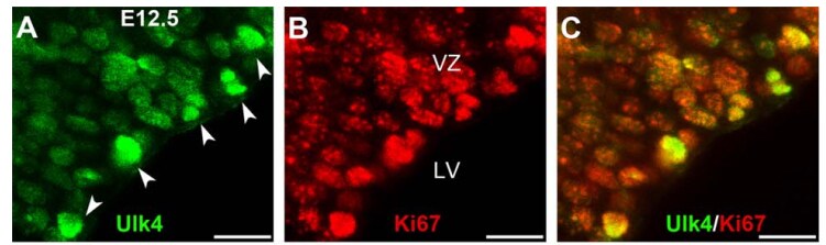
Immunocytochemistry/Immunofluorescence: ULK4 Antibody [NBP1-20229] - At E12.5, Ulk4 (green) was expressed in Ki67 positive cells (red). This image was submitted via customer Review.Applications for ULK4 Antibody
Immunocytochemistry/ Immunofluorescence
Immunohistochemistry
Western Blot
Reviewed Applications
Read 2 reviews rated 4 using NBP1-20229 in the following applications:
Formulation, Preparation, and Storage
Purification
Reconstitution
Formulation
Preservative
Concentration
Shipping
Stability & Storage
Calculators
Background: ULK4
Alternate Names
Gene Symbol
UniProt
Additional ULK4 Products
Product Documents for ULK4 Antibody
Certificate of Analysis
To download a Certificate of Analysis, please enter a lot or batch number in the search box below.
Product Specific Notices for ULK4 Antibody
This product is for research use only and is not approved for use in humans or in clinical diagnosis. Primary Antibodies are guaranteed for 1 year from date of receipt.
Citations for ULK4 Antibody
 Powered by Bioz
Customer Reviews for ULK4 Antibody (2)
Have you used ULK4 Antibody?
Submit a review and receive an Amazon gift card!
$25/€18/£15/$25CAN/¥2500 Yen for a review with an image
$10/€7/£6/$10CAN/¥1110 Yen for a review without an image
Submit a review
Customer Images

-
Application: Immunohistochemistry-ParaffinSample Tested: Mouse cortexSpecies: MouseVerified Customer | Posted 10/19/2017

-
Application: ImmunocytochemistrySample Tested: Mouse brainSpecies: MouseVerified Customer | Posted 05/12/2017At E12.5, Ulk4 (green) was expressed in Ki67 positive cells (red).
There are no reviews that match your criteria.
Protocols
Find general support by application which include: protocols, troubleshooting, illustrated assays, videos and webinars.
- Antigen Retrieval Protocol (PIER)
- Antigen Retrieval for Frozen Sections Protocol
- Appropriate Fixation of IHC/ICC Samples
- Cellular Response to Hypoxia Protocols
- Chromogenic IHC Staining of Formalin-Fixed Paraffin-Embedded (FFPE) Tissue Protocol
- Chromogenic Immunohistochemistry Staining of Frozen Tissue
- ClariTSA™ Fluorophore Kits
- Detection & Visualization of Antibody Binding
- Fluorescent IHC Staining of Frozen Tissue Protocol
- Graphic Protocol for Heat-induced Epitope Retrieval
- Graphic Protocol for the Preparation and Fluorescent IHC Staining of Frozen Tissue Sections
- Graphic Protocol for the Preparation and Fluorescent IHC Staining of Paraffin-embedded Tissue Sections
- Graphic Protocol for the Preparation of Gelatin-coated Slides for Histological Tissue Sections
- ICC Cell Smear Protocol for Suspension Cells
- ICC Immunocytochemistry Protocol Videos
- ICC for Adherent Cells
- IHC Sample Preparation (Frozen sections vs Paraffin)
- Immunocytochemistry (ICC) Protocol
- Immunocytochemistry Troubleshooting
- Immunofluorescence of Organoids Embedded in Cultrex Basement Membrane Extract
- Immunofluorescent IHC Staining of Formalin-Fixed Paraffin-Embedded (FFPE) Tissue Protocol
- Immunohistochemistry (IHC) and Immunocytochemistry (ICC) Protocols
- Immunohistochemistry Frozen Troubleshooting
- Immunohistochemistry Paraffin Troubleshooting
- Preparing Samples for IHC/ICC Experiments
- Preventing Non-Specific Staining (Non-Specific Binding)
- Primary Antibody Selection & Optimization
- Protocol for Heat-Induced Epitope Retrieval (HIER)
- Protocol for Making a 4% Formaldehyde Solution in PBS
- Protocol for VisUCyte™ HRP Polymer Detection Reagent
- Protocol for the Fluorescent ICC Staining of Cell Smears - Graphic
- Protocol for the Fluorescent ICC Staining of Cultured Cells on Coverslips - Graphic
- Protocol for the Preparation & Fixation of Cells on Coverslips
- Protocol for the Preparation and Chromogenic IHC Staining of Frozen Tissue Sections
- Protocol for the Preparation and Chromogenic IHC Staining of Frozen Tissue Sections - Graphic
- Protocol for the Preparation and Chromogenic IHC Staining of Paraffin-embedded Tissue Sections
- Protocol for the Preparation and Chromogenic IHC Staining of Paraffin-embedded Tissue Sections - Graphic
- Protocol for the Preparation and Fluorescent ICC Staining of Cells on Coverslips
- Protocol for the Preparation and Fluorescent ICC Staining of Non-adherent Cells
- Protocol for the Preparation and Fluorescent ICC Staining of Stem Cells on Coverslips
- Protocol for the Preparation and Fluorescent IHC Staining of Frozen Tissue Sections
- Protocol for the Preparation and Fluorescent IHC Staining of Paraffin-embedded Tissue Sections
- Protocol for the Preparation of Gelatin-coated Slides for Histological Tissue Sections
- Protocol for the Preparation of a Cell Smear for Non-adherent Cell ICC - Graphic
- R&D Systems Quality Control Western Blot Protocol
- TUNEL and Active Caspase-3 Detection by IHC/ICC Protocol
- The Importance of IHC/ICC Controls
- Troubleshooting Guide: Immunohistochemistry
- Troubleshooting Guide: Western Blot Figures
- Western Blot Conditions
- Western Blot Protocol
- Western Blot Protocol for Cell Lysates
- Western Blot Troubleshooting
- Western Blot Troubleshooting Guide
- View all Protocols, Troubleshooting, Illustrated assays and Webinars
FAQs for ULK4 Antibody
-
Q: I want to ask the molecular size of ULK4?
A: In Western blot our ULK4 antibodies detect the target protein at 65kD.
-
Q: What is the molecular size for ULK4 antibody (NBP1-20229)?
A: ULK4 has a molecular weight of approximately 142 kDa (UniProt Q96C45).
-
Q: I want to ask the molecular size of ULK4?
A: In Western blot our ULK4 antibodies detect the target protein at 65kD.
-
Q: What is the molecular size for ULK4 antibody (NBP1-20229)?
A: ULK4 has a molecular weight of approximately 142 kDa (UniProt Q96C45).