USP22 Antibody - BSA Free
Novus Biologicals | Catalog # NBP1-49644
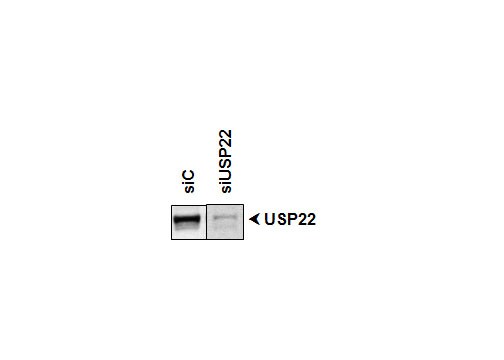
![Western Blot: USP22 AntibodyBSA Free [NBP1-49644]](https://resources.rndsystems.com/images/products/USP22-Antibody-Western-Blot-NBP1-49644-img0001.jpg "Western Blot: USP22 AntibodyBSA Free [NBP1-49644]")
Key Product Details
Species Reactivity
Validated:
Human, Mouse
Cited:
Human, Mouse
Applications
Validated:
Immunohistochemistry, Immunohistochemistry-Paraffin, Western Blot, Simple Western, Chromatin Immunoprecipitation, Chromatin Immunoprecipitation (ChIP), Proximity Ligation Assay, Gel Super Shift Assays
Cited:
Immunohistochemistry-Paraffin, Western Blot, Immunoprecipitation, Chemotaxis
Label
Unconjugated
Antibody Source
Polyclonal Rabbit IgG
Format
BSA Free
Loading...
Product Specifications
Immunogen
A genomic peptide made to an internal region of the human USP22 protein (within residues 100-205). [Swiss-Prot Q9H3R0]
Localization
Nucleus.
Clonality
Polyclonal
Host
Rabbit
Isotype
IgG
Scientific Data Images for USP22 Antibody - BSA Free
Western Blot: USP22 AntibodyBSA Free [NBP1-49644]
Western Blot: USP22 Antibody [NBP1-49644] - Western blot analysis of Usp22 in HeLa whole cell extracts.![Western Blot: USP22 AntibodyBSA Free [NBP1-49644]](https://resources.rndsystems.com/images/products/USP22-Antibody-Western-Blot-NBP1-49644-img0002.jpg "Western Blot: USP22 AntibodyBSA Free [NBP1-49644]")
Western Blot: USP22 AntibodyBSA Free [NBP1-49644]
Western Blot: USP22 Antibody [NBP1-49644] - analysis of USP22 in HeLa whole cell lysate using anti-USP22 antibody. The primary antibody was used at a dilution of 1:500 and incubated overnight at 4C. Image from verified customer review.![Simple Western: USP22 AntibodyBSA Free [NBP1-49644]](https://resources.rndsystems.com/images/products/USP22-Antibody-Simple-Western-NBP1-49644-img0003.jpg "Simple Western: USP22 AntibodyBSA Free [NBP1-49644]")
Simple Western: USP22 AntibodyBSA Free [NBP1-49644]
Simple Western: USP22 Antibody [NBP1-49644] - Simple Western lane view shows a specific band for USP22 in 1.0 mg/ml of HeLa lysate. This experiment was performed under reducing conditions using the 12-230 kDa separation system.
Western Blot: USP22 Antibody - BSA Free [NBP1-49644] -
Catalytic activity of USP22 is not a barrier to reprogramming.A Diagram showing USP22 domains and mutated amino acid positions. B Western blot image showing USP22 protein levels after USP22 overexpressions in both non-targeting and USP22 targeting gRNA expressing fibroblasts. Actin was used as a loading control. C Western blot image showing H2Bub protein levels after USP22 overexpressions in both non-targeting and USP22 targeting gRNA expressing fibroblasts. Histones H3 and H2B were used as loading controls. D Fold change in reprogramming efficiency upon USP22 overexpressions in both wild-type and USP22 knockout background. Triangular images above the bars are sections of representative Tra-1-60-stained wells. Error bars indicate the error of mean. n = 3, independent experiments for KR mutations, n = 4 for CA and KQ mutations in USP22 knockout background, and n = 5 for other comparisons. Two-sided t-test was performed between sgNT1 and USP22 sg1 without any overexpression and p-value is 0.0153. Two-sided t-test was performed between USP22 sg1 expressing cells without overexpression and WT, C185A, K129Q, and K129R and p-value is 0.0129, 0.0054, 0.0264, and 0.0052, respectively. E Western blot image showing H2Bub and H2Aub protein levels after ATXN7L3 or ENY2 knockouts in fibroblasts. H2B was used as a loading control. F Fold change in reprogramming efficiency upon ATXN7L3, ENY2, USP27X, or USP51 knockouts. Triangular images above the bars are sections of representative Tra-1-60-stained wells. Error bars indicate the error of mean. n = 5, independent experiments. Two-sided t-test shows p-value smaller than 0.0001 for ENY2 sg1 and sg2 compared to sgNT1. Image collected and cropped by CiteAb from the following open publication (https://pubmed.ncbi.nlm.nih.gov/40102626), licensed under a CC-BY license. Not internally tested by Novus Biologicals.Applications for USP22 Antibody - BSA Free
Application
Recommended Usage
Chromatin Immunoprecipitation
reported in scientific literature (PMID 34155658)
Gel Super Shift Assays
reported in scientific literature
Immunohistochemistry-Paraffin
reported in scientific literature (PMID 24197134)
Proximity Ligation Assay
reported in scientific literature (PMID 33198416)
Simple Western
1:25
Western Blot
2 ug/ml
Application Notes
In Western blot, a band is seen ~59 kDa.
In Simple Western only 10 - 15 uL of the recommended dilution is used per data point.
See Simple Western Antibody Database for Simple Western validation: Tested in HeLa lysate 1.0 mg/mL, separated by Size, antibody dilution of 1:25, apparent MW was 65 kDa. Separated by Size-Wes, Sally Sue/Peggy Sue.
Reviewed Applications
Read 1 review rated 5 using NBP1-49644 in the following applications:
Formulation, Preparation, and Storage
Purification
Immunogen affinity purified
Formulation
PBS
Format
BSA Free
Preservative
0.05% Sodium Azide
Concentration
1 mg/ml
Shipping
The product is shipped with polar packs. Upon receipt, store it immediately at the temperature recommended below.
Stability & Storage
Store at 4C short term. Aliquot and store at -20C long term. Avoid freeze-thaw cycles.
Background: USP22
Long Name
Ubiquitin Specific Protease 22
Alternate Names
Ubiquitin Thioesterase 22, USP3L
Gene Symbol
USP22
UniProt
Additional USP22 Products
Product Documents for USP22 Antibody - BSA Free
Certificate of Analysis
To download a Certificate of Analysis, please enter a lot or batch number in the search box below.
Product Specific Notices for USP22 Antibody - BSA Free
Manufactured by Genomic Antibody Technology™. GAT FAQs
This product is for research use only and is not approved for use in humans or in clinical diagnosis. Primary Antibodies are guaranteed for 1 year from date of receipt.
Related Research Areas
Citations for USP22 Antibody - BSA Free
 Powered by Bioz
Customer Reviews for USP22 Antibody - BSA Free (1)
5 out of 5
1 Customer Rating
Have you used USP22 Antibody - BSA Free?
Submit a review and receive an Amazon gift card!
$25/€18/£15/$25CAN/¥2500 Yen for a review with an image
$10/€7/£6/$10CAN/¥1110 Yen for a review without an image
Submit a review
Customer Images
Showing
1
-
1 of
1 review
Showing All
Filter By:
-
Application: Western BlotSample Tested:Species: HumanVerified Customer | Posted 11/25/2014

There are no reviews that match your criteria.
Protocols
View specific protocols for USP22 Antibody - BSA Free (NBP1-49644):
Western Blot Protocol
1. Perform SDS-PAGE on samples to be analyzed, loading 40 ug of total protein per lane.
2. Transfer proteins to membrane according to the instructions provided by the manufacturer of the membrane and transfer apparatus.
3. Stain according to standard Ponceau S procedure (or similar product) to assess transfer success, and mark molecular weight standards where appropriate.
4. Rinse the blot.
5. Block the membrane using standard blocking buffer for at least 1 hour.
6. Wash the membrane in wash buffer three times for 10 minutes each.
7. Dilute primary antibody in blocking buffer and incubate 1 hour at room temperature.
8. Wash the membrane in wash buffer three times for 10 minutes each.
9. Apply the diluted HRP conjugated secondary antibody in blocking buffer (as per manufacturers instructions) and incubate 1 hour at room temperature.
10. Wash the blot in wash buffer three times for 10 minutes each (this step can be repeated as required to reduce background).
11. Apply the detection reagent of choice in accordance with the manufacturers instructions.
*Note: Tween-20 can be added to the blocking or antibody dilution buffer at a final concentration of 0.05-0.2%.
Find general support by application which include: protocols, troubleshooting, illustrated assays, videos and webinars.
- Antigen Retrieval Protocol (PIER)
- Antigen Retrieval for Frozen Sections Protocol
- Appropriate Fixation of IHC/ICC Samples
- Cellular Response to Hypoxia Protocols
- ChIP Protocol Video
- Chromatin Immunoprecipitation (ChIP) Protocol
- Chromatin Immunoprecipitation Protocol
- Chromogenic IHC Staining of Formalin-Fixed Paraffin-Embedded (FFPE) Tissue Protocol
- Chromogenic Immunohistochemistry Staining of Frozen Tissue
- ClariTSA™ Fluorophore Kits
- Detection & Visualization of Antibody Binding
- Fluorescent IHC Staining of Frozen Tissue Protocol
- Graphic Protocol for Heat-induced Epitope Retrieval
- Graphic Protocol for the Preparation and Fluorescent IHC Staining of Frozen Tissue Sections
- Graphic Protocol for the Preparation and Fluorescent IHC Staining of Paraffin-embedded Tissue Sections
- Graphic Protocol for the Preparation of Gelatin-coated Slides for Histological Tissue Sections
- IHC Sample Preparation (Frozen sections vs Paraffin)
- Immunofluorescent IHC Staining of Formalin-Fixed Paraffin-Embedded (FFPE) Tissue Protocol
- Immunohistochemistry (IHC) and Immunocytochemistry (ICC) Protocols
- Immunohistochemistry Frozen Troubleshooting
- Immunohistochemistry Paraffin Troubleshooting
- Preparing Samples for IHC/ICC Experiments
- Preventing Non-Specific Staining (Non-Specific Binding)
- Primary Antibody Selection & Optimization
- Protocol for Heat-Induced Epitope Retrieval (HIER)
- Protocol for Making a 4% Formaldehyde Solution in PBS
- Protocol for VisUCyte™ HRP Polymer Detection Reagent
- Protocol for the Preparation & Fixation of Cells on Coverslips
- Protocol for the Preparation and Chromogenic IHC Staining of Frozen Tissue Sections
- Protocol for the Preparation and Chromogenic IHC Staining of Frozen Tissue Sections - Graphic
- Protocol for the Preparation and Chromogenic IHC Staining of Paraffin-embedded Tissue Sections
- Protocol for the Preparation and Chromogenic IHC Staining of Paraffin-embedded Tissue Sections - Graphic
- Protocol for the Preparation and Fluorescent IHC Staining of Frozen Tissue Sections
- Protocol for the Preparation and Fluorescent IHC Staining of Paraffin-embedded Tissue Sections
- Protocol for the Preparation of Gelatin-coated Slides for Histological Tissue Sections
- R&D Systems Quality Control Western Blot Protocol
- TUNEL and Active Caspase-3 Detection by IHC/ICC Protocol
- The Importance of IHC/ICC Controls
- Troubleshooting Guide: Immunohistochemistry
- Troubleshooting Guide: Western Blot Figures
- Western Blot Conditions
- Western Blot Protocol
- Western Blot Protocol for Cell Lysates
- Western Blot Troubleshooting
- Western Blot Troubleshooting Guide
- View all Protocols, Troubleshooting, Illustrated assays and Webinars
Loading...