![Western Blot: USP9x Antibody [NBP1-48321]](https://resources.rndsystems.com/images/products/USP9x-Antibody-Western-Blot-NBP1-48321-img0010.jpg "Western Blot: USP9x Antibody [NBP1-48321]")
Loading...
Key Product Details
Species Reactivity
Validated:
Human, Mouse
Cited:
Human, Mouse
Predicted:
Rat (95%). Backed by our 100% Guarantee.
Applications
Validated:
Immunohistochemistry, Immunohistochemistry-Paraffin, Western Blot, Immunocytochemistry/ Immunofluorescence
Cited:
Western Blot, Immunocytochemistry/ Immunofluorescence, IF/IHC
Label
Unconjugated
Antibody Source
Polyclonal Rabbit IgG
Loading...
Product Specifications
Immunogen
Genomic peptide made to an internal region of the human USP9x protein (within residues 1150-1300). [Swiss-Prot Q93008]
Reactivity Notes
Immunogen displays the following percentage of sequence identity for non-tested species: rat (95%) and zebrafish (80%).
Localization
Cytoplasm
Clonality
Polyclonal
Host
Rabbit
Isotype
IgG
Theoretical MW
270 kDa.
Disclaimer note: The observed molecular weight of the protein may vary from the listed predicted molecular weight due to post translational modifications, post translation cleavages, relative charges, and other experimental factors.
Disclaimer note: The observed molecular weight of the protein may vary from the listed predicted molecular weight due to post translational modifications, post translation cleavages, relative charges, and other experimental factors.
Scientific Data Images for USP9x Antibody
Western Blot: USP9x Antibody [NBP1-48321]
USP9x-Antibody-Western-Blot-NBP1-48321-img0010.jpg![Immunocytochemistry/ Immunofluorescence: USP9x Antibody [NBP1-48321]](https://resources.rndsystems.com/images/products/USP9x-Antibody-Immunocytochemistry-Immunofluorescence-NBP1-48321-img0005.jpg "Immunocytochemistry/ Immunofluorescence: USP9x Antibody [NBP1-48321]")
Immunocytochemistry/ Immunofluorescence: USP9x Antibody [NBP1-48321]
Immunocytochemistry/Immunofluorescence: USP9x Antibody [NBP1-48321] - Immunocytochemical analysis of USP9X in NTERA-2 cells![Immunohistochemistry: USP9x Antibody [NBP1-48321]](https://resources.rndsystems.com/images/products/USP9x-Antibody-Immunohistochemistry-NBP1-48321-img0006.jpg "Immunohistochemistry: USP9x Antibody [NBP1-48321]")
Immunohistochemistry: USP9x Antibody [NBP1-48321]
Immunohistochemistry: USP9x Antibody [NBP1-48321] - IHC staining of Usp9x in brain tissue.![Western Blot: USP9x Antibody [NBP1-48321]](https://resources.rndsystems.com/images/products/USP9x-Antibody-Western-Blot-NBP1-48321-img0007.jpg "Western Blot: USP9x Antibody [NBP1-48321]")
Western Blot: USP9x Antibody [NBP1-48321]
Western Blot: USP9x Antibody [NBP1-48321] - Analysis of USP9X in Caco-2 whole cell lysates.![Western Blot: USP9x Antibody [NBP1-48321]](https://resources.rndsystems.com/images/products/USP9x-Antibody-Western-Blot-NBP1-48321-img0008.jpg "Western Blot: USP9x Antibody [NBP1-48321]")
Western Blot: USP9x Antibody [NBP1-48321]
Western Blot: USP9x Antibody [NBP1-48321] - Analysis of USP9X in NIH/3T3 whole cell lysate.![Immunocytochemistry/ Immunofluorescence: USP9x Antibody [NBP1-48321]](https://resources.rndsystems.com/images/products/USP9x-Antibody-Immunocytochemistry-Immunofluorescence-NBP1-48321-img0009.jpg "Immunocytochemistry/ Immunofluorescence: USP9x Antibody [NBP1-48321]")
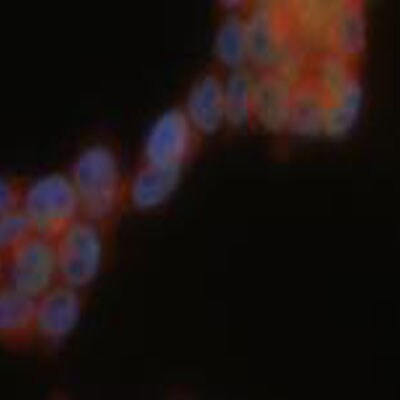
Immunocytochemistry/ Immunofluorescence: USP9x Antibody [NBP1-48321]
Immunocytochemistry/Immunofluorescence: USP9x Antibody [NBP1-48321] - ICC/IC analysis of USP9x in mouse ES cell line 129. Image courtesy of anonymous customer product review.Applications for USP9x Antibody
Application
Recommended Usage
Immunocytochemistry/ Immunofluorescence
1:25-1:200
Immunohistochemistry
1:50
Immunohistochemistry-Paraffin
1:50
Western Blot
1:1000
Application Notes
This USP9x antibody is useful for Immunocytochemistry/Immunofluorescence, Immunohistochemistry paraffin embedded sections and Western blot, where a band is seen ~270 kDa. Prior to immunostaining paraffin tissues, antigen retrieval with sodium citrate buffer (pH 6.0) is recommended. The observed molecular weight of the protein may vary from the listed predicted molecular weight due to post translational modifications, post translation cleavages, relative charges, and other experimental factors.
Reviewed Applications
Read 1 review rated 5 using NBP1-48321 in the following applications:
Formulation, Preparation, and Storage
Purification
Immunogen affinity purified
Formulation
PBS, 0.1% BSA, and 30% Glycerol
Preservative
0.05% Sodium Azide
Concentration
0.2 mg/ml
Shipping
The product is shipped with polar packs. Upon receipt, store it immediately at the temperature recommended below.
Stability & Storage
Store at -20C. Avoid freeze-thaw cycles.
Background: USP9x
Long Name
Ubiquitin-specific Protease 9, X-linked
Alternate Names
DFFRX, FAF, FAM
Gene Symbol
USP9X
UniProt
Additional USP9x Products
Product Documents for USP9x Antibody
Certificate of Analysis
To download a Certificate of Analysis, please enter a lot or batch number in the search box below.
Product Specific Notices for USP9x Antibody
Manufactured by Genomic Antibody Technology™. GAT FAQs
This product is for research use only and is not approved for use in humans or in clinical diagnosis. Primary Antibodies are guaranteed for 1 year from date of receipt.
Related Research Areas
Citations for USP9x Antibody
 Powered by Bioz
Customer Reviews for USP9x Antibody (1)
5 out of 5
1 Customer Rating
Have you used USP9x Antibody?
Submit a review and receive an Amazon gift card!
$25/€18/£15/$25CAN/¥2500 Yen for a review with an image
$10/€7/£6/$10CAN/¥1110 Yen for a review without an image
Submit a review
Customer Images
Showing
1
-
1 of
1 review
Showing All
Filter By:
-
Application: ImmunofluorescenceSample Tested: Mouse ES cell line (129)Species: MouseVerified Customer | Posted 08/15/2011

There are no reviews that match your criteria.
Protocols
View specific protocols for USP9x Antibody (NBP1-48321):
USP9x Antibody:
Western Blot Protocol
1. Perform SDS-PAGE (4-12% MOPS) on samples to be analyzed, loading 40 ug of total protein per lane.
2. Transfer proteins to Nitrocellulose according to the instructions provided by the manufacturer of the transfer
apparatus.
3. Rinse membrane with dH2O and then stain the blot using Ponceau S for 1-2 minutes to access the transfer of
proteins onto the nitrocellulose membrane. Rinse the blot in water to remove excess stain and mark the lane locations
and locations of molecular weight markers using a pencil.
4. Rinse the blot in TBS for approximately 5 minutes.
5. Block the membrane using 5% BSA in TBS + Tween, 1 hour at RT.
6. Rinse the membrane in dH2O and then wash the membrane in wash buffer [TBS + 0.1% Tween] 3 times for 10
minutes each.
7. Dilute the rabbit anti-Usp9x primary antibody (NBP1-48321) in blocking buffer and incubate 1 hour at room
temperature.
8. Rinse the membrane in dH2O and then wash the membrane in wash buffer [TBS + 0.1% Tween] 3 times for 10
minutes each.
9. Apply the diluted rabbit-IgG HRP-conjugated secondary antibody in blocking buffer (as per manufacturers
instructions) and incubate 1 hour at room temperature.
10. Wash the blot in wash buffer [TBS + 0.1% Tween] 3 times for 10 minutes each (this step can be repeated as
required to reduce background).
11. Apply the detection reagent of choice in accordance with the manufacturers instructions (Pierce ECL).
Note: Tween-20 can be added to the blocking or antibody dilution buffer at a final concentration of 0.05-0.2%, provided
it does not interfere with antibody-antigen binding.
Find general support by application which include: protocols, troubleshooting, illustrated assays, videos and webinars.
- Antigen Retrieval Protocol (PIER)
- Antigen Retrieval for Frozen Sections Protocol
- Appropriate Fixation of IHC/ICC Samples
- Cellular Response to Hypoxia Protocols
- Chromogenic IHC Staining of Formalin-Fixed Paraffin-Embedded (FFPE) Tissue Protocol
- Chromogenic Immunohistochemistry Staining of Frozen Tissue
- ClariTSA™ Fluorophore Kits
- Detection & Visualization of Antibody Binding
- Fluorescent IHC Staining of Frozen Tissue Protocol
- Graphic Protocol for Heat-induced Epitope Retrieval
- Graphic Protocol for the Preparation and Fluorescent IHC Staining of Frozen Tissue Sections
- Graphic Protocol for the Preparation and Fluorescent IHC Staining of Paraffin-embedded Tissue Sections
- Graphic Protocol for the Preparation of Gelatin-coated Slides for Histological Tissue Sections
- ICC Cell Smear Protocol for Suspension Cells
- ICC Immunocytochemistry Protocol Videos
- ICC for Adherent Cells
- IHC Sample Preparation (Frozen sections vs Paraffin)
- Immunocytochemistry (ICC) Protocol
- Immunocytochemistry Troubleshooting
- Immunofluorescence of Organoids Embedded in Cultrex Basement Membrane Extract
- Immunofluorescent IHC Staining of Formalin-Fixed Paraffin-Embedded (FFPE) Tissue Protocol
- Immunohistochemistry (IHC) and Immunocytochemistry (ICC) Protocols
- Immunohistochemistry Frozen Troubleshooting
- Immunohistochemistry Paraffin Troubleshooting
- Preparing Samples for IHC/ICC Experiments
- Preventing Non-Specific Staining (Non-Specific Binding)
- Primary Antibody Selection & Optimization
- Protocol for Heat-Induced Epitope Retrieval (HIER)
- Protocol for Making a 4% Formaldehyde Solution in PBS
- Protocol for VisUCyte™ HRP Polymer Detection Reagent
- Protocol for the Fluorescent ICC Staining of Cell Smears - Graphic
- Protocol for the Fluorescent ICC Staining of Cultured Cells on Coverslips - Graphic
- Protocol for the Preparation & Fixation of Cells on Coverslips
- Protocol for the Preparation and Chromogenic IHC Staining of Frozen Tissue Sections
- Protocol for the Preparation and Chromogenic IHC Staining of Frozen Tissue Sections - Graphic
- Protocol for the Preparation and Chromogenic IHC Staining of Paraffin-embedded Tissue Sections
- Protocol for the Preparation and Chromogenic IHC Staining of Paraffin-embedded Tissue Sections - Graphic
- Protocol for the Preparation and Fluorescent ICC Staining of Cells on Coverslips
- Protocol for the Preparation and Fluorescent ICC Staining of Non-adherent Cells
- Protocol for the Preparation and Fluorescent ICC Staining of Stem Cells on Coverslips
- Protocol for the Preparation and Fluorescent IHC Staining of Frozen Tissue Sections
- Protocol for the Preparation and Fluorescent IHC Staining of Paraffin-embedded Tissue Sections
- Protocol for the Preparation of Gelatin-coated Slides for Histological Tissue Sections
- Protocol for the Preparation of a Cell Smear for Non-adherent Cell ICC - Graphic
- R&D Systems Quality Control Western Blot Protocol
- TUNEL and Active Caspase-3 Detection by IHC/ICC Protocol
- The Importance of IHC/ICC Controls
- Troubleshooting Guide: Immunohistochemistry
- Troubleshooting Guide: Western Blot Figures
- Western Blot Conditions
- Western Blot Protocol
- Western Blot Protocol for Cell Lysates
- Western Blot Troubleshooting
- Western Blot Troubleshooting Guide
- View all Protocols, Troubleshooting, Illustrated assays and Webinars
Loading...
Associated Pathways
