Vanilloid R1/TRPV1 Antibody - BSA Free
Novus Biologicals | Catalog # NBP1-71774
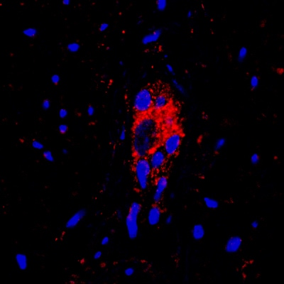
![Immunohistochemistry: Vanilloid R1/TRPV1 Antibody - BSA Free [NBP1-71774]](https://resources.rndsystems.com/images/products/Vanilloid-R1-TRPV1-Antibody-Immunohistochemistry-NBP1-71774-img0010.jpg "Immunohistochemistry: Vanilloid R1/TRPV1 Antibody - BSA Free [NBP1-71774]")
Key Product Details
Species Reactivity
Validated:
Human, Mouse
Cited:
Human, Mouse, Primate, Primate - Callithrix jacchus (Common Marmoset)
Applications
Validated:
Immunohistochemistry, Immunohistochemistry-Paraffin, Immunocytochemistry/ Immunofluorescence
Cited:
Immunohistochemistry, Immunohistochemistry-Paraffin, Immunohistochemistry-Frozen, Western Blot, IF/IHC
Label
Unconjugated
Antibody Source
Polyclonal Rabbit IgG
Format
BSA Free
Loading...
Product Specifications
Immunogen
A genomic peptide made to a C-terminal region of the human TRPV1 protein (within residues 725-839). [Swiss-Prot# Q8NER1]
Localization
Cell junction: synapse. Multi-pass membrane protein.
Clonality
Polyclonal
Host
Rabbit
Isotype
IgG
Scientific Data Images for Vanilloid R1/TRPV1 Antibody - BSA Free
Immunohistochemistry: Vanilloid R1/TRPV1 Antibody - BSA Free [NBP1-71774]
Vanilloid-R1-TRPV1-Antibody-Immunohistochemistry-NBP1-71774-img0010.jpg![Immunocytochemistry/ Immunofluorescence: Vanilloid R1/TRPV1 Antibody - BSA Free [NBP1-71774]](https://resources.rndsystems.com/images/products/Vanilloid-R1-TRPV1-Antibody-Immunocytochemistry-Immunofluorescence-NBP1-71774-img0007.jpg "Immunocytochemistry/ Immunofluorescence: Vanilloid R1/TRPV1 Antibody - BSA Free [NBP1-71774]")
Immunocytochemistry/ Immunofluorescence: Vanilloid R1/TRPV1 Antibody - BSA Free [NBP1-71774]
Immunocytochemistry/Immunofluorescence: Vanilloid R1/TRPV1 Antibody [NBP1-71774] - TRPV1 antibody was tested at 1:50 in HeLa cells with FITC (green). Nuclei were counterstained with DAPI (blue).![Immunohistochemistry: Vanilloid R1/TRPV1 Antibody - BSA Free [NBP1-71774]](https://resources.rndsystems.com/images/products/Vanilloid-R1-TRPV1-Antibody-Immunohistochemistry-NBP1-71774-img0008.jpg "Immunohistochemistry: Vanilloid R1/TRPV1 Antibody - BSA Free [NBP1-71774]")
Immunohistochemistry: Vanilloid R1/TRPV1 Antibody - BSA Free [NBP1-71774]
Immunohistochemistry: Vanilloid R1/TRPV1 Antibody [NBP1-71774] - Analysis of TRPV1 in mouse dorsal root ganglion using DAB with hematoxylin counterstain.![Immunohistochemistry-Paraffin: Vanilloid R1/TRPV1 Antibody - BSA Free [NBP1-71774]](https://resources.rndsystems.com/images/products/Vanilloid-R1-TRPV1-Antibody-Immunohistochemistry-Paraffin-NBP1-71774-img0009.jpg "Immunohistochemistry-Paraffin: Vanilloid R1/TRPV1 Antibody - BSA Free [NBP1-71774]")
Immunohistochemistry-Paraffin: Vanilloid R1/TRPV1 Antibody - BSA Free [NBP1-71774]
Immunohistochemistry-Paraffin: Vanilloid R1/TRPV1 Antibody [NBP1-71774] - Human stomach tissue. Positive staining can be observed in gastric neurons. Image from verified customer review.
Immunohistochemistry: Vanilloid R1/TRPV1 Antibody - BSA Free [NBP1-71774] -
The TRPV1+ and TRPV1+CD4+ immune cells in patients with allergic rhinitisA. Immunohistochemical staining of TRPV1 (single staining) and CD4/TRPV1 (double staining) in patients with allergic rhinitis compared with control subjects B. Counts of TRPV1 + cells and TRPV1+ CD4+ cells in each groups. The statistical P values are presented as * (P < 0.05), and ** (P < 0.01).Applications for Vanilloid R1/TRPV1 Antibody - BSA Free
Application
Recommended Usage
Immunocytochemistry/ Immunofluorescence
1:50-1:100
Immunohistochemistry
1:200
Immunohistochemistry-Paraffin
1:200
Application Notes
This TRPV1 antibody is useful for ICC/IF and IHC-paraffin embedded tissues. Prior to immunostaining paraffin tissues, antigen retrieval with sodium citrate buffer (pH 6.0) is recommended.
Reviewed Applications
Read 1 review rated 5 using NBP1-71774 in the following applications:
Formulation, Preparation, and Storage
Purification
Immunogen affinity purified
Formulation
PBS
Format
BSA Free
Preservative
0.05% Sodium Azide
Concentration
1.0 mg/ml
Shipping
The product is shipped with polar packs. Upon receipt, store it immediately at the temperature recommended below.
Stability & Storage
Store at 4C short term. Aliquot and store at -20C long term. Avoid freeze-thaw cycles.
Background: Vanilloid R1/TRPV1
Long Name
Vanilloid Receptor 1/Transient Receptor Potential Cation Channel 1
Alternate Names
Capsaicin Receptor, TRPV1, VanilloidR1, VR1
Entrez Gene IDs
7442 (Human)
Gene Symbol
TRPV1
UniProt
Additional Vanilloid R1/TRPV1 Products
Product Documents for Vanilloid R1/TRPV1 Antibody - BSA Free
Certificate of Analysis
To download a Certificate of Analysis, please enter a lot or batch number in the search box below.
Product Specific Notices for Vanilloid R1/TRPV1 Antibody - BSA Free
Manufactured by Genomic Antibody Technology™. GAT FAQs
This product is for research use only and is not approved for use in humans or in clinical diagnosis. Primary Antibodies are guaranteed for 1 year from date of receipt.
Related Research Areas
Citations for Vanilloid R1/TRPV1 Antibody - BSA Free
 Powered by Bioz
Customer Reviews for Vanilloid R1/TRPV1 Antibody - BSA Free (1)
5 out of 5
1 Customer Rating
Have you used Vanilloid R1/TRPV1 Antibody - BSA Free?
Submit a review and receive an Amazon gift card!
$25/€18/£15/$25CAN/¥2500 Yen for a review with an image
$10/€7/£6/$10CAN/¥1110 Yen for a review without an image
Submit a review
Customer Images
Showing
1
-
1 of
1 review
Showing All
Filter By:
-
Application: Immunohistochemistry-ParaffinSample Tested: Stomach tissueSpecies: HumanVerified Customer | Posted 01/23/2019Positive staining can be observed in gastric neurons. This antibody is very good for immunostaining.

There are no reviews that match your criteria.
Protocols
View specific protocols for Vanilloid R1/TRPV1 Antibody - BSA Free (NBP1-71774):
Vanilloid R1/TRPV1 Antibody:
Immunocytochemistry Protocol
Culture cells to appropriate density in 35 mm culture dishes or 6-well plates.
1. Remove culture medium and add 10% formalin to the dish. Fix at room temperature for 30 minutes.
2. Remove the formalin and add ice cold methanol. Incubate for 5-10 minutes.
3. Remove methanol and add washing solution (i.e. PBS). Be sure to not let the specimen dry out. Wash three times for 10 minutes.
4. To block nonspecific antibody binding incubate in 10% normal goat serum from 1 hour to overnight at room temperature.
5. Add primary antibody at appropriate dilution and incubate at room temperature from 2 hours to overnight at room temperature.
6. Remove primary antibody and replace with washing solution. Wash three times for 10 minutes.
7. Add secondary antibody at appropriate dilution. Incubate for 1 hour at room temperature.
8. Remove antibody and replace with wash solution, then wash for 10 minutes. Add Hoechst 33258 to wash solution at 1:25,0000 and incubate for 10 minutes. Wash a third time for 10 minutes.
9. Cells can be viewed directly after washing. The plates can also be stored in PBS containing Azide covered in Parafilm (TM). Cells can also be cover-slipped using Fluoromount, with appropriate sealing.
*The above information is only intended as a guide. The researcher should determine what protocol best meets their needs. Please follow safe laboratory procedures."
Vanilloid R1/TRPV1 Antibody:
Immunohistochemistry-Paraffin Embedded Sections
Antigen Unmasking:
Bring slides to a boil in 10 mM sodium citrate buffer (pH 6.0) then maintain at a sub-boiling temperature for 10 minutes. Cool slides on bench-top for 30 minutes.
Staining:
1. Wash sections in deionized water three times for 5 minutes each.
2. Wash sections in wash buffer for 5 minutes.
3. Block each section with 100-400 ul blocking solution for 1 hour at room temperature.
4. Remove blocking solution and add 100-400 ul diluted primary antibody. Incubate overnight at 4C.
5. Remove antibody solution and wash sections in wash buffer three times for 5 minutes each.
6. Add 100-400 ul biotinylated diluted secondary antibody. Incubate 30 minutes at room temperature.
7. Remove secondary antibody solution and wash sections three times with wash buffer for 5 minutes each.
8. Add 100-400 ul Streptavidin-HRP reagent to each section and incubate for 30 minutes at room temperature.
9. Wash sections three times in wash buffer for 5 minutes each.
10. Add 100-400 ul DAB substrate to each section and monitor staining closely.
11. As soon as the sections develop, immerse slides in deionized water.
12. Counterstain sections in hematoxylin.
13. Wash sections in deionized water two times for 5 minutes each.
14. Dehydrate sections.
15. Mount coverslips.
Find general support by application which include: protocols, troubleshooting, illustrated assays, videos and webinars.
- Antigen Retrieval Protocol (PIER)
- Antigen Retrieval for Frozen Sections Protocol
- Appropriate Fixation of IHC/ICC Samples
- Cellular Response to Hypoxia Protocols
- Chromogenic IHC Staining of Formalin-Fixed Paraffin-Embedded (FFPE) Tissue Protocol
- Chromogenic Immunohistochemistry Staining of Frozen Tissue
- ClariTSA™ Fluorophore Kits
- Detection & Visualization of Antibody Binding
- Fluorescent IHC Staining of Frozen Tissue Protocol
- Graphic Protocol for Heat-induced Epitope Retrieval
- Graphic Protocol for the Preparation and Fluorescent IHC Staining of Frozen Tissue Sections
- Graphic Protocol for the Preparation and Fluorescent IHC Staining of Paraffin-embedded Tissue Sections
- Graphic Protocol for the Preparation of Gelatin-coated Slides for Histological Tissue Sections
- ICC Cell Smear Protocol for Suspension Cells
- ICC Immunocytochemistry Protocol Videos
- ICC for Adherent Cells
- IHC Sample Preparation (Frozen sections vs Paraffin)
- Immunocytochemistry (ICC) Protocol
- Immunocytochemistry Troubleshooting
- Immunofluorescence of Organoids Embedded in Cultrex Basement Membrane Extract
- Immunofluorescent IHC Staining of Formalin-Fixed Paraffin-Embedded (FFPE) Tissue Protocol
- Immunohistochemistry (IHC) and Immunocytochemistry (ICC) Protocols
- Immunohistochemistry Frozen Troubleshooting
- Immunohistochemistry Paraffin Troubleshooting
- Preparing Samples for IHC/ICC Experiments
- Preventing Non-Specific Staining (Non-Specific Binding)
- Primary Antibody Selection & Optimization
- Protocol for Heat-Induced Epitope Retrieval (HIER)
- Protocol for Making a 4% Formaldehyde Solution in PBS
- Protocol for VisUCyte™ HRP Polymer Detection Reagent
- Protocol for the Fluorescent ICC Staining of Cell Smears - Graphic
- Protocol for the Fluorescent ICC Staining of Cultured Cells on Coverslips - Graphic
- Protocol for the Preparation & Fixation of Cells on Coverslips
- Protocol for the Preparation and Chromogenic IHC Staining of Frozen Tissue Sections
- Protocol for the Preparation and Chromogenic IHC Staining of Frozen Tissue Sections - Graphic
- Protocol for the Preparation and Chromogenic IHC Staining of Paraffin-embedded Tissue Sections
- Protocol for the Preparation and Chromogenic IHC Staining of Paraffin-embedded Tissue Sections - Graphic
- Protocol for the Preparation and Fluorescent ICC Staining of Cells on Coverslips
- Protocol for the Preparation and Fluorescent ICC Staining of Non-adherent Cells
- Protocol for the Preparation and Fluorescent ICC Staining of Stem Cells on Coverslips
- Protocol for the Preparation and Fluorescent IHC Staining of Frozen Tissue Sections
- Protocol for the Preparation and Fluorescent IHC Staining of Paraffin-embedded Tissue Sections
- Protocol for the Preparation of Gelatin-coated Slides for Histological Tissue Sections
- Protocol for the Preparation of a Cell Smear for Non-adherent Cell ICC - Graphic
- TUNEL and Active Caspase-3 Detection by IHC/ICC Protocol
- The Importance of IHC/ICC Controls
- Troubleshooting Guide: Immunohistochemistry
- View all Protocols, Troubleshooting, Illustrated assays and Webinars
Loading...