![Immunohistochemistry-Paraffin: VAP-1/AOC3 Antibody [NB100-57090]](https://resources.rndsystems.com/images/products/VAP-1-AOC3-Antibody-Immunohistochemistry-Paraffin-NB100-57090-img0002.jpg "Immunohistochemistry-Paraffin: VAP-1/AOC3 Antibody [NB100-57090]")
Loading...
Key Product Details
Species Reactivity
Validated:
Human
Predicted:
Mouse (100%), Rat (100%). Backed by our 100% Guarantee.
Applications
Immunohistochemistry, Immunohistochemistry-Paraffin, Peptide ELISA
Label
Unconjugated
Antibody Source
Polyclonal Goat IgG
Loading...
Product Specifications
Immunogen
Peptide with sequence C-DEDPSFYSADSIY corresponding to internal region (near C-Terminus) according to NP_003725.1.
Epitope
DEDPSFYSADSIY
Clonality
Polyclonal
Host
Goat
Isotype
IgG
Scientific Data Images for VAP-1/AOC3 Antibody
Immunohistochemistry-Paraffin: VAP-1/AOC3 Antibody [NB100-57090]
Immunohistochemistry-Paraffin: VAP-1/AOC3 Antibody [NB100-57090] - (3.8ug/ml) staining of paraffin embedded Human Liver. Steamed antigen retrieval with citrate buffer pH 6, AP-staining.Applications for VAP-1/AOC3 Antibody
Application
Recommended Usage
Immunohistochemistry
3 - 5 ug/ml
Immunohistochemistry-Paraffin
3 - 5 ug/ml
Peptide ELISA
Detection limit 1:64000
Application Notes
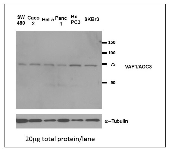
WB: Preliminary experiments gave an approx. 55 kDa band in human adipose and lung lysates after 0.01 ug/ml antibody staining. Please note that currently we cannot find an explanation in the literature for the band we observe given the calculated size of 84.6 kDa band according to NP_003725.1. The 55 kDa band was successfully blocked by incubation with the immunizing peptide. IHC-P: Human liver shows membranous staining in sinusoid lining cells surrounding hepatocytes.
Reviewed Applications
Read 1 review rated 4 using NB100-57090 in the following applications:
Formulation, Preparation, and Storage
Purification
Immunogen affinity purified
Formulation
Tris saline (20 mM Tris pH 7.3, 150 mM NaCl), 0.5% BSA
Preservative
0.02% Sodium Azide
Concentration
0.5 mg/ml
Shipping
The product is shipped with polar packs. Upon receipt, store it immediately at the temperature recommended below.
Stability & Storage
Store at -20C. Avoid freeze-thaw cycles.
Background: VAP-1/AOC3
Long Name
Vascular Adhesion Protein-1
Alternate Names
AOC3, HPAO, SSAO, VAP1
Gene Symbol
AOC3
UniProt
Additional VAP-1/AOC3 Products
Product Documents for VAP-1/AOC3 Antibody
Certificate of Analysis
To download a Certificate of Analysis, please enter a lot or batch number in the search box below.
Product Specific Notices for VAP-1/AOC3 Antibody
This product is for research use only and is not approved for use in humans or in clinical diagnosis. Primary Antibodies are guaranteed for 1 year from date of receipt.
Related Research Areas
Citations for VAP-1/AOC3 Antibody
 Powered by Bioz
Customer Reviews for VAP-1/AOC3 Antibody (1)
4 out of 5
1 Customer Rating
Have you used VAP-1/AOC3 Antibody?
Submit a review and receive an Amazon gift card!
$25/€18/£15/$25CAN/¥2500 Yen for a review with an image
$10/€7/£6/$10CAN/¥1110 Yen for a review without an image
Submit a review
Customer Images
Showing
1
-
1 of
1 review
Showing All
Filter By:
-
Application: Western BlotSample Tested:Species: HumanVerified Customer | Posted 01/07/2015Immunoblots (human cell lines)

There are no reviews that match your criteria.
Protocols
Find general support by application which include: protocols, troubleshooting, illustrated assays, videos and webinars.
- Antigen Retrieval Protocol (PIER)
- Antigen Retrieval for Frozen Sections Protocol
- Appropriate Fixation of IHC/ICC Samples
- Cellular Response to Hypoxia Protocols
- Chromogenic IHC Staining of Formalin-Fixed Paraffin-Embedded (FFPE) Tissue Protocol
- Chromogenic Immunohistochemistry Staining of Frozen Tissue
- ClariTSA™ Fluorophore Kits
- Detection & Visualization of Antibody Binding
- ELISA Sample Preparation & Collection Guide
- ELISA Troubleshooting Guide
- Fluorescent IHC Staining of Frozen Tissue Protocol
- Graphic Protocol for Heat-induced Epitope Retrieval
- Graphic Protocol for the Preparation and Fluorescent IHC Staining of Frozen Tissue Sections
- Graphic Protocol for the Preparation and Fluorescent IHC Staining of Paraffin-embedded Tissue Sections
- Graphic Protocol for the Preparation of Gelatin-coated Slides for Histological Tissue Sections
- How to Run an R&D Systems DuoSet ELISA
- How to Run an R&D Systems Quantikine ELISA
- How to Run an R&D Systems Quantikine™ QuicKit™ ELISA
- IHC Sample Preparation (Frozen sections vs Paraffin)
- Immunofluorescent IHC Staining of Formalin-Fixed Paraffin-Embedded (FFPE) Tissue Protocol
- Immunohistochemistry (IHC) and Immunocytochemistry (ICC) Protocols
- Immunohistochemistry Frozen Troubleshooting
- Immunohistochemistry Paraffin Troubleshooting
- Preparing Samples for IHC/ICC Experiments
- Preventing Non-Specific Staining (Non-Specific Binding)
- Primary Antibody Selection & Optimization
- Protocol for Heat-Induced Epitope Retrieval (HIER)
- Protocol for Making a 4% Formaldehyde Solution in PBS
- Protocol for VisUCyte™ HRP Polymer Detection Reagent
- Protocol for the Preparation & Fixation of Cells on Coverslips
- Protocol for the Preparation and Chromogenic IHC Staining of Frozen Tissue Sections
- Protocol for the Preparation and Chromogenic IHC Staining of Frozen Tissue Sections - Graphic
- Protocol for the Preparation and Chromogenic IHC Staining of Paraffin-embedded Tissue Sections
- Protocol for the Preparation and Chromogenic IHC Staining of Paraffin-embedded Tissue Sections - Graphic
- Protocol for the Preparation and Fluorescent IHC Staining of Frozen Tissue Sections
- Protocol for the Preparation and Fluorescent IHC Staining of Paraffin-embedded Tissue Sections
- Protocol for the Preparation of Gelatin-coated Slides for Histological Tissue Sections
- Quantikine HS ELISA Kit Assay Principle, Alkaline Phosphatase
- Quantikine HS ELISA Kit Principle, Streptavidin-HRP Polymer
- Sandwich ELISA (Colorimetric) – Biotin/Streptavidin Detection Protocol
- Sandwich ELISA (Colorimetric) – Direct Detection Protocol
- TUNEL and Active Caspase-3 Detection by IHC/ICC Protocol
- The Importance of IHC/ICC Controls
- Troubleshooting Guide: ELISA
- Troubleshooting Guide: Immunohistochemistry
- View all Protocols, Troubleshooting, Illustrated assays and Webinars
Loading...