Vimentin Antibody (2D1) - BSA Free
Novus Biologicals | Catalog # NBP1-92687
![Knockout Validated: Vimentin Antibody (2D1) [NBP1-92687]](https://resources.rndsystems.com/images/products/Vimentin-Antibody-2D1-Knockdown-Validated-NBP1-92687-img0011.jpg "Western Blot: Vimentin Antibody (2D1) [NBP1-92687]")
Key Product Details
Validated by
Knockout/Knockdown
Species Reactivity
Validated:
Human, Rat, Mouse (Negative)
Cited:
Human, Rat
Applications
Validated:
Knockout Validated, Immunohistochemistry, Immunohistochemistry-Paraffin, Western Blot, Immunocytochemistry/ Immunofluorescence, Simple Western
Cited:
Immunohistochemistry-Paraffin, Western Blot, Immunocytochemistry/ Immunofluorescence, IF/IHC
Label
Unconjugated
Antibody Source
Monoclonal Mouse IgG2A Clone # 2D1
Format
BSA Free
Loading...
Product Specifications
Immunogen
Full length recombinant human Vimentin Antibody expressed in and purified from E. coli. [UniProt# P08670]
Epitope
amino acids 409-425 of human Vimentin (UniProt Accession P08670): SRISLPLPNFSSLNREL
Reactivity Notes
Clones 2D1 (NBP1-92687) and 2A52 (NBP1-92688) both failed to detect the target in mouse tissues although they work well on human and rat samples. This allowed us to firmly map the epitope for both antibodies to the peptide SRISLPLPNFSSLNREL, amino acids 409-425 of the human sequence. This peptide is located at the beginning of the non-helical "tail" region of the molecule and the peptide is totally conserved between human and rat and in most mammalian species, including cow, pig, horse, camel, and many monkeys. Interestingly mouse has the peptide SRISLPLPTFSSLNREL divergent by one amino acid, and neither clones bind this peptide. As a result these antibodies can be used to identify human or rat cells in mouse cultures or tissues and may work with other species that also contain this peptide.
Localization
Cytoplasm
Marker
Mesenchymal Cells Marker
Clonality
Monoclonal
Host
Mouse
Isotype
IgG2A
Theoretical MW
53.6 kDa.
Disclaimer note: The observed molecular weight of the protein may vary from the listed predicted molecular weight due to post translational modifications, post translation cleavages, relative charges, and other experimental factors.
Disclaimer note: The observed molecular weight of the protein may vary from the listed predicted molecular weight due to post translational modifications, post translation cleavages, relative charges, and other experimental factors.
Scientific Data Images for Vimentin Antibody (2D1) - BSA Free
Western Blot: Vimentin Antibody (2D1) [NBP1-92687]
Western Blot: Vimentin Antibody (2D1) [NBP1-92687] - Western blot shows lysates of K562 human Chronic Myelogenous Leukemia parental cell line and Vimentin knockout (KO) K562 cell line. PVDF membrane was probed with 1:10,000 of Mouse Anti-Human Vimentin Monoclonal Antibody (Catalog # NBP1-92687) followed by HRP-conjugated Anti-Mouse IgG Secondary Antibody (Catalog #HAF018). Specific band was detected for Vimentin at approximately 55 kDa (as indicated) in the parental K562 cell line, but is not detectable in the knockout K562 cell line. This experiment was conducted under reducing conditions.![Immunocytochemistry/ Immunofluorescence: Vimentin Antibody (2D1) [NBP1-92687]](https://resources.rndsystems.com/images/products/Vimentin-Antibody-2D1-Immunocytochemistry-Immunofluorescence-NBP1-92687-img0003.jpg "Immunocytochemistry/ Immunofluorescence: Vimentin Antibody (2D1) [NBP1-92687]")
Immunocytochemistry/ Immunofluorescence: Vimentin Antibody (2D1) [NBP1-92687]
Immunocytochemistry/Immunofluorescence: Vimentin Antibody (2D1) [NBP1-92687] - View of mixed neuron/glial cultures stained with NBP1-92687 (green) and the GFAP rabbit polyclonal (NB300-141, red). Vimentin is expressed alone in fibroblastic and endothelial cells, which are the flattened cells in the middle of the image which appear green. Astrocytes may express primarily GFAP, or GFAP and Vimentin, and so appear red (GFAP only) or golden yellow (GFAP and Vimentin). In cells which express both GFAP and Vimentin, the two proteins assemble to produce heteropolymer filaments.![Western Blot: Vimentin Antibody (2D1) [NBP1-92687]](https://resources.rndsystems.com/images/products/Vimentin-Antibody-2D1-Western-Blot-NBP1-92687-img0009.jpg "Western Blot: Vimentin Antibody (2D1) [NBP1-92687]")
Western Blot: Vimentin Antibody (2D1) [NBP1-92687]
Western Blot: Vimentin Antibody (2D1) [NBP1-92687] - Analysis of different cell lysates using mouse mAb to vimentin, NBP1-92687, dilution 1:10,000 in green: [1] protein standard (red), [2] HEK293, [3] HeLa, [4] SH-SY5Y, [5] COS-1, and [6] C6 cells. The band at about 50kDa mark corresponds to the vimentin protein.![Immunocytochemistry/ Immunofluorescence: Vimentin Antibody (2D1) [NBP1-92687]](https://resources.rndsystems.com/images/products/Vimentin-Antibody-2D1-Immunocytochemistry-Immunofluorescence-NBP1-92687-img0010.jpg "Immunocytochemistry/ Immunofluorescence: Vimentin Antibody (2D1) [NBP1-92687]")
Immunocytochemistry/ Immunofluorescence: Vimentin Antibody (2D1) [NBP1-92687]
Immunocytochemistry/Immunofluorescence: Vimentin Antibody (2D1) [NBP1-92687] - Analysis of cortical neuron-glial cell cultures from E20 rat stained with mouse mAb to vimentin, NBP1-92687, dilution 1:2,000 in red, and costained with chicken pAb to glial fibrillary acidic protein (GFAP), dilution 1:5,000, in green. The blue is DAPI staining of nuclear DNA. Fibroblastic and other developing cells express only vimentin and appear red. Astrocytes that express GFAP only are green while those that express both GFAP and vimentin appear golden yellow.![Immunohistochemistry-Paraffin: Vimentin Antibody (2D1) [NBP1-92687]](https://resources.rndsystems.com/images/products/Vimentin-Antibody-2D1-Immunohistochemistry-Paraffin-NBP1-92687-img0012.jpg "Immunohistochemistry-Paraffin: Vimentin Antibody (2D1) [NBP1-92687]")
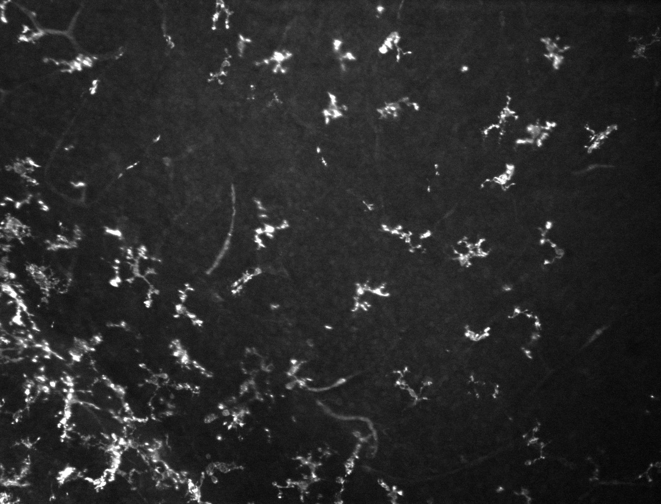
Immunohistochemistry-Paraffin: Vimentin Antibody (2D1) [NBP1-92687]
Immunohistochemistry-Paraffin: Vimentin Antibody (2D1) [NBP1-92687] - Human breast cancer tissue stained for Vimentin (white) and countersatined with DAPI (blue). Alexa Fluor 750 version of antibody used (NBP1-92687AF750). Image from verified customer review.![Immunohistochemistry: Vimentin Antibody (2D1) [NBP1-92687]](https://resources.rndsystems.com/images/products/Vimentin-Antibody-2D1-Immunohistochemistry-NBP1-92687-img0005.jpg "Immunohistochemistry: Vimentin Antibody (2D1) [NBP1-92687]")
Immunohistochemistry: Vimentin Antibody (2D1) [NBP1-92687]
Immunohistochemistry: Vimentin Antibody (2D1) [NBP1-92687] - Mouse cortex microslice section (from postnatal day 3) was immunostained with (1:500 dil.). This image was submitted via customer Review.![Simple Western: Vimentin Antibody (2D1) [NBP1-92687]](https://resources.rndsystems.com/images/products/Vimentin-Antibody-2D1-Simple-Western-NBP1-92687-img0004.jpg "Simple Western: Vimentin Antibody (2D1) [NBP1-92687]")
Simple Western: Vimentin Antibody (2D1) [NBP1-92687]
Simple Western: Vimentin Antibody (2D1) [NBP1-92687] - Simple Western lane view shows a specific band for Vimentin in 0.5 mg/ml of HeLa lysate. This experiment was performed under reducing conditions using the 12-230 kDa separation system. [NBP1-92687] -")
Western Blot: Vimentin Antibody (2D1) [NBP1-92687] -
Western Blot: Vimentin Antibody (2D1) [NBP1-92687] - High glucose (HG) concentrations induced epithelial-to-mesenchymal transition protein expression & enhanced migration activity in colorectal cancer (CRC) cells. SW480 (low metastatic potential) & SW620 (high metastatic potential) cells were cultured in different concentrations of glucose (normal: NG; HG; & osmotic control: NG + l-glucose). (A) Morphological change occurred from epithelial to mesenchymal type in the HG-concentration group. (B) HG concentration caused downregulation of E-cadherin & upregulation of N-cadherin, beta CTN, & vimentin, but c-myc was unchanged, as detected using Western blotting. beta -actin was evaluated as an internal control. (C,D) Wound healing assay showed that HG concentration promoted cell motility in SW480 & SW620 CRC cells after 48 & 72 h of culture, compared with the NG & NG + l-glucose groups. (E) In a Transwell migration assay, 3.5 × 10⁵ SW480 & SW620 CRC cells were plated onto a 24-well plate & cultured in NG & HG-concentration medium for 96 h. HG concentration promoted cell motility in SW480 & SW620 cells. NG + l-glucose cells were evaluated as ostomic controls. (F) These data show that HG concentration caused upregulation of p-IGF1R in CRC. In addition, HG concentration promoted IGF1R downstream signaling, including p-Src & p-ERK; these proteins were increased when CRC cells were cultured in HG-concentration medium. Levels of beta -actin were evaluated as loading controls. Statistically significant differences between the two groups were judged using Student’s t-tests; * p < 0.05, ** p < 0.005, *** p < 0.001; n.s. = nonsignificant. Image collected & cropped by CiteAb from the following publication (https://pubmed.ncbi.nlm.nih.gov/30965609), licensed under a CC-BY license. Not internally tested by Novus Biologicals.Applications for Vimentin Antibody (2D1) - BSA Free
Application
Recommended Usage
Immunocytochemistry/ Immunofluorescence
1:1000
Immunohistochemistry
1:1000
Simple Western
1:100
Western Blot
1:10000
Application Notes
This Vimentin (2D1) antibody is useful for Immunocytochemistry/Immunofluorescence, Immunohistochemistry, and Western blot, where a band can be seen at approximately 50 kDa. Use in IHC-P was reported in scientific literature (PMID: 30327566).
In Simple Western only 10 - 15 uL of the recommended dilution is used per data point.
See Simple Western Antibody Database for Simple Western validation: Tested in HeLa lysate 0.5 mg/mL, separated by Size, antibody dilution of 1:100, apparent MW was 59 kDa. Separated by Size-Wes, Sally Sue/Peggy Sue.
Reviewed Applications
Read 1 review rated 5 using NBP1-92687 in the following applications:
Formulation, Preparation, and Storage
Purification
Protein G purified
Formulation
50% PBS, 50% glycerol
Format
BSA Free
Preservative
5mM Sodium Azide
Concentration
1 mg/ml
Shipping
The product is shipped with polar packs. Upon receipt, store it immediately at the temperature recommended below.
Stability & Storage
Store at 4C short term. Aliquot and store at -20C long term. Avoid freeze-thaw cycles.
Background: Vimentin
Activated macrophages have been shown to secrete phosphorylated vimentin which can be stimulated by a variety of pathophysiological factors including oxidized low-density lipoproteins and TNF-alpha or inhibited by IL-10 (1). The vimentin protein is often expressed at the cell surface playing a role in cell-cell interactions, tissue damage and repair, immune response, and pathogen recognition (1). Vimentin functions in many cytoskeletal processes including cell migration, which is highlighted by its upregulation during epithelial-to-mesenchymal transition (EMT) (4,5). Vimentin is a commonly used marker for EMT and is expressed by many tumor types (5). For example, high metastasis of oral squamous cell carcinomas also showed high vimentin positive expression in immunohistochemical staining analysis (5). A number of vimentin targeting compounds are in cancer-related clinical trials, however, given the multifunctional role of vimentin, the effect of inhibition on non-malignant cells needs to be thoroughly examined (5).
References
1. Ramos, I., Stamatakis, K., Oeste, C. L., & Perez-Sala, D. (2020). Vimentin as a Multifaceted Player and Potential Therapeutic Target in Viral Infections. International Journal of Molecular Sciences. https://doi.org/10.3390/ijms21134675
2. Uniprot (P08670)
3. Morrow, C. S., & Moore, D. L. (2020). Vimentin's side gig: Regulating cellular proteostasis in mammalian systems. Cytoskeleton (Hoboken, N.J.). https://doi.org/10.1002/cm.21645
4. van Bodegraven, E. J., & Etienne-Manneville, S. (2020). Intermediate filaments against actomyosin: the david and goliath of cell migration. Current Opinion in Cell Biology. https://doi.org/10.1016/j.ceb.2020.05.006
5. Strouhalova, K., Prechova, M., Gandalovicova, A., Brabek, J., Gregor, M., & Rosel, D. (2020). Vimentin Intermediate Filaments as Potential Target for Cancer Treatment. Cancers. https://doi.org/10.3390/cancers12010184
Additional Vimentin Products
Product Documents for Vimentin Antibody (2D1) - BSA Free
Certificate of Analysis
To download a Certificate of Analysis, please enter a lot or batch number in the search box below.
Product Specific Notices for Vimentin Antibody (2D1) - BSA Free
This product is for research use only and is not approved for use in humans or in clinical diagnosis. Primary Antibodies are guaranteed for 1 year from date of receipt.
Citations for Vimentin Antibody (2D1) - BSA Free
 Powered by Bioz
Customer Reviews for Vimentin Antibody (2D1) - BSA Free (1)
5 out of 5
1 Customer Rating
Have you used Vimentin Antibody (2D1) - BSA Free?
Submit a review and receive an Amazon gift card!
$25/€18/£15/$25CAN/¥2500 Yen for a review with an image
$10/€7/£6/$10CAN/¥1110 Yen for a review without an image
Submit a review
Customer Images
Showing
1
-
1 of
1 review
Showing All
Filter By:
-
Application: Immunohistochemistry-microsliceSample Tested: Mouse cortexSpecies: MouseVerified Customer | Posted 07/27/2017Mouse cortex microslice section (from postnatal day 3) was immunostained with Vimentin Antibody (2D1) (1:500 dil.)

There are no reviews that match your criteria.
Protocols
Find general support by application which include: protocols, troubleshooting, illustrated assays, videos and webinars.
- Antigen Retrieval Protocol (PIER)
- Antigen Retrieval for Frozen Sections Protocol
- Appropriate Fixation of IHC/ICC Samples
- Cellular Response to Hypoxia Protocols
- Chromogenic IHC Staining of Formalin-Fixed Paraffin-Embedded (FFPE) Tissue Protocol
- Chromogenic Immunohistochemistry Staining of Frozen Tissue
- ClariTSA™ Fluorophore Kits
- Detection & Visualization of Antibody Binding
- Fluorescent IHC Staining of Frozen Tissue Protocol
- Graphic Protocol for Heat-induced Epitope Retrieval
- Graphic Protocol for the Preparation and Fluorescent IHC Staining of Frozen Tissue Sections
- Graphic Protocol for the Preparation and Fluorescent IHC Staining of Paraffin-embedded Tissue Sections
- Graphic Protocol for the Preparation of Gelatin-coated Slides for Histological Tissue Sections
- ICC Cell Smear Protocol for Suspension Cells
- ICC Immunocytochemistry Protocol Videos
- ICC for Adherent Cells
- IHC Sample Preparation (Frozen sections vs Paraffin)
- Immunocytochemistry (ICC) Protocol
- Immunocytochemistry Troubleshooting
- Immunofluorescence of Organoids Embedded in Cultrex Basement Membrane Extract
- Immunofluorescent IHC Staining of Formalin-Fixed Paraffin-Embedded (FFPE) Tissue Protocol
- Immunohistochemistry (IHC) and Immunocytochemistry (ICC) Protocols
- Immunohistochemistry Frozen Troubleshooting
- Immunohistochemistry Paraffin Troubleshooting
- Preparing Samples for IHC/ICC Experiments
- Preventing Non-Specific Staining (Non-Specific Binding)
- Primary Antibody Selection & Optimization
- Protocol for Heat-Induced Epitope Retrieval (HIER)
- Protocol for Making a 4% Formaldehyde Solution in PBS
- Protocol for VisUCyte™ HRP Polymer Detection Reagent
- Protocol for the Fluorescent ICC Staining of Cell Smears - Graphic
- Protocol for the Fluorescent ICC Staining of Cultured Cells on Coverslips - Graphic
- Protocol for the Preparation & Fixation of Cells on Coverslips
- Protocol for the Preparation and Chromogenic IHC Staining of Frozen Tissue Sections
- Protocol for the Preparation and Chromogenic IHC Staining of Frozen Tissue Sections - Graphic
- Protocol for the Preparation and Chromogenic IHC Staining of Paraffin-embedded Tissue Sections
- Protocol for the Preparation and Chromogenic IHC Staining of Paraffin-embedded Tissue Sections - Graphic
- Protocol for the Preparation and Fluorescent ICC Staining of Cells on Coverslips
- Protocol for the Preparation and Fluorescent ICC Staining of Non-adherent Cells
- Protocol for the Preparation and Fluorescent ICC Staining of Stem Cells on Coverslips
- Protocol for the Preparation and Fluorescent IHC Staining of Frozen Tissue Sections
- Protocol for the Preparation and Fluorescent IHC Staining of Paraffin-embedded Tissue Sections
- Protocol for the Preparation of Gelatin-coated Slides for Histological Tissue Sections
- Protocol for the Preparation of a Cell Smear for Non-adherent Cell ICC - Graphic
- R&D Systems Quality Control Western Blot Protocol
- TUNEL and Active Caspase-3 Detection by IHC/ICC Protocol
- The Importance of IHC/ICC Controls
- Troubleshooting Guide: Immunohistochemistry
- Troubleshooting Guide: Western Blot Figures
- Western Blot Conditions
- Western Blot Protocol
- Western Blot Protocol for Cell Lysates
- Western Blot Troubleshooting
- Western Blot Troubleshooting Guide
- View all Protocols, Troubleshooting, Illustrated assays and Webinars
FAQs for Vimentin Antibody (2D1) - BSA Free
Showing
1
-
2 of
2 FAQs
Showing All
-
Q: Hi, what exactly does Mu(-) refer to?
A: (-) refers to a negative species reactivity. In this case, the antibody reacts to human and rat Vimentin but not to mouse Vimentin
-
Q: I am planning on using mouse monoclonal Vimentin Antibody (2D1), NBP1-92687 on mouse fibroblasts. Has the antibody been tested for cross-reactivity with endogenous IgG? Do I have to expect strong background?
A: If the proposed samples are mouse fibroblasts grown in tissue culture, there should be no problem as any endogenous mouse IgG would be diluted out after a few days in culture, as the cells are fed. Western blotting or IHC of pancreatic tissue would be more of a problem, as this would contain some endogenous IgG, though much of this can be removed by perfusing the animal with PBS prior to taking out the pancreas. The endogenous mouse IgG is mostly in blood vessels and may show up in IHC, though various kits are available which are supposed to address this issue. On western blots, the mouse IgG usually shows up as a somewhat diffuse band at about 55 kDa (the IgG heavy chains; most anti-mouse reagents bind primarily the heavy chain) and in some cases a faint band at about 20 kDa (the IgG light chains). Vimentin should appear as a sharp and clean single band at 50 kDa, so should be easily distinguished from any endogenous mouse IgG. This issue could be avoided entirely by using an alternative anti-vimentin antibody with a different host.
-
Q: Hi, what exactly does Mu(-) refer to?
A: (-) refers to a negative species reactivity. In this case, the antibody reacts to human and rat Vimentin but not to mouse Vimentin
-
Q: I am planning on using mouse monoclonal Vimentin Antibody (2D1), NBP1-92687 on mouse fibroblasts. Has the antibody been tested for cross-reactivity with endogenous IgG? Do I have to expect strong background?
A: If the proposed samples are mouse fibroblasts grown in tissue culture, there should be no problem as any endogenous mouse IgG would be diluted out after a few days in culture, as the cells are fed. Western blotting or IHC of pancreatic tissue would be more of a problem, as this would contain some endogenous IgG, though much of this can be removed by perfusing the animal with PBS prior to taking out the pancreas. The endogenous mouse IgG is mostly in blood vessels and may show up in IHC, though various kits are available which are supposed to address this issue. On western blots, the mouse IgG usually shows up as a somewhat diffuse band at about 55 kDa (the IgG heavy chains; most anti-mouse reagents bind primarily the heavy chain) and in some cases a faint band at about 20 kDa (the IgG light chains). Vimentin should appear as a sharp and clean single band at 50 kDa, so should be easily distinguished from any endogenous mouse IgG. This issue could be avoided entirely by using an alternative anti-vimentin antibody with a different host.

