![Western Blot: Vimentin Antibody [NB300-223]](https://resources.rndsystems.com/images/products/Vimentin-Antibody-Western-Blot-NB300-223-img0002.jpg "Western Blot: Vimentin Antibody [NB300-223]")
Loading...
Key Product Details
Species Reactivity
Validated:
Human, Mouse, Rat, Porcine, Bovine, Canine, Chicken, Equine
Cited:
Human, Mouse, Rat, Canine
Applications
Validated:
Immunohistochemistry, Immunohistochemistry-Paraffin, Immunohistochemistry-Frozen, Western Blot, Immunocytochemistry/ Immunofluorescence
Cited:
Immunohistochemistry, Immunohistochemistry-Paraffin, Immunohistochemistry-Frozen, Western Blot, Block/Neutralize, Immunocytochemistry/ Immunofluorescence, IF/IHC, Electron Microscopy, Knockdown Validated
Label
Unconjugated
Antibody Source
Polyclonal Chicken IgY
Loading...
Product Specifications
Immunogen
Full length recombinant human Vimentin Antibody expressed in and purified from E. coli. [Swiss-Prot# P08670]
Reactivity Notes
Use in Mouse reported in scientific literature (PMID:33675257).
Marker
Mesenchymal Cells Marker
Clonality
Polyclonal
Host
Chicken
Isotype
IgY
Theoretical MW
53.6 kDa.
Disclaimer note: The observed molecular weight of the protein may vary from the listed predicted molecular weight due to post translational modifications, post translation cleavages, relative charges, and other experimental factors.
Disclaimer note: The observed molecular weight of the protein may vary from the listed predicted molecular weight due to post translational modifications, post translation cleavages, relative charges, and other experimental factors.
Description
This antibody is supplied as a concentrated total IgY preparation derived from egg yolk. Production involves an initial organic extraction to remove lipids and lipoproteins, followed by salt fractionation of the aqueous phase to precipitate and enrich the IgY fraction. The material is then extensively dialyzed against PBS, and preservative is added during final formulation.
Please note that the exact concentration of target-specific IgY cannot be determined, as the final preparation contains both antigen-specific IgY and non-immune IgY.
Please note that the exact concentration of target-specific IgY cannot be determined, as the final preparation contains both antigen-specific IgY and non-immune IgY.
Scientific Data Images for Vimentin Antibody
Western Blot: Vimentin Antibody [NB300-223]
Western Blot: Vimentin Antibody [NB300-223] - Analysis of tissue and cell lysates. Antibody at 1:5000 in red. [1] protein standard (red), [2] rat whole brain lysate, [3] HeLa, [4] SH-SY5Y, [5] HEK293, and [6] NIH-3T3 cell lysates. NB300-223 binds to the vimentin protein showing a single band at ~50 kDa. The blot was simultaneously probed with mouse mAb to MAP2C/D, dilution 1:5000 in green, revealing multiple bands around 280 kDa that correspond to full length MAP2A/2B isotypes, and ~70 kDa bands which are MAP2C/D isotypes. MAP2 isotypes are seen only in extracts containing neuronal lineage cells.![Immunohistochemistry: Vimentin Antibody [NB300-223]](https://resources.rndsystems.com/images/products/Vimentin-Antibody-Immunohistochemistry-NB300-223-img0006.jpg "Immunohistochemistry: Vimentin Antibody [NB300-223]")
Immunohistochemistry: Vimentin Antibody [NB300-223]
Vimentin-Antibody-Immunohistochemistry-NB300-223-img0006.jpg![Immunocytochemistry/ Immunofluorescence: Vimentin Antibody [NB300-223]](https://resources.rndsystems.com/images/products/Vimentin-Antibody-Immunocytochemistry-Immunofluorescence-NB300-223-img0004.jpg "Immunocytochemistry/ Immunofluorescence: Vimentin Antibody [NB300-223]")
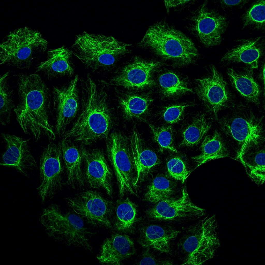
Immunocytochemistry/ Immunofluorescence: Vimentin Antibody [NB300-223]
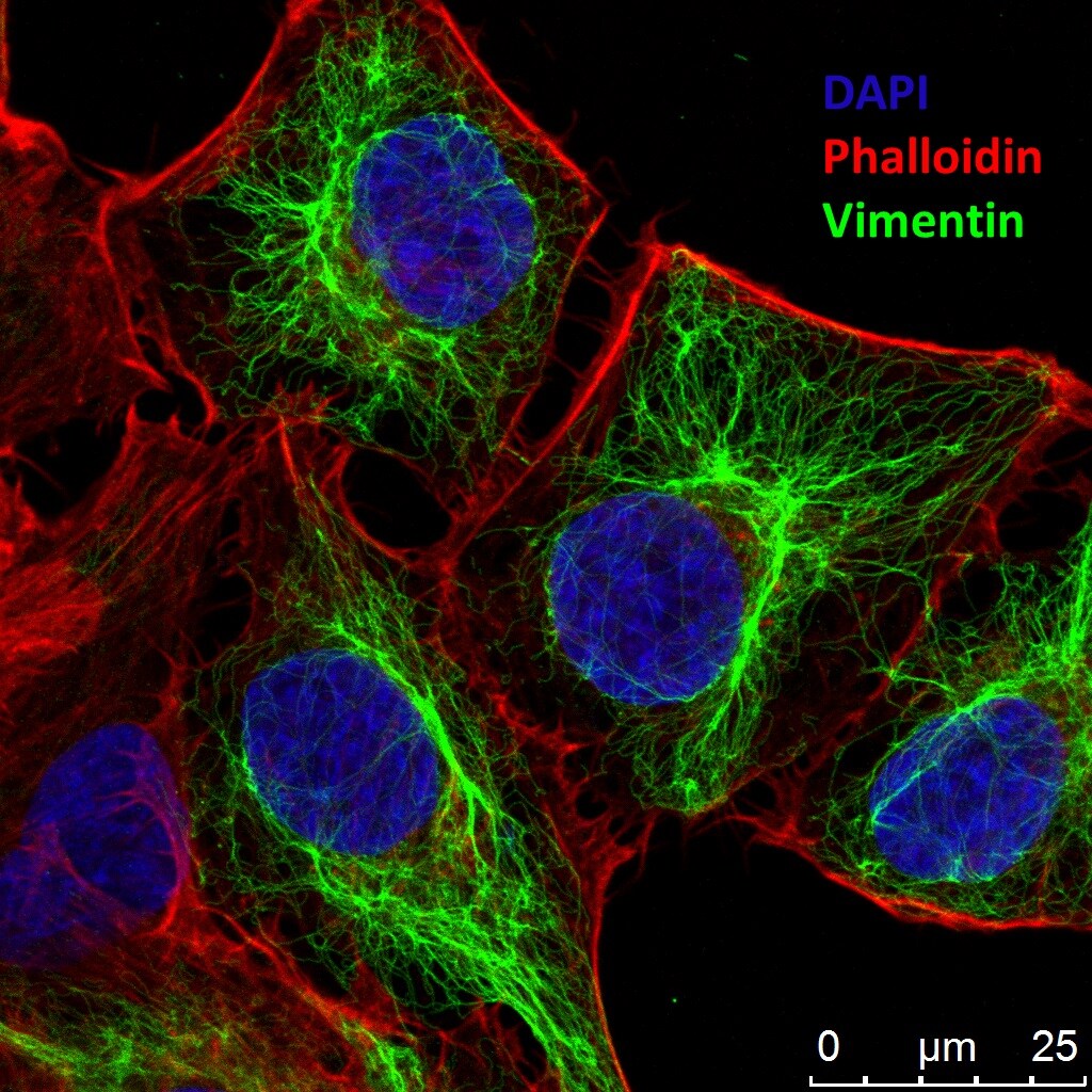
Immunocytochemistry/Immunofluorescence: Vimentin Antibody [NB300-223] - Human A7 cells. Cells were fixed with 10% formalin and permeabilized with 0.1% TritonX-100. After blocking, cells were incubated with primary antibody (1:600) at 4C O/N followed with Alexa Fluor 488 labeled secondary antibody and counterstained with DAPI and Phalloidin for nuclei and F-actin respectively. Imaged with Leica TCS SP5 confocal microscope. ICC/IF image submitted by a verified customer review.![Immunohistochemistry: Vimentin Antibody [NB300-223]](https://resources.rndsystems.com/images/products/Vimentin-Antibody-Immunohistochemistry-NB300-223-img0007.jpg "Immunohistochemistry: Vimentin Antibody [NB300-223]")
Immunohistochemistry: Vimentin Antibody [NB300-223]
Vimentin-Antibody-Immunohistochemistry-NB300-223-img0007.jpg![Immunocytochemistry/ Immunofluorescence: Vimentin Antibody [NB300-223]](https://resources.rndsystems.com/images/products/Vimentin-Antibody-Immunocytochemistry-Immunofluorescence-NB300-223-img0008.jpg "Immunocytochemistry/ Immunofluorescence: Vimentin Antibody [NB300-223]")
Immunocytochemistry/ Immunofluorescence: Vimentin Antibody [NB300-223]
Vimentin-Antibody-Immunocytochemistry-Immunofluorescence-NB300-223-img0008.jpg![Immunocytochemistry/ Immunofluorescence: Vimentin Antibody [NB300-223]](https://resources.rndsystems.com/images/products/Vimentin-Antibody-Immunocytochemistry-Immunofluorescence-NB300-223-img0001.jpg "Immunocytochemistry/ Immunofluorescence: Vimentin Antibody [NB300-223]")
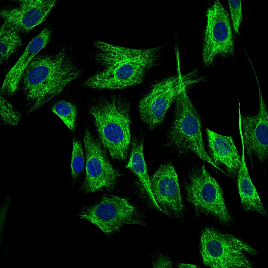
Immunocytochemistry/ Immunofluorescence: Vimentin Antibody [NB300-223]
Immunocytochemistry/Immunofluorescence: Vimentin Antibody [NB300-223] - Analysis of HeLa cell culture. Antibody at 1:10,000 in green, and costained with mouse mAb to actin, dilution 1:500 in red. The blue is DAPI staining of nuclear DNA. The vimentin antibody stains the intermediate filament network while the actin antibody labels the submembranous cytoskeleton, stress fibers, and bundles of actin associated with cell adhesion sites.![Immunohistochemistry-Frozen: Vimentin Antibody [NB300-223]](https://resources.rndsystems.com/images/products/Vimentin-Antibody-Immunohistochemistry-Frozen-NB300-223-img0003.jpg "Immunohistochemistry-Frozen: Vimentin Antibody [NB300-223]")
Immunohistochemistry-Frozen: Vimentin Antibody [NB300-223]
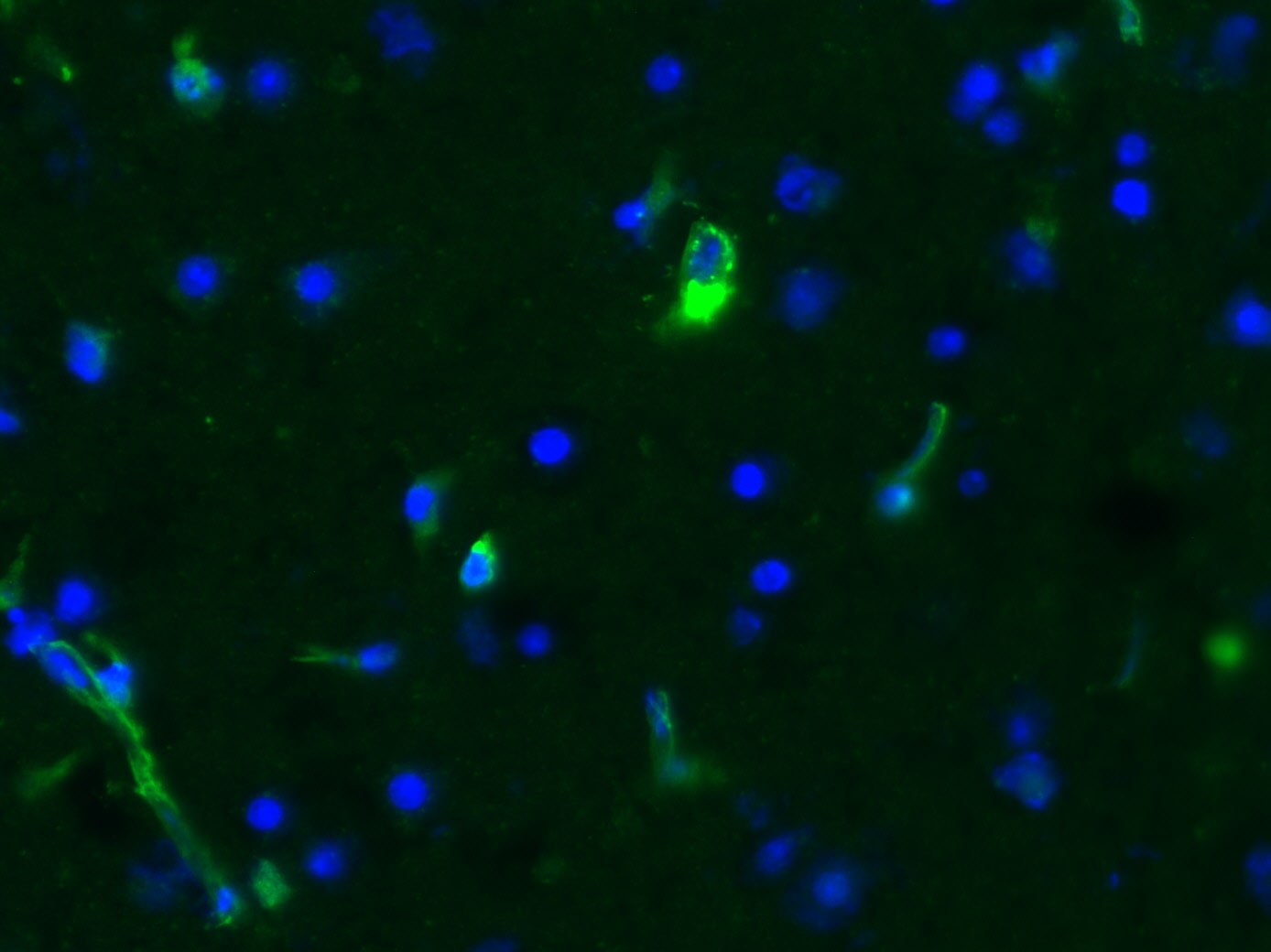
Immunohistochemistry-Frozen: Vimentin Antibody [NB300-223] - 10 um Cryosections from PFA fixed murine brain acute slices stained with Vimentin antibody (1:600) and Alexa Fluor 488 anti chicken IgG. Antibody showed specific staining, also of blood vessels. IHC-Fr image submitted by a verified customer review.![Immunohistochemistry-Frozen: Vimentin Antibody [NB300-223]](https://resources.rndsystems.com/images/products/Vimentin-Antibody-Immunohistochemistry-Frozen-NB300-223-img0005.jpg "Immunohistochemistry-Frozen: Vimentin Antibody [NB300-223]")
Immunohistochemistry-Frozen: Vimentin Antibody [NB300-223]
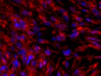
Immunohistochemistry-Frozen: Vimentin Antibody [NB300-223] - Infarcted mouse heart frozen sections stained with vimentin antibody at 1:200. Secondary antibody at 1:500. Fixation with 4% PFA. IHC-Fr image submitted by a verified customer review.
Immunocytochemistry/ Immunofluorescence: Vimentin Antibody [NB300-223] -
Immunocytochemistry/ Immunofluorescence: Vimentin Antibody [NB300-223] - Increased numbers of differentiating NSPCs in SVZ of G93A-SOD1 brain. (A) Confocal microscopy images of dorsal & ventral regions of SVZ in G93A-SOD1 & Wt-SOD1 brain at postnatal week 18, stained for nestin (green), GFAP (red), & vimentin (blue). Scale bar = 50 μm. (B) Quantification of nestin-, GFAP-, & vimentin-positive cells in SVZ of G93A-SOD1 & Wt-SOD1 mice. Data are means ± SD of 3 mice per group. **p < 0.001; limma moderated t-test. (C) Hematoxylin & eosin staining of SVZ sections adjacent to those analyzed by confocal microscopy. Scale bar = 50 μm. D: dorsal. V: ventral. (D) Confocal microscopy images showing Dlx2-stained cells in ventral SVZ in G93A-SOD1 & Wt-SOD1 brain. Scale bar = 50 μm. (E) Quantification of Dlx2-positive cells in G93A-SOD1 & Wt-SOD1 SVZ. Data are means ± SD of 3 mice per group. **p < 0.001; limma moderated t-test. Image collected & cropped by CiteAb from the following publication (https://molecularbrain.biomedcentral.com/articles/10.1186/s13041-015-00…), licensed under a CC-BY license. Not internally tested by Novus Biologicals.
Immunocytochemistry/ Immunofluorescence: Vimentin Antibody [NB300-223] -
Immunocytochemistry/ Immunofluorescence: Vimentin Antibody [NB300-223] - Attenuated reactive astrocytosis after stroke in Smad1 cKO mice.(A) Images of IHC for reactive astrocyte marker GFAP on ipsilateral hemisphere at 7 days (top two panels) or 3 months (bottom panels) post-stroke. The border between the stroke core & peri-infarct area is outlined by dotted lines. Arrows point to striatal infarct core. (B) Enlarged IHC images of boxed areas in (A) at the cortical peri-infarct area (blue box in D) with the indicated reactive astrocyte markers GFAP, Nestin, & Vimentin. Enlarged images of GFAP IHC highlight the hypertrophic morphology of GFAP+ astrocytes in mutants. (C) Quantification of the number of GFAP+ astrocytes & the intensity of GFAP immunoreactivity (IR) at the peri-infarct area shown in (B). n = 4, one-way ANOVA for the number of astrocytes, unpaired Student’s t-test for GFAP intensity, ***p<0.001, Ipsi, ipsilateral; Ctra, contralateral cortex. (D) Diagram of infarct territory affected by tMCAO (blue) & peri-infarct cortical area (blue box). (E) Reactive astrocytosis was similarly attenuated in the ipsilateral hippocampus of Smad1 cKO mice. Scale, 500 μm (A), 100 μm (B), & 200 μm (E). Image collected & cropped by CiteAb from the following publication (https://dx.plos.org/10.1371/journal.pone.0136967), licensed under a CC-BY license. Not internally tested by Novus Biologicals.
Immunocytochemistry/ Immunofluorescence: Vimentin Antibody [NB300-223] -
Immunocytochemistry/ Immunofluorescence: Vimentin Antibody [NB300-223] - Tumor formation & metastasis by PyMT-1099 cells. (A) PyMT-1099 cells untreated or treated with TGF beta for >20 days (PyMT-1099 LT) were injected orthotopically into mammary fat pads of NSG mice. The graph represents tumor growth in PyMT-1099 & PyMT-1099 LT group of mice. (B) Histological tumor sections from tumors of PyMT-1099 or PyMT-1099 LT cells described in (A) were stained with H&E to assess the morphology of primary tumors. Representative microphotographs are shown from tumors of 2 out of the 6 mice used in the experiment. (C) Immunofluorescence analysis was performed to assess the expression of the EMT markers FN1, VIM, E-CAD & N-CAD in tumors formed by PyMT-1099 or PyMT-1099 LT cells in the experiment described in (A). DAPI was used as a nuclear counterstain. Representative pictures are shown from tumors of 2 out of the 6 mice used in the experiment. Scale bar, 100 μm. (D) The graph represents the number of lung metastases formed in NSG mice orthotopically transplanted with PyMT-1099 or PyMT-1099 LT cells; n = 6. (E) The graph represents the number of lung metastases formed in NSG mice injected with PyMT-1099 or PyMT-1099 LT cells through the tail vein; n = 6. The mice were sacrificed 8 weeks post-injection, & lungs were resected for the analysis of cancer cell colonization/ metastases formation. Image collected & cropped by CiteAb from the following publication (https://pubmed.ncbi.nlm.nih.gov/30108334), licensed under a CC-BY license. Not internally tested by Novus Biologicals.
Western Blot: Vimentin Antibody [NB300-223] -
DNM1P35 promotes EMT of ovarian cancer cells. (A,B) The expression of EMT markers were assessed by qPCR (A) and Western blotting (B) in SK-OV-3 and OVCAR-3 cells expressing shCTRL and shDNM1P35. (C,D) The expression of EMT markers were assessed by qPCR (C) and Western blotting (D) in SK-OV-3 and OVCAR-3 cells expressing vector and DNM1P35. (E,F) The expression of EMT markers were assessed by qPCR (E) and Western blotting (F) in SK-OV-3 and OVCAR-3 cells expressing shCTRL and shZEB1. (G,H) The expression of EMT markers were assessed by qPCR (G) and Western blotting (H) in SK-OV-3 and OVCAR-3 cells expressing shCTRL and shZEB1 and overexpression of DNM1P35. ***, p < 0.001, ****, p < 0.0001. ns, not significant. Image collected and cropped by CiteAb from the following open publication (https://pubmed.ncbi.nlm.nih.gov/39732940), licensed under a CC-BY license. Not internally tested by Novus Biologicals.
Western Blot: Vimentin Antibody [NB300-223] -
DNM1P35 targets miR-326 to promote ovarian cancer progression and EMT. (A) The potential target of DNM1P35 was predicted by the STAR (database. (B) Dual-luciferase activity assay in HEK293T cells expressing WT and MUT DNM1P35 luciferase reporter and treated with miR-NC or miR-326. (C) The expression of ZEB1 in SK-OV-3 and OVCAR-3 cells transfected with miR-NC and miR-326 was determined by qPCR. (D) The expression of EMT markers in SK-OV-3 and OVCAR-3 cells transfected with miR-NC and miR-326 was determined by Western blotting. (E) The growth of ovarian cancer cells was assessed by CCK-8 assay. (F) SK-OV-3 and OVCAR-3 cell colonies were stained by crystal violet. (G) Migration of SK-OV-3 and OVCAR-3 cells was measured by wound healing assay. (H) Invasion of SK-OV-3 and OVCAR-3 cells was measured by trans-well assay. ***, p < 0.001. Image collected and cropped by CiteAb from the following open publication (https://pubmed.ncbi.nlm.nih.gov/39732940), licensed under a CC-BY license. Not internally tested by Novus Biologicals.
Western Blot: Vimentin Antibody [NB300-223] -
DNM1P35 promotes EMT of ovarian cancer cells. (A,B) The expression of EMT markers were assessed by qPCR (A) and Western blotting (B) in SK-OV-3 and OVCAR-3 cells expressing shCTRL and shDNM1P35. (C,D) The expression of EMT markers were assessed by qPCR (C) and Western blotting (D) in SK-OV-3 and OVCAR-3 cells expressing vector and DNM1P35. (E,F) The expression of EMT markers were assessed by qPCR (E) and Western blotting (F) in SK-OV-3 and OVCAR-3 cells expressing shCTRL and shZEB1. (G,H) The expression of EMT markers were assessed by qPCR (G) and Western blotting (H) in SK-OV-3 and OVCAR-3 cells expressing shCTRL and shZEB1 and overexpression of DNM1P35. ***, p < 0.001, ****, p < 0.0001. ns, not significant. Image collected and cropped by CiteAb from the following open publication (https://pubmed.ncbi.nlm.nih.gov/39732940), licensed under a CC-BY license. Not internally tested by Novus Biologicals.Applications for Vimentin Antibody
Application
Recommended Usage
Immunocytochemistry/ Immunofluorescence
1:5000
Immunohistochemistry
1:150
Immunohistochemistry-Paraffin
1:150
Western Blot
1:10000-1:20000
Application Notes
Use in IHC-P and IHC-Fr reported in scientific literature (PMID: 23752194 and 25626686).
Reviewed Applications
Read 6 reviews rated 5 using NB300-223 in the following applications:
Formulation, Preparation, and Storage
Purification
IgY purified
Formulation
Supplied as a concentrated total IgY preparation from egg yolk, dialyzed against PBS with added preservative.
Preservative
0.035% Sodium Azide
Concentration
Please see the vial label for concentration. If unlisted please contact technical services.
Shipping
The product is shipped with polar packs. Upon receipt, store it immediately at the temperature recommended below.
Stability & Storage
Store at 4C short term. Aliquot and store at -20C long term. Avoid freeze-thaw cycles.
Background: Vimentin
Activated macrophages have been shown to secrete phosphorylated vimentin which can be stimulated by a variety of pathophysiological factors including oxidized low-density lipoproteins and TNF-alpha or inhibited by IL-10 (1). The vimentin protein is often expressed at the cell surface playing a role in cell-cell interactions, tissue damage and repair, immune response, and pathogen recognition (1). Vimentin functions in many cytoskeletal processes including cell migration, which is highlighted by its upregulation during epithelial-to-mesenchymal transition (EMT) (4,5). Vimentin is a commonly used marker for EMT and is expressed by many tumor types (5). For example, high metastasis of oral squamous cell carcinomas also showed high vimentin positive expression in immunohistochemical staining analysis (5). A number of vimentin targeting compounds are in cancer-related clinical trials, however, given the multifunctional role of vimentin, the effect of inhibition on non-malignant cells needs to be thoroughly examined (5).
References
1. Ramos, I., Stamatakis, K., Oeste, C. L., & Perez-Sala, D. (2020). Vimentin as a Multifaceted Player and Potential Therapeutic Target in Viral Infections. International Journal of Molecular Sciences. https://doi.org/10.3390/ijms21134675
2. Uniprot (P08670)
3. Morrow, C. S., & Moore, D. L. (2020). Vimentin's side gig: Regulating cellular proteostasis in mammalian systems. Cytoskeleton (Hoboken, N.J.). https://doi.org/10.1002/cm.21645
4. van Bodegraven, E. J., & Etienne-Manneville, S. (2020). Intermediate filaments against actomyosin: the david and goliath of cell migration. Current Opinion in Cell Biology. https://doi.org/10.1016/j.ceb.2020.05.006
5. Strouhalova, K., Prechova, M., Gandalovicova, A., Brabek, J., Gregor, M., & Rosel, D. (2020). Vimentin Intermediate Filaments as Potential Target for Cancer Treatment. Cancers. https://doi.org/10.3390/cancers12010184
Alternate Names
VIM
Gene Symbol
VIM
UniProt
Additional Vimentin Products
Product Documents for Vimentin Antibody
Certificate of Analysis
To download a Certificate of Analysis, please enter a lot or batch number in the search box below.
Product Specific Notices for Vimentin Antibody
Chicken products cannot be exported to Canada.
This product is for research use only and is not approved for use in humans or in clinical diagnosis. Primary Antibodies are guaranteed for 1 year from date of receipt.
Citations for Vimentin Antibody
 Powered by Bioz
Customer Reviews for Vimentin Antibody (6)
5 out of 5
6 Customer Ratings
Have you used Vimentin Antibody?
Submit a review and receive an Amazon gift card!
$25/€18/£15/$25CAN/¥2500 Yen for a review with an image
$10/€7/£6/$10CAN/¥1110 Yen for a review without an image
Submit a review
Customer Images
Showing
1
-
5 of
6 reviews
Showing All
Filter By:
-
Application: Immunohistochemistry-FrozenSample Tested: Mouse heart frozen sections and mouse frozen heartSpecies: MouseVerified Customer | Posted 01/30/2020Infarcted mouse heart sections staining with vimentinMouse heart after myocardial infarction Frozen section 4% PFA fixation Primary antibody concentration: 1:200 Secondary antibody concentration: 1:500

-
Application: ImmunocytochemistrySample Tested: A7 cellsSpecies: HumanVerified Customer | Posted 04/22/2019Cells were fixed with 10% formalin and permeabilized with 0.1% TritonX-100. After blocking, cells were incubated with primary antibody (1:600) at 4℃ O/N followed with Alexa Fluor-488 labeled secondary antibody and counterstained with DAPI and Phalloidin for nuclei and F-actin respectively. Results were imaged with Leica TCS SP5 confocal microscope.
-
Application: Immunohistochemistry-FrozenSample Tested: Mouse brainSpecies: MouseVerified Customer | Posted 03/11/201910 µm Cryosections from PFA fixed murine brain acute slices stained with Vimentin antibody (1:600) and Alexa Fluor 488 anti chicken IgG. Antibody showed specific staining, also of blood vessels.
-
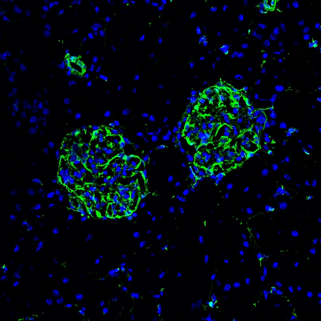
Application: Immunohistochemistry-FrozenSample Tested: Rat Kidney Sections (Fresh Frozen)Species: RatVerified Customer | Posted 06/15/2017
-
Application: ImmunocytochemistrySample Tested: NRK52ESpecies: RatVerified Customer | Posted 06/15/2017
-
Application: ImmunocytochemistrySample Tested: NIH-3T3 mouse embryonic fibroblast cell lineSpecies: MouseVerified Customer | Posted 06/15/2017
There are no reviews that match your criteria.
Protocols
View specific protocols for Vimentin Antibody (NB300-223):
Immunostaining of cells in tissue culture
The purpose of fixation is to denature the components of cells enough so that they stay on the dish and can be bound by antibodies, hopefully without destroying cellular morphology. Fixatives such as formalin, paraformaldehyde and glutaraldehyde chemically cross-link proteins, by binding to amino acid side chains. This chemical modification can also have the consequence of blocking antibody binding sites. Substances such as acetone and methonal are not true fixatives but are denaturants, which precipitate proteins without covalently modifying them. We routinely use a combination of mild formalin fixation followed by cold methanol for neurons, mixed neuron/glial cultures and most used cell lines. The formalin preserves the cellular morphology quite well, while the methanol further denatures the proteins of the cells and helps keep what is left of the cell adherent to the dish. For soluble proteins it may be necessary to skip the methanol step, but in this case you have to be very careful with the washing steps, as the cells tend to wash off the dish. Certain antibodies may be very sensitive to formalin fixation, so you may have to experiment a little, perhaps skipping that step. The following procedure work for antibodies to most cytoskeletal and signaling molecules. This procedure is good for cells in 6 well dishes or in 35mm tissue culture plates.
1. Draw the culture medium with aspirator and add 1 ml of
3.7 % formalin in PBS solution to the dish (from 10mls Fisher 37% formalin plus 90mls PBS, the Fisher formalin contains 37% formaldehyde plus about 1% methanol which may be relevant sometimes). Let sit at room temp for 1 minute (can add 0.1% Tween 20 to PBS used here and all subsequent steps to reduce background; probably best not to do this first time around, though, as it may extract your antigen or help wash your cells off the dish).
2. Take off the formalin/PBS and add 1ml of cold methanol (-20C, kept in well sealed bottle in fridge). Let sit for no more than 1 minute.
3. Take off methanol and add 1ml of PBS, not letting the specimen dry out. To block nonspecific antibody binding can add ~10ml (=1%) of goat serum (Sigma), and can incubate for 30 minutes. Then add antibody reagents. Typically 100ml of hybridoma tissue culture supernatent or 1ml of mouse ascites fluid or crude serum. Incubate for 1 hour at room temp. (or 37C for 30 minutes to 1 hour, or 4C overnight, exact time not too critical). Shake gently for well adherent cell lines (3T3, HEK293, etc.).
4. Remove primary antibody and replace with 1 ml of PBS. Let sit for 5-10 minutes, replace PBS and repeat twice, to give three washes in PBS.
5. Add 0.5 mls of secondary antibody. These are fluorescently labeled goat anti-chicken antibodies and are conjugated to ALEXA dyes and are from Molecular probes (Eugene Oregon, the ALEXA dyes are sulphonated rhodamine compounds and are much more stable to UV than FITC, TRITC, Texas red etc.). Typically make 1:2,000 dilutions of these secondaries in PBS plus 1% goat serum, BSA or non fat milk carrier. Incubate for 1 hour at room temp. (or 37C for 30 minutes to 1 hour, or 4C overnight). Shake gently for well adherent cell lines (3T3, HEK293, etc.).
6. Remove secondary antibody and replace with 1 ml of PBS. Let sit for 5-10 minutes, replace PBS and repeat twice, to give three washes in PBS.
7. Drop one drop of Fisher mounting medium onto dish and apply 22mm square coverslip. View in the microscope.
Find general support by application which include: protocols, troubleshooting, illustrated assays, videos and webinars.
- Antigen Retrieval Protocol (PIER)
- Antigen Retrieval for Frozen Sections Protocol
- Appropriate Fixation of IHC/ICC Samples
- Cellular Response to Hypoxia Protocols
- Chromogenic IHC Staining of Formalin-Fixed Paraffin-Embedded (FFPE) Tissue Protocol
- Chromogenic Immunohistochemistry Staining of Frozen Tissue
- ClariTSA™ Fluorophore Kits
- Detection & Visualization of Antibody Binding
- Fluorescent IHC Staining of Frozen Tissue Protocol
- Graphic Protocol for Heat-induced Epitope Retrieval
- Graphic Protocol for the Preparation and Fluorescent IHC Staining of Frozen Tissue Sections
- Graphic Protocol for the Preparation and Fluorescent IHC Staining of Paraffin-embedded Tissue Sections
- Graphic Protocol for the Preparation of Gelatin-coated Slides for Histological Tissue Sections
- ICC Cell Smear Protocol for Suspension Cells
- ICC Immunocytochemistry Protocol Videos
- ICC for Adherent Cells
- IHC Sample Preparation (Frozen sections vs Paraffin)
- Immunocytochemistry (ICC) Protocol
- Immunocytochemistry Troubleshooting
- Immunofluorescence of Organoids Embedded in Cultrex Basement Membrane Extract
- Immunofluorescent IHC Staining of Formalin-Fixed Paraffin-Embedded (FFPE) Tissue Protocol
- Immunohistochemistry (IHC) and Immunocytochemistry (ICC) Protocols
- Immunohistochemistry Frozen Troubleshooting
- Immunohistochemistry Paraffin Troubleshooting
- Preparing Samples for IHC/ICC Experiments
- Preventing Non-Specific Staining (Non-Specific Binding)
- Primary Antibody Selection & Optimization
- Protocol for Heat-Induced Epitope Retrieval (HIER)
- Protocol for Making a 4% Formaldehyde Solution in PBS
- Protocol for VisUCyte™ HRP Polymer Detection Reagent
- Protocol for the Fluorescent ICC Staining of Cell Smears - Graphic
- Protocol for the Fluorescent ICC Staining of Cultured Cells on Coverslips - Graphic
- Protocol for the Preparation & Fixation of Cells on Coverslips
- Protocol for the Preparation and Chromogenic IHC Staining of Frozen Tissue Sections
- Protocol for the Preparation and Chromogenic IHC Staining of Frozen Tissue Sections - Graphic
- Protocol for the Preparation and Chromogenic IHC Staining of Paraffin-embedded Tissue Sections
- Protocol for the Preparation and Chromogenic IHC Staining of Paraffin-embedded Tissue Sections - Graphic
- Protocol for the Preparation and Fluorescent ICC Staining of Cells on Coverslips
- Protocol for the Preparation and Fluorescent ICC Staining of Non-adherent Cells
- Protocol for the Preparation and Fluorescent ICC Staining of Stem Cells on Coverslips
- Protocol for the Preparation and Fluorescent IHC Staining of Frozen Tissue Sections
- Protocol for the Preparation and Fluorescent IHC Staining of Paraffin-embedded Tissue Sections
- Protocol for the Preparation of Gelatin-coated Slides for Histological Tissue Sections
- Protocol for the Preparation of a Cell Smear for Non-adherent Cell ICC - Graphic
- R&D Systems Quality Control Western Blot Protocol
- TUNEL and Active Caspase-3 Detection by IHC/ICC Protocol
- The Importance of IHC/ICC Controls
- Troubleshooting Guide: Immunohistochemistry
- Troubleshooting Guide: Western Blot Figures
- Western Blot Conditions
- Western Blot Protocol
- Western Blot Protocol for Cell Lysates
- Western Blot Troubleshooting
- Western Blot Troubleshooting Guide
- View all Protocols, Troubleshooting, Illustrated assays and Webinars
FAQs for Vimentin Antibody
Showing
1
-
2 of
2 FAQs
Showing All
-
Q: Could you please tell me if this antibody is IgG or IgY? Also, have any of your secondary Cy5 antibodies been tested with it?
A: NB300-223 is a polyclonal antibody, and as such the isotype has not been determined. I recommend using an anti-chicken IgG secondary. We have not tested any Cy5-conjugated secondaries with this primary, but we do guarantee all of our products 100% for the species and applications listed on the data sheets.
-
Q: We have a customer who is looking for a chicken antibody for co-IF staining of mouse heart tissues. I found NB300-223. May I ask what species was used for the IF staining seen in the image on the data sheet? Do you know if the antibody has been specifically tested with mouse tissues in IF or IHC?
A: The image on our datasheet was obtained from rat neurons. This antibody doesn't look like it has been specifically tested on mouse tissues, but the rat and mouse proteins are 99.5% identical, and the human and mouse proteins are 98% identical so I see no reason why this antibody wouldn't work on mouse tissue.
-
Q: Could you please tell me if this antibody is IgG or IgY? Also, have any of your secondary Cy5 antibodies been tested with it?
A: NB300-223 is a polyclonal antibody, and as such the isotype has not been determined. I recommend using an anti-chicken IgG secondary. We have not tested any Cy5-conjugated secondaries with this primary, but we do guarantee all of our products 100% for the species and applications listed on the data sheets.
-
Q: We have a customer who is looking for a chicken antibody for co-IF staining of mouse heart tissues. I found NB300-223. May I ask what species was used for the IF staining seen in the image on the data sheet? Do you know if the antibody has been specifically tested with mouse tissues in IF or IHC?
A: The image on our datasheet was obtained from rat neurons. This antibody doesn't look like it has been specifically tested on mouse tissues, but the rat and mouse proteins are 99.5% identical, and the human and mouse proteins are 98% identical so I see no reason why this antibody wouldn't work on mouse tissue.

