Vinculin Antibody - Azide Free
Novus Biologicals | Catalog # NBP2-20859
![Immunocytochemistry/ Immunofluorescence: Vinculin Antibody [NBP2-20859]](https://resources.rndsystems.com/images/products/Vinculin-Antibody-Immunocytochemistry-Immunofluorescence-NBP2-20859-img0013.jpg "Immunocytochemistry/ Immunofluorescence: Vinculin Antibody [NBP2-20859]")
Key Product Details
Species Reactivity
Validated:
Cited:
Predicted:
Applications
Validated:
Cited:
Label
Antibody Source
Format
Product Specifications
Immunogen
Localization
Marker
Clonality
Host
Isotype
Theoretical MW
Disclaimer note: The observed molecular weight of the protein may vary from the listed predicted molecular weight due to post translational modifications, post translation cleavages, relative charges, and other experimental factors.
Scientific Data Images for Vinculin Antibody - Azide Free
Immunocytochemistry/ Immunofluorescence: Vinculin Antibody [NBP2-20859]
Immunocytochemistry/Immunofluorescence: Vinculin Antibody [NBP2-20859] - Vinculin antibody detects Vinculin protein at focal adhesion by immunofluorescent analysis. Sample: HeLa cells were fixed in ice-cold MeOH for 5 min. Green: Vinculin stained by Vinculin antibody diluted at 1:500. Blue: Fluoroshield with DAPI. Scale bar= 10 um.![Immunohistochemistry-Paraffin: Vinculin Antibody [NBP2-20859]](https://resources.rndsystems.com/images/products/Vinculin-Antibody-Immunohistochemistry-Paraffin-NBP2-20859-img0010.jpg "Immunohistochemistry-Paraffin: Vinculin Antibody [NBP2-20859]")
Immunohistochemistry-Paraffin: Vinculin Antibody [NBP2-20859]
Immunohistochemistry-Paraffin: Vinculin Antibody [NBP2-20859] - Human hepatoma, using Vinculin antibody at 1:500 dilution. Antigen Retrieval: TrilogyTM (EDTA based) buffer, 15min![Western Blot: Vinculin Antibody [NBP2-20859]](https://resources.rndsystems.com/images/products/Vinculin-Antibody-Western-Blot-NBP2-20859-img0011.jpg "Western Blot: Vinculin Antibody [NBP2-20859]")
Western Blot: Vinculin Antibody [NBP2-20859]
Western Blot: Vinculin Antibody [NBP2-20859] - Various whole cell extracts (30 ug) were separated by 7.5% SDS-PAGE, and the membrane was blotted with Vinculin antibody diluted at 1:1000. HRP-conjugated anti-rabbit IgG antibody was used to detect the primary antibody.![Western Blot: Vinculin Antibody [NBP2-20859]](https://resources.rndsystems.com/images/products/Vinculin-Antibody-Western-Blot-NBP2-20859-img0012.jpg "Western Blot: Vinculin Antibody [NBP2-20859]")
Western Blot: Vinculin Antibody [NBP2-20859]
Western Blot: Vinculin Antibody [NBP2-20859] - Various whole cell extracts (30 ug) were separated by 5% SDS-PAGE, and the membrane was blotted with Vinculin antibody diluted at 1:1000. HRP-conjugated anti-rabbit IgG antibody was used to detect the primary antibody.![Immunohistochemistry-Paraffin: Vinculin Antibody [NBP2-20859]](https://resources.rndsystems.com/images/products/Vinculin-Antibody-Immunohistochemistry-Paraffin-NBP2-20859-img0009.jpg "Immunohistochemistry-Paraffin: Vinculin Antibody [NBP2-20859]")
Immunohistochemistry-Paraffin: Vinculin Antibody [NBP2-20859]
Immunohistochemistry-Paraffin: Vinculin Antibody [NBP2-20859] - Mouse testis. Vinculin antibody [N1N3] diluted at 1:500. Antigen Retrieval: Citrate buffer, pH 6.0, 15 min.
Western Blot: Vinculin Antibody [NBP2-20859] -
Mouse tissue extract (50 ug) was separated by 5% SDS-PAGE, and the membrane was blotted with Vinculin antibody [N1N3] (NBP2-20859) diluted at 1:1000. The HRP-conjugated anti-rabbit IgG antibody was used to detect the primary antibody.
Western Blot: Vinculin Antibody [NBP2-20859] -
Various whole cell extracts (50 ug) were separated by 7.5% SDS-PAGE, and the membrane was blotted with Vinculin antibody [N1N3] (NBP2-20859) diluted at 1:1000. The HRP-conjugated anti-rabbit IgG antibody was used to detect the primary antibody.
Western Blot: Vinculin Antibody [NBP2-20859] -
Vinculin antibody detects Vinculin protein by western blot analysis. Whole cell extracts (30 ug) was separated by 7.5% SDS-PAGE, and the membrane was blotted with Vinculin antibody (NBP2-20859) diluted at 1:1000. The HRP-conjugated anti-rabbit IgG antibody was used to detect the primary antibody.
Western Blot: Vinculin Antibody [NBP2-20859] -
Various whole cell extracts (30 ug) were separated by 5% SDS-PAGE, and the membrane was blotted with Vinculin antibody [N1N3] (NBP2-20859) diluted at 1:10000. The HRP-conjugated anti-rabbit IgG antibody was used to detect the primary antibody.
Western Blot: Vinculin Antibody - Azide Free [NBP2-20859] -
Effects of erastin (2.5 uM) on various oxidative and ferroptosis-related genes in OVCAR-8 (A) and NCI/ADR-RES cells (B) cells following treatment with erastin for 4 h and 24 h. Protein levels for GPX4, NrF2, and NOX4 following treatment with 2.5 uM for 4 and 24 h in OVCAR-8 and NCI/ADR-RES cells (C). * p < 0.05, ** and *** p values < 0.005 and 0.001, respectively, compared to control ( beta -Actin at 4 h and 24 h, respectively). Image collected and cropped by CiteAb from the following open publication (https://www.mdpi.com/1422-0067/25/16/8666), licensed under a CC-BY license. Not internally tested by Novus Biologicals.Applications for Vinculin Antibody - Azide Free
Immunocytochemistry/ Immunofluorescence
Immunohistochemistry
Immunohistochemistry-Paraffin
Western Blot
Reviewed Applications
Read 1 review rated 4 using NBP2-20859 in the following applications:
Formulation, Preparation, and Storage
Purification
Formulation
Format
Preservative
Concentration
Shipping
Stability & Storage
Background: Vinculin
Additional Vinculin Products
Product Documents for Vinculin Antibody - Azide Free
Certificate of Analysis
To download a Certificate of Analysis, please enter a lot or batch number in the search box below.
Product Specific Notices for Vinculin Antibody - Azide Free
This product is for research use only and is not approved for use in humans or in clinical diagnosis. Primary Antibodies are guaranteed for 1 year from date of receipt.
Related Research Areas
Citations for Vinculin Antibody - Azide Free
 Powered by Bioz
Customer Reviews for Vinculin Antibody - Azide Free (1)
Have you used Vinculin Antibody - Azide Free?
Submit a review and receive an Amazon gift card!
$25/€18/£15/$25CAN/¥2500 Yen for a review with an image
$10/€7/£6/$10CAN/¥1110 Yen for a review without an image
Submit a review
Customer Images
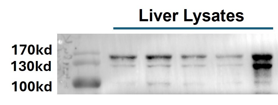
-
Application: Western BlotSample Tested: Liver tissueSpecies: MouseVerified Customer | Posted 12/10/2024WB_Liver lysate_VinculinThe antibody was diluted 1:3000 in 1X TBST. Multiple bands were observed.

There are no reviews that match your criteria.
Protocols
Find general support by application which include: protocols, troubleshooting, illustrated assays, videos and webinars.
- Antigen Retrieval Protocol (PIER)
- Antigen Retrieval for Frozen Sections Protocol
- Appropriate Fixation of IHC/ICC Samples
- Cellular Response to Hypoxia Protocols
- Chromogenic IHC Staining of Formalin-Fixed Paraffin-Embedded (FFPE) Tissue Protocol
- Chromogenic Immunohistochemistry Staining of Frozen Tissue
- ClariTSA™ Fluorophore Kits
- Detection & Visualization of Antibody Binding
- Fluorescent IHC Staining of Frozen Tissue Protocol
- Graphic Protocol for Heat-induced Epitope Retrieval
- Graphic Protocol for the Preparation and Fluorescent IHC Staining of Frozen Tissue Sections
- Graphic Protocol for the Preparation and Fluorescent IHC Staining of Paraffin-embedded Tissue Sections
- Graphic Protocol for the Preparation of Gelatin-coated Slides for Histological Tissue Sections
- ICC Cell Smear Protocol for Suspension Cells
- ICC Immunocytochemistry Protocol Videos
- ICC for Adherent Cells
- IHC Sample Preparation (Frozen sections vs Paraffin)
- Immunocytochemistry (ICC) Protocol
- Immunocytochemistry Troubleshooting
- Immunofluorescence of Organoids Embedded in Cultrex Basement Membrane Extract
- Immunofluorescent IHC Staining of Formalin-Fixed Paraffin-Embedded (FFPE) Tissue Protocol
- Immunohistochemistry (IHC) and Immunocytochemistry (ICC) Protocols
- Immunohistochemistry Frozen Troubleshooting
- Immunohistochemistry Paraffin Troubleshooting
- Preparing Samples for IHC/ICC Experiments
- Preventing Non-Specific Staining (Non-Specific Binding)
- Primary Antibody Selection & Optimization
- Protocol for Heat-Induced Epitope Retrieval (HIER)
- Protocol for Making a 4% Formaldehyde Solution in PBS
- Protocol for VisUCyte™ HRP Polymer Detection Reagent
- Protocol for the Fluorescent ICC Staining of Cell Smears - Graphic
- Protocol for the Fluorescent ICC Staining of Cultured Cells on Coverslips - Graphic
- Protocol for the Preparation & Fixation of Cells on Coverslips
- Protocol for the Preparation and Chromogenic IHC Staining of Frozen Tissue Sections
- Protocol for the Preparation and Chromogenic IHC Staining of Frozen Tissue Sections - Graphic
- Protocol for the Preparation and Chromogenic IHC Staining of Paraffin-embedded Tissue Sections
- Protocol for the Preparation and Chromogenic IHC Staining of Paraffin-embedded Tissue Sections - Graphic
- Protocol for the Preparation and Fluorescent ICC Staining of Cells on Coverslips
- Protocol for the Preparation and Fluorescent ICC Staining of Non-adherent Cells
- Protocol for the Preparation and Fluorescent ICC Staining of Stem Cells on Coverslips
- Protocol for the Preparation and Fluorescent IHC Staining of Frozen Tissue Sections
- Protocol for the Preparation and Fluorescent IHC Staining of Paraffin-embedded Tissue Sections
- Protocol for the Preparation of Gelatin-coated Slides for Histological Tissue Sections
- Protocol for the Preparation of a Cell Smear for Non-adherent Cell ICC - Graphic
- R&D Systems Quality Control Western Blot Protocol
- TUNEL and Active Caspase-3 Detection by IHC/ICC Protocol
- The Importance of IHC/ICC Controls
- Troubleshooting Guide: Immunohistochemistry
- Troubleshooting Guide: Western Blot Figures
- Western Blot Conditions
- Western Blot Protocol
- Western Blot Protocol for Cell Lysates
- Western Blot Troubleshooting
- Western Blot Troubleshooting Guide
- View all Protocols, Troubleshooting, Illustrated assays and Webinars
FAQs for Vinculin Antibody - Azide Free
-
Q: Do you have any 100 coda or bigger controls?
A:
I can recommend a couple of high weight antibodies that are conserved in most samples. Please verify the presence of the protein before using as a loading control. PARP1 or PARP is approximately 113kDa, and you may view the products we offer to PARP using this link. Vinculin is approximately 116kDa, and you may view the products we offer to Vinculin using this link.