Vitamin D BP Antibody - BSA Free
Novus Biologicals | Catalog # NBP1-88027
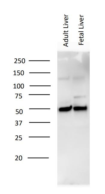
![Western Blot: Vitamin D BP Antibody [NBP1-88027]](https://resources.rndsystems.com/images/products/Vitamin-D-BP-Antibody-Western-Blot-NBP1-88027-img0009.jpg "Western Blot: Vitamin D BP Antibody [NBP1-88027]")
Key Product Details
Validated by
Species Reactivity
Applications
Label
Antibody Source
Format
Product Specifications
Immunogen
Reactivity Notes
Clonality
Host
Isotype
Scientific Data Images for Vitamin D BP Antibody - BSA Free
Western Blot: Vitamin D BP Antibody [NBP1-88027]
Western Blot: Vitamin D BP Antibody [NBP1-88027] - Equine adult and embryonic liver cell lysates. WB image submitted by a verified customer review.![Immunohistochemistry-Paraffin: Vitamin D BP Antibody [NBP1-88027]](https://resources.rndsystems.com/images/products/Vitamin-D-BP-Antibody-Immunohistochemistry-Paraffin-NBP1-88027-img0006.jpg "Immunohistochemistry-Paraffin: Vitamin D BP Antibody [NBP1-88027]")
Immunohistochemistry-Paraffin: Vitamin D BP Antibody [NBP1-88027]
Immunohistochemistry-Paraffin: Vitamin D BP Antibody [NBP1-88027] - Staining of human liver shows cytoplasmic positivity in hepatocytes.![Vitamin D BP Antibody - BSA Free Western Blot: Vitamin D BP Antibody - BSA Free [NBP1-88027]](https://resources.rndsystems.com/images/products/nbp1-88027_rabbit-polyclonal-vitamin-d-bp-antibody-65202516285524.jpg "Western Blot: Vitamin D BP Antibody - BSA Free [NBP1-88027]")
Western Blot: Vitamin D BP Antibody - BSA Free [NBP1-88027]
Analysis in mouse cell line NIH-3T3 and rat cell line NBT-II.![Vitamin D BP Antibody - BSA Free Western Blot: Vitamin D BP Antibody - BSA Free [NBP1-88027]](https://resources.rndsystems.com/images/products/nbp1-88027_rabbit-polyclonal-vitamin-d-bp-antibody-652025162720.jpg "Western Blot: Vitamin D BP Antibody - BSA Free [NBP1-88027]")

Western Blot: Vitamin D BP Antibody - BSA Free [NBP1-88027] -
Legumain is required for VDBP processing and regulation. (A) Purified VDBP from human plasma (1.9 μM) was incubated in legumain assay buffer (pH 5.8) at 37 C with or without purified active bovine legumain (2 μM) for 5 h before gel electrophoresis and immunoblotting of VDBP (n = 1). (B–H) Wild-type (Lgmn+/+) and legumain-deficient (Lgmn−/−) mice were treated with 50 ug/kg 25(OH)D3 (n = 6–7) or an equal volume vehicle (n = 7, control) subcutaneously every two to three days (four times in total). Tissues were harvested 24 h after the final injection (day 8). (B) One representative immunoblot of VDBP and GAPDH (housekeeping) in kidney and liver (n = 4). (C–F) Quantification of VDBP immunoband (IB) intensity as arbitrary units (ARBU) relative to GAPDH in immunoblots represented in (B) (n = 4). (C) Hepatic VDBP 45 kDa immunoband. (D) Renal VDBP 45 kDa immunoband. (E) Hepatic VDBP 55 kDa immunoband. (F) Renal VDBP 55 kDa immunoband. (G) Plasma VDBP concentration (μg/mL) was measured by ELISA (n = 6–7). (H) Hepatic VDBP mRNA expression relative to the geometric mean of CT values of four housekeeping controls (2− delta delta CT, n = 5). (C–H) Data represent mean +/- SEM. Two-way ANOVA. # p < 0.05, ## p < 0.01, ### p < 0.001 vs. different genotype, same treatment. Numbers (n) represent individual biological replicates. Image collected and cropped by CiteAb from the following open publication (https://www.mdpi.com/2073-4409/13/1/36), licensed under a CC-BY license. Not internally tested by Novus Biologicals.Applications for Vitamin D BP Antibody - BSA Free
Immunohistochemistry
Immunohistochemistry-Paraffin
Western Blot
See Simple Western Antibody Database for Simple Western validation: separated by Size
Reviewed Applications
Read 2 reviews rated 5 using NBP1-88027 in the following applications:
Formulation, Preparation, and Storage
Purification
Formulation
Format
Preservative
Concentration
Shipping
Stability & Storage
Background: Vitamin D BP
Long Name
Alternate Names
Gene Symbol
Additional Vitamin D BP Products
Product Documents for Vitamin D BP Antibody - BSA Free
Certificate of Analysis
To download a Certificate of Analysis, please enter a lot or batch number in the search box below.
Product Specific Notices for Vitamin D BP Antibody - BSA Free
This product is for research use only and is not approved for use in humans or in clinical diagnosis. Primary Antibodies are guaranteed for 1 year from date of receipt.
Citations for Vitamin D BP Antibody - BSA Free
 Powered by Bioz
Customer Reviews for Vitamin D BP Antibody - BSA Free (2)
Have you used Vitamin D BP Antibody - BSA Free?
Submit a review and receive an Amazon gift card!
$25/€18/£15/$25CAN/¥2500 Yen for a review with an image
$10/€7/£6/$10CAN/¥1110 Yen for a review without an image
Submit a review
Customer Images
-
Application: Western BlotSample Tested: Liver and Embryonic liverSpecies: EquineVerified Customer | Posted 02/02/2020!:3000 dilution

-
Application: Simple WesternSample Tested: SerumSpecies: RatVerified Customer | Posted 05/25/2017
There are no reviews that match your criteria.
Protocols
Find general support by application which include: protocols, troubleshooting, illustrated assays, videos and webinars.
- Antigen Retrieval Protocol (PIER)
- Antigen Retrieval for Frozen Sections Protocol
- Appropriate Fixation of IHC/ICC Samples
- Cellular Response to Hypoxia Protocols
- Chromogenic IHC Staining of Formalin-Fixed Paraffin-Embedded (FFPE) Tissue Protocol
- Chromogenic Immunohistochemistry Staining of Frozen Tissue
- ClariTSA™ Fluorophore Kits
- Detection & Visualization of Antibody Binding
- Fluorescent IHC Staining of Frozen Tissue Protocol
- Graphic Protocol for Heat-induced Epitope Retrieval
- Graphic Protocol for the Preparation and Fluorescent IHC Staining of Frozen Tissue Sections
- Graphic Protocol for the Preparation and Fluorescent IHC Staining of Paraffin-embedded Tissue Sections
- Graphic Protocol for the Preparation of Gelatin-coated Slides for Histological Tissue Sections
- IHC Sample Preparation (Frozen sections vs Paraffin)
- Immunofluorescent IHC Staining of Formalin-Fixed Paraffin-Embedded (FFPE) Tissue Protocol
- Immunohistochemistry (IHC) and Immunocytochemistry (ICC) Protocols
- Immunohistochemistry Frozen Troubleshooting
- Immunohistochemistry Paraffin Troubleshooting
- Preparing Samples for IHC/ICC Experiments
- Preventing Non-Specific Staining (Non-Specific Binding)
- Primary Antibody Selection & Optimization
- Protocol for Heat-Induced Epitope Retrieval (HIER)
- Protocol for Making a 4% Formaldehyde Solution in PBS
- Protocol for VisUCyte™ HRP Polymer Detection Reagent
- Protocol for the Preparation & Fixation of Cells on Coverslips
- Protocol for the Preparation and Chromogenic IHC Staining of Frozen Tissue Sections
- Protocol for the Preparation and Chromogenic IHC Staining of Frozen Tissue Sections - Graphic
- Protocol for the Preparation and Chromogenic IHC Staining of Paraffin-embedded Tissue Sections
- Protocol for the Preparation and Chromogenic IHC Staining of Paraffin-embedded Tissue Sections - Graphic
- Protocol for the Preparation and Fluorescent IHC Staining of Frozen Tissue Sections
- Protocol for the Preparation and Fluorescent IHC Staining of Paraffin-embedded Tissue Sections
- Protocol for the Preparation of Gelatin-coated Slides for Histological Tissue Sections
- R&D Systems Quality Control Western Blot Protocol
- TUNEL and Active Caspase-3 Detection by IHC/ICC Protocol
- The Importance of IHC/ICC Controls
- Troubleshooting Guide: Immunohistochemistry
- Troubleshooting Guide: Western Blot Figures
- Western Blot Conditions
- Western Blot Protocol
- Western Blot Protocol for Cell Lysates
- Western Blot Troubleshooting
- Western Blot Troubleshooting Guide
- View all Protocols, Troubleshooting, Illustrated assays and Webinars
FAQs for Vitamin D BP Antibody - BSA Free
-
Q: Does vitamin d binding protein have both nuclear and cytoplasmic staining?
A: From the image on our website, NBP1-88027 shows cytoplasmic staining. Furthermore, as Vitamin D binding protein is secreted, nuclear staining would not be expected.