Von Willebrand Factor Antibody - BSA Free
Novus Biologicals | Catalog # NB600-586
Key Product Details
Validated by
Species Reactivity
Validated:
Cited:
Applications
Validated:
Cited:
Label
Antibody Source
Format
Product Specifications
Immunogen
Reactivity Notes
Localization
Marker
Specificity
Clonality
Host
Isotype
Scientific Data Images for Von Willebrand Factor Antibody - BSA Free
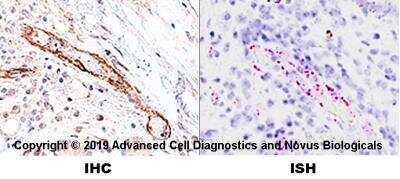
![Immunohistochemistry: Von Willebrand Factor Antibody [NB600-586]](https://resources.rndsystems.com/images/products/Von-Willebrand-Factor-Antibody-Immunohistochemistry-NB600-586-img0001.jpg "Immunohistochemistry: Von Willebrand Factor Antibody [NB600-586]")
Immunohistochemistry: Von Willebrand Factor Antibody [NB600-586]
Immunohistochemistry: Von Willebrand Factor Antibody [NB600-586] - Formalin fixed paraffin embedded human tonsil stained with Factor VIII antibody.![Immunohistochemistry-Paraffin: Von Willebrand Factor Antibody [NB600-586]](https://resources.rndsystems.com/images/products/Von-Willebrand-Factor-Antibody-Immunohistochemistry-Paraffin-NB600-586-img0005.jpg "Immunohistochemistry-Paraffin: Von Willebrand Factor Antibody [NB600-586]")
Immunohistochemistry-Paraffin: Von Willebrand Factor Antibody [NB600-586]
Immunohistochemistry-Paraffin: Von Willebrand Factor Antibody [NB600-586] - Staining of formalin-fixed, paraffin-embedded Human Kidney sections with 1:2,000 Rabbit Anti-von Willebrand Factor.![Immunohistochemistry-Frozen: Von Willebrand Factor Antibody [NB600-586]](https://resources.rndsystems.com/images/products/Von-Willebrand-Factor-Antibody-Immunohistochemistry-Frozen-NB600-586-img0008.jpg "Immunohistochemistry-Frozen: Von Willebrand Factor Antibody [NB600-586]")
Immunohistochemistry-Frozen: Von Willebrand Factor Antibody [NB600-586]
Immunohistochemistry-Frozen: Von Willebrand Factor Antibody [NB600-586] - Canine brain/spinal cord. VWF antibody in green. A pericyte marker in red. Alexa Fluor conjugated secondary antibodies were used for visualization. IHC-Fr image submitted by a verified customer review.![Immunohistochemistry-Paraffin: Von Willebrand Factor Antibody [NB600-586]](https://resources.rndsystems.com/images/products/Von-Willebrand-Factor-Antibody-Immunohistochemistry-Paraffin-NB600-586-img0006.jpg "Immunohistochemistry-Paraffin: Von Willebrand Factor Antibody [NB600-586]")
Immunohistochemistry-Paraffin: Von Willebrand Factor Antibody [NB600-586]
Immunohistochemistry-Paraffin: Von Willebrand Factor Antibody [NB600-586] - Staining of formalin-fixed, paraffin-embedded Human Kidney sections with 1:2,000 Rabbit Anti-von Willebrand Factor.Applications for Von Willebrand Factor Antibody - BSA Free
Immunocytochemistry/ Immunofluorescence
Immunohistochemistry
Immunohistochemistry-Paraffin
Reviewed Applications
Read 3 reviews rated 4.3 using NB600-586 in the following applications:
Formulation, Preparation, and Storage
Purification
Formulation
Format
Preservative
Concentration
Shipping
Stability & Storage
Background: Von Willebrand Factor
Alternate Names
Entrez Gene IDs
Gene Symbol
UniProt
Additional Von Willebrand Factor Products
Product Documents for Von Willebrand Factor Antibody - BSA Free
Certificate of Analysis
To download a Certificate of Analysis, please enter a lot or batch number in the search box below.
Product Specific Notices for Von Willebrand Factor Antibody - BSA Free
This product is for research use only and is not approved for use in humans or in clinical diagnosis. Primary Antibodies are guaranteed for 1 year from date of receipt.
Citations for Von Willebrand Factor Antibody - BSA Free
 Powered by Bioz
Customer Reviews for Von Willebrand Factor Antibody - BSA Free (3)
Have you used Von Willebrand Factor Antibody - BSA Free?
Submit a review and receive an Amazon gift card!
$25/€18/£15/$25CAN/¥2500 Yen for a review with an image
$10/€7/£6/$10CAN/¥1110 Yen for a review without an image
Submit a review
Customer Images


-
Application: Immunohistochemistry-FrozenSample Tested: brain and spinal cordSpecies: CanineVerified Customer | Posted 12/16/2019This multi-immunolabeled image was acquired by fluorescence microscopy. vWF antibody (NB600-586) (green), a pericyte marker (red).Alexa Fluor conjugated secondary antibodies were used.

-
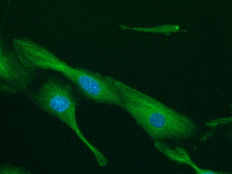
Application: ImmunofluorescenceSample Tested: EndothelialSpecies: MouseVerified Customer | Posted 01/05/2018vWF staning is detected in a mouse vascular endothelial cell line, UV♀2 (dilution: 1:50 [green], DAPI: blue).
-
Application: Immunohistochemistry-FrozenSample Tested: Connective tissueSpecies: FelineVerified Customer | Posted 08/01/2017Immunofluorescence on 4%PFA fixed section. Green-vWF(NB600-586), Blue-DAPI. Vessels are immunolabeled.
There are no reviews that match your criteria.
Protocols
Find general support by application which include: protocols, troubleshooting, illustrated assays, videos and webinars.
- Antigen Retrieval Protocol (PIER)
- Antigen Retrieval for Frozen Sections Protocol
- Appropriate Fixation of IHC/ICC Samples
- Cellular Response to Hypoxia Protocols
- Chromogenic IHC Staining of Formalin-Fixed Paraffin-Embedded (FFPE) Tissue Protocol
- Chromogenic Immunohistochemistry Staining of Frozen Tissue
- ClariTSA™ Fluorophore Kits
- Detection & Visualization of Antibody Binding
- Fluorescent IHC Staining of Frozen Tissue Protocol
- Graphic Protocol for Heat-induced Epitope Retrieval
- Graphic Protocol for the Preparation and Fluorescent IHC Staining of Frozen Tissue Sections
- Graphic Protocol for the Preparation and Fluorescent IHC Staining of Paraffin-embedded Tissue Sections
- Graphic Protocol for the Preparation of Gelatin-coated Slides for Histological Tissue Sections
- ICC Cell Smear Protocol for Suspension Cells
- ICC Immunocytochemistry Protocol Videos
- ICC for Adherent Cells
- IHC Sample Preparation (Frozen sections vs Paraffin)
- ISH-IHC Protocol for Chromogenic Detection on Formalin Fixed Paraffin Embedded (FFPE) Tissue
- Immunocytochemistry (ICC) Protocol
- Immunocytochemistry Troubleshooting
- Immunofluorescence of Organoids Embedded in Cultrex Basement Membrane Extract
- Immunofluorescent IHC Staining of Formalin-Fixed Paraffin-Embedded (FFPE) Tissue Protocol
- Immunohistochemistry (IHC) and Immunocytochemistry (ICC) Protocols
- Immunohistochemistry Frozen Troubleshooting
- Immunohistochemistry Paraffin Troubleshooting
- Preparing Samples for IHC/ICC Experiments
- Preventing Non-Specific Staining (Non-Specific Binding)
- Primary Antibody Selection & Optimization
- Protocol for Heat-Induced Epitope Retrieval (HIER)
- Protocol for Making a 4% Formaldehyde Solution in PBS
- Protocol for VisUCyte™ HRP Polymer Detection Reagent
- Protocol for the Fluorescent ICC Staining of Cell Smears - Graphic
- Protocol for the Fluorescent ICC Staining of Cultured Cells on Coverslips - Graphic
- Protocol for the Preparation & Fixation of Cells on Coverslips
- Protocol for the Preparation and Chromogenic IHC Staining of Frozen Tissue Sections
- Protocol for the Preparation and Chromogenic IHC Staining of Frozen Tissue Sections - Graphic
- Protocol for the Preparation and Chromogenic IHC Staining of Paraffin-embedded Tissue Sections
- Protocol for the Preparation and Chromogenic IHC Staining of Paraffin-embedded Tissue Sections - Graphic
- Protocol for the Preparation and Fluorescent ICC Staining of Cells on Coverslips
- Protocol for the Preparation and Fluorescent ICC Staining of Non-adherent Cells
- Protocol for the Preparation and Fluorescent ICC Staining of Stem Cells on Coverslips
- Protocol for the Preparation and Fluorescent IHC Staining of Frozen Tissue Sections
- Protocol for the Preparation and Fluorescent IHC Staining of Paraffin-embedded Tissue Sections
- Protocol for the Preparation of Gelatin-coated Slides for Histological Tissue Sections
- Protocol for the Preparation of a Cell Smear for Non-adherent Cell ICC - Graphic
- TUNEL and Active Caspase-3 Detection by IHC/ICC Protocol
- The Importance of IHC/ICC Controls
- Troubleshooting Guide: Immunohistochemistry
- View all Protocols, Troubleshooting, Illustrated assays and Webinars
FAQs for Von Willebrand Factor Antibody - BSA Free
-
Q: I have Von Willebrand Factor Antibody (NB600-586) being stored at -20 degrees Celsius and was wondering what the proper way to thaw them prior to use?
A: There is no special method. You can just take it out of the freezer and let it thaw on the lab bench. We suggest you could check it in an hour; it's not a large volume so shouldn't take very long.