![Simple Western: VPS34 Antibody [NB110-87320]](https://resources.rndsystems.com/images/products/VPS34-Antibody-Simple-Western-NB110-87320-img0006.jpg "Simple Western: VPS34 Antibody [NB110-87320]")
Loading...
Key Product Details
Validated by
Knockout/Knockdown
Species Reactivity
Validated:
Human, Mouse, Rat, Porcine
Cited:
Human, Mouse
Applications
Validated:
Western Blot, Immunocytochemistry/ Immunofluorescence, Simple Western, Knockdown Validated
Cited:
Western Blot, Flow Cytometry
Label
Unconjugated
Antibody Source
Polyclonal Rabbit IgG
Loading...
Product Specifications
Immunogen
Synthetic peptide made to an internal portion of the human VPS34 protein (within residues 700-850). [Swiss-Prot# Q8NEB9]
Reactivity Notes
Immunogen displays the following percentage of sequence identity for non-tested species: Xenopus (87%).
Clonality
Polyclonal
Host
Rabbit
Isotype
IgG
Theoretical MW
100 kDa.
Disclaimer note: The observed molecular weight of the protein may vary from the listed predicted molecular weight due to post translational modifications, post translation cleavages, relative charges, and other experimental factors.
Disclaimer note: The observed molecular weight of the protein may vary from the listed predicted molecular weight due to post translational modifications, post translation cleavages, relative charges, and other experimental factors.
Scientific Data Images for VPS34 Antibody
Simple Western: VPS34 Antibody [NB110-87320]
Simple Western: VPS34 Antibody [NB110-87320] - Simple Western lane view shows a specific band for VPS34 in 0.5 mg/ml of HepG2 lysate. This experiment was performed under reducing conditions using the 12-230 kDa separation system.![Knockdown Validated: VPS34 Antibody [NB110-87320]](https://resources.rndsystems.com/images/products/VPS34-Antibody-Knockdown-Validated-NB110-87320-img0007.jpg "Western Blot: VPS34 Antibody [NB110-87320]")
![Western Blot: VPS34 Antibody [NB110-87320]](https://resources.rndsystems.com/images/products/VPS34-Antibody-Western-Blot-NB110-87320-img0004.jpg "Western Blot: VPS34 Antibody [NB110-87320]")
Western Blot: VPS34 Antibody [NB110-87320]
Western Blot: VPS34 Antibody [NB110-87320] - HepG2 whole cell lysates.![Immunocytochemistry/ Immunofluorescence: VPS34 Antibody [NB110-87320]](https://resources.rndsystems.com/images/products/VPS34-Antibody-Immunocytochemistry-Immunofluorescence-NB110-87320-img0005.jpg "Immunocytochemistry/ Immunofluorescence: VPS34 Antibody [NB110-87320]")
Immunocytochemistry/ Immunofluorescence: VPS34 Antibody [NB110-87320]
Immunocytochemistry/Immunofluorescence: VPS34 Antibody [NB110-87320] - VPS34 antibody was tested at 1:200 in HeLa cells with FITC (green). Nuclei (Blue) were counterstained with Dapi (blue).
Western Blot: VPS34 Antibody [NB110-87320] -
Western Blot: VPS34 Antibody [NB110-87320] - Celastrol regulates autophagy- & mitophagy-related gene expressions in neurons. SH-SY5Y cells treated with 0.1–1 μM celastrol for 4 h. (A) Real-time quantitative PCR results show that 1 μM celastrol treatment for 4 h enhanced mRNA expressions of phagophore induction genes Beclin 1 & Ambra1, phagophore elongation genes Atg7 & Atg12, & vacuole formation genes LC3A & Atg4B. (B) Western blot results of SH-SY5Y treated with 100–500 nM celastrol show celastrol (500 nM) increased in protein expressions of Beclin 1 & Vps34 (phagophore induction), Atg7 (phagophore elongation), LC3-II (vacuole formation), DJ-1, & PINK1 (mitophagy). Celastrol (500 nM) suppressed LRRK2 expression. (C) MPTP (10 mg/kg/day for 3 days, i.p.) but not celastrol (3 mg/kg/day for 3 days, i.p.) caused dopaminergic nerve terminal degeneration (tyrosine hydroxylase↓) & mitophagy inactivation (DJ-1↓ & PINK1↓) in the striatum of mice as compared to saline control. Celastrol cotreatment with MPTP reversed it as compared to MPTP. Data are given as the mean ± SEM (n = 3–5). p-value was determined using the Kruskal–Wallis test followed by Dunn’s multiple comparison post hoc test. * p < 0.05 compared with controls. Image collected & cropped by CiteAb from the following publication (https://pubmed.ncbi.nlm.nih.gov/31906147), licensed under a CC-BY license. Not internally tested by Novus Biologicals.Applications for VPS34 Antibody
Application
Recommended Usage
Immunocytochemistry/ Immunofluorescence
1:100
Simple Western
1:20
Western Blot
1:1000
Application Notes
This VPS34 antibody is useful for Immunocytochemistry/Immunofluorescence and Western blot, where a band is seen approx. 100 kDa.
In Simple Western only 10 - 15 uL of the recommended dilution is used per data point.
See Simple Western Antibody Database for Simple Western validation: Tested in HepG2 lysate 0.5 mg/mL, separated by Size, antibody dilution of 1:20, apparent MW was 102 kDa. Separated by Size-Wes, Sally Sue/Peggy Sue.
Reviewed Applications
Read 1 review rated 4 using NB110-87320 in the following applications:
Formulation, Preparation, and Storage
Purification
Immunogen affinity purified
Formulation
PBS, 0.1% BSA, and 50% Glycerol
Preservative
0.05% Sodium Azide
Concentration
0.2 mg/ml
Shipping
The product is shipped with polar packs. Upon receipt, store it immediately at the temperature recommended below.
Stability & Storage
Store at 4C short term. Aliquot and store at -20C long term. Avoid freeze-thaw cycles.
Background: VPS34/PIK3C3
Alternate Names
PI 3-Kinase C3, PI3K type 3, PIK3C3
Gene Symbol
PIK3C3
UniProt
Additional VPS34/PIK3C3 Products
Product Documents for VPS34 Antibody
Certificate of Analysis
To download a Certificate of Analysis, please enter a lot or batch number in the search box below.
Product Specific Notices for VPS34 Antibody
This product is for research use only and is not approved for use in humans or in clinical diagnosis. Primary Antibodies are guaranteed for 1 year from date of receipt.
Related Research Areas
Citations for VPS34 Antibody
 Powered by Bioz
Customer Reviews for VPS34 Antibody (1)
4 out of 5
1 Customer Rating
Have you used VPS34 Antibody?
Submit a review and receive an Amazon gift card!
$25/€18/£15/$25CAN/¥2500 Yen for a review with an image
$10/€7/£6/$10CAN/¥1110 Yen for a review without an image
Submit a review
Customer Images
Showing
1
-
1 of
1 review
Showing All
Filter By:
-
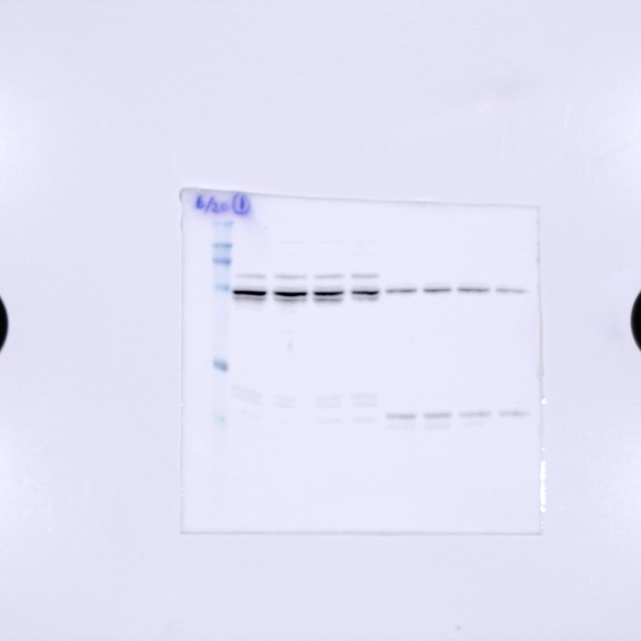
Application: Western BlotSample Tested: Skeletal muscle satellite cells (cultured in vitro)Species: MouseVerified Customer | Posted 07/27/2017Lane 1 - 4 : Cytoplasmic fraction Lane 5 - 8 : Membrane fraction

There are no reviews that match your criteria.
Protocols
View specific protocols for VPS34 Antibody (NB110-87320):
VPS34 Antibody:
Western Blot Protocol
1. Perform SDS-PAGE (4-12% MOPS) on samples to be analyzed, loading 40 ug of total protein per lane.
2. Transfer proteins to Nitrocellulose according to the instructions provided by the manufacturer of the transfer
apparatus.
3. Rinse membrane with dH2O and then stain the blot using Ponceau S for 1-2 minutes to access the transfer of proteins onto the nitrocellulose membrane. Rinse the blot in water to remove excess stain and mark the lane locations and locations of molecular weight markers using a pencil.
4. Rinse the blot in TBS for approximately 5 minutes.
5. Block the membrane using 5% BSA in TBS + Tween, 1 hour at RT.
6. Rinse the membrane in dH2O and then wash the membrane in wash buffer [TBS + 0.1% Tween] 3 times for 10 minutes each.
7. Dilute the rabbit anti-VPS34 primary antibody (NB 110-87320) in blocking buffer and incubate 1 hour at room temperature.
8. Rinse the membrane in dH2O and then wash the membrane in wash buffer [TBS + 0.1% Tween] 3 times for 10 minutes each.
9. Apply the diluted rabbit-IgG HRP-conjugated secondary antibody in blocking buffer (as per manufacturers
instructions) and incubate 1 hour at room temperature.
10. Wash the blot in wash buffer [TBS + 0.1% Tween] 3 times for 10 minutes each (this step can be repeated as required to reduce background).
11. Apply the detection reagent of choice in accordance with the manufacturers instructions (Pierce ECL).
Note: Tween-20 can be added to the blocking or antibody dilution buffer at a final concentration of 0.05-0.2%, provided it does not interfere with antibody-antigen binding.
Find general support by application which include: protocols, troubleshooting, illustrated assays, videos and webinars.
- Appropriate Fixation of IHC/ICC Samples
- Cellular Response to Hypoxia Protocols
- ClariTSA™ Fluorophore Kits
- Detection & Visualization of Antibody Binding
- ICC Cell Smear Protocol for Suspension Cells
- ICC Immunocytochemistry Protocol Videos
- ICC for Adherent Cells
- Immunocytochemistry (ICC) Protocol
- Immunocytochemistry Troubleshooting
- Immunofluorescence of Organoids Embedded in Cultrex Basement Membrane Extract
- Immunohistochemistry (IHC) and Immunocytochemistry (ICC) Protocols
- Preparing Samples for IHC/ICC Experiments
- Preventing Non-Specific Staining (Non-Specific Binding)
- Primary Antibody Selection & Optimization
- Protocol for VisUCyte™ HRP Polymer Detection Reagent
- Protocol for the Fluorescent ICC Staining of Cell Smears - Graphic
- Protocol for the Fluorescent ICC Staining of Cultured Cells on Coverslips - Graphic
- Protocol for the Preparation and Fluorescent ICC Staining of Cells on Coverslips
- Protocol for the Preparation and Fluorescent ICC Staining of Non-adherent Cells
- Protocol for the Preparation and Fluorescent ICC Staining of Stem Cells on Coverslips
- Protocol for the Preparation of a Cell Smear for Non-adherent Cell ICC - Graphic
- R&D Systems Quality Control Western Blot Protocol
- TUNEL and Active Caspase-3 Detection by IHC/ICC Protocol
- The Importance of IHC/ICC Controls
- Troubleshooting Guide: Western Blot Figures
- Western Blot Conditions
- Western Blot Protocol
- Western Blot Protocol for Cell Lysates
- Western Blot Troubleshooting
- Western Blot Troubleshooting Guide
- View all Protocols, Troubleshooting, Illustrated assays and Webinars
Loading...