ZEB2 Antibody - BSA Free
Novus Biologicals | Catalog # NBP1-82991
![Western Blot: ZEB2 Antibody [NBP1-82991]](https://resources.rndsystems.com/images/products/ZEB2-Antibody-Western-Blot-NBP1-82991-img0013.jpg "Western Blot: ZEB2 Antibody [NBP1-82991]")
Loading...
Key Product Details
Validated by
Orthogonal Validation
Species Reactivity
Validated:
Human
Cited:
Human, Mouse
Predicted:
Mouse (94%), Rat (95%). Backed by our 100% Guarantee.
Applications
Validated:
Immunohistochemistry, Immunohistochemistry-Paraffin, Western Blot, Chromatin Immunoprecipitation-exo-Seq
Cited:
Immunohistochemistry-Paraffin, Western Blot, Flow Cytometry, Immunocytochemistry/ Immunofluorescence, IF/IHC, Knockdown Validated
Label
Unconjugated
Antibody Source
Polyclonal Rabbit IgG
Format
BSA Free
Loading...
Product Specifications
Immunogen
This antibody was developed against Recombinant Protein corresponding to amino acids: SPVKPMDSITSPSIAELHNSVTNCDPPLRLTKPSHFTNIKPVEKLDHSRSNTPSPLNLSSTSSKNSHSSSYTPNSFSSEELQAEPLDLSLPKQMKEPKSIIATKNKTKASSISLDHNSVSSSSE
Clonality
Polyclonal
Host
Rabbit
Isotype
IgG
Scientific Data Images for ZEB2 Antibody - BSA Free
![Western Blot: ZEB2 Antibody [NBP1-82991]](https://resources.rndsystems.com/images/products/ZEB2-Antibody-Western-Blot-NBP1-82991-img0018.jpg "Western Blot: ZEB2 Antibody [NBP1-82991]")
Western Blot: ZEB2 Antibody [NBP1-82991]
ZEB2-Antibody-Western-Blot-NBP1-82991-img0018.jpg![Immunohistochemistry-Paraffin: ZEB2 Antibody [NBP1-82991]](https://resources.rndsystems.com/images/products/ZEB2-Antibody-Immunohistochemistry-Paraffin-NBP1-82991-img0017.jpg "Immunohistochemistry-Paraffin: ZEB2 Antibody [NBP1-82991]")
Immunohistochemistry-Paraffin: ZEB2 Antibody [NBP1-82991]
Immunohistochemistry-Paraffin: ZEB2 Antibody [NBP1-82991] - Staining of human pancreas shows no positivity as expected.![Immunohistochemistry-Paraffin: ZEB2 Antibody [NBP1-82991]](https://resources.rndsystems.com/images/products/ZEB2-Antibody-Immunohistochemistry-Paraffin-NBP1-82991-img0014.jpg "Immunohistochemistry-Paraffin: ZEB2 Antibody [NBP1-82991]")
Immunohistochemistry-Paraffin: ZEB2 Antibody [NBP1-82991]
Immunohistochemistry-Paraffin: ZEB2 Antibody [NBP1-82991] - Staining of human cerebral cortex shows moderate nuclear positivity in glial cells.![Immunohistochemistry-Paraffin: ZEB2 Antibody [NBP1-82991]](https://resources.rndsystems.com/images/products/ZEB2-Antibody-Immunohistochemistry-Paraffin-NBP1-82991-img0015.jpg "Immunohistochemistry-Paraffin: ZEB2 Antibody [NBP1-82991]")
Immunohistochemistry-Paraffin: ZEB2 Antibody [NBP1-82991]
Immunohistochemistry-Paraffin: ZEB2 Antibody [NBP1-82991] - Staining of human colon shows moderate nuclear positivity in a subset of lymphoid cells.![Immunohistochemistry-Paraffin: ZEB2 Antibody [NBP1-82991]](https://resources.rndsystems.com/images/products/ZEB2-Antibody-Immunohistochemistry-Paraffin-NBP1-82991-img0016.jpg "Immunohistochemistry-Paraffin: ZEB2 Antibody [NBP1-82991]")
Immunohistochemistry-Paraffin: ZEB2 Antibody [NBP1-82991]
Immunohistochemistry-Paraffin: ZEB2 Antibody [NBP1-82991] - Staining of human glioma shows moderate nuclear positivity in tumor cells.
Western Blot: ZEB2 Antibody [NBP1-82991] -
SU5402, FGFR1 inhibitor, affects the EMT transcription factors.(A, B) ERK1/2 phosphorylation (p-ERK1/2) was determined by immunoblotting in HSC-4 & OTC-04 treated for 30 min with 30 ng/ml FGF2 or 30 ng/ml FGF7 in presence of 10% FBS (A) & in TSU & HOC313 cells treated for 1 h with 30 ng/ml FGF2 or 30 μM SU5402 in absence of FBS (B). F2, FGF2; F7, FGF7; SU, SU5402. (C) We have previously reported that, after treatment with TGF-beta, NMuMG cells underwent EMT with the IIIc-isoform of FGFR1[17]. After NMuMG cells pretreated with TGF-beta were transfected with mouse Fgfr1 siRNA or treated with SU5402, the cells were further incubated in culture medium (CM) from TSU cells. SU, SU5402; siFR1, siRNA against mouse Fgfr1. (D)FGF2 mRNA levels were determined by RT-qPCR analyses. The ratio of FGF2 mRNA to GAPDH mRNA in HSC-4 cells was indicated as “1”. Each value represents the mean ± SD of triplicate determinations from a representative experiment. Similar results obtained in at least three independent experiments. (E) ERK1/2 phosphorylation (p-ERK1/2) in TSU & HOC313 cells were monitored in presence of indicated concentration of SU5402 for 1 h under serum-free culture conditions, followed by immunoblot analysis. (F, G) Expression of indicated genes in TSU cells under serum-free culture conditions was determined by RT-qPCR (F) & immunoblot (G) analyses, following treatment with 10 μM SU5402. Each value represents the mean ± SD of triplicate determinations from a representative experiment. Similar results obtained from at least three independent experiments. p values were determined by Student’s t-test. **p < 0.01. (H) TSU cells treated with 10 μM U0126 in absence of FBS were subjected to immunoblotting with the indicated antibodies. alpha -tubulin was used as a loading control (A, B, C, E, G, & H). Image collected & cropped by CiteAb from the following publication (https://pubmed.ncbi.nlm.nih.gov/31682640), licensed under a CC-BY license. Not internally tested by Novus Biologicals.
Western Blot: ZEB2 Antibody [NBP1-82991] -
Western Blot: ZEB2 Antibody [NBP1-82991] - ZEB2 is a downstream target of TRIM14. a Western blot analysis of TRIM14, EMT related biomarks (Snail1, Twist1, ZEB1, ZEB2 & SLUG), & beta -actin in LN229 & U251 cells transfected with shTRIM14–1, shTRIM14–2 or control shRNA. b Western blot analysis of TRIM14, N-cadherin, E-cadherin, Vimentin & beta -actin in LN229 & U251 cells transduced with the indicated plasmid. c Representative images of transwell invasion assay using LN229 & U251 cells transfected with the indicated plasmid. Quantification of transwell invasion assay is shown. ***p < 0.001; n = 3 experiment. d Representative images of wound healing assay using LN229 & U251 cells with the indicated plasmid. Quantification of wound healing assay is shown. ***p < 0.001, n = 3 experiments. e Representative images of 3D spheroid BME cell invasion assays using LN229 & U251 cells with the indicated plasmid. Quantification of 3D spheroid BME cell invasion assays is shown. ***p < 0.001, n = 3 experiments Image collected & cropped by CiteAb from the following publication (https://pubmed.ncbi.nlm.nih.gov/30728039), licensed under a CC-BY license. Not internally tested by Novus Biologicals.
Western Blot: ZEB2 Antibody [NBP1-82991] -
Western Blot: ZEB2 Antibody [NBP1-82991] - TRIM14 function was demonstrated in vivo & IHC. a ShCtrl-transplanted & shTRIM14-transplanted LN229 cells were counted & transplanted in orthotopic nude mice model respectively, n = 6. b Survival curve of shCtrl-transplanted or shTRIM14-transplanted intracranial xenografts. ***, P < 0.001, n = 6. c Representative pseudocolour bioluminescence images of orthotopic tumours bearing control or TRIM14-depleted LN229 cells on the days as indicated. Quantification of tumor volume is shown. ***p < 0.001; n = 3 experiment. d The HE assay was performed to show tumor cytostructure. e Western blot analysis of TRIM14 & ZEB2 extracted from four pairs mice from shCtrl-transplanted group & shCtrl-transplanted group. Equal loading is confirmed by beta -actin levels. f IHC staining of TRIM14 & ZEB2 in four representative GBM specimens Image collected & cropped by CiteAb from the following publication (https://pubmed.ncbi.nlm.nih.gov/30728039), licensed under a CC-BY license. Not internally tested by Novus Biologicals.
Western Blot: ZEB2 Antibody [NBP1-82991] -
Western Blot: ZEB2 Antibody [NBP1-82991] - ZEB2 is involved in the function of TRIM14. a Western blot analysis of TRIM14, EMT related biomarks (Snail1, Twist1, ZEB1, ZEB2 & SLUG), & beta -actin in HS683 & U87 cells transfected with vector or TRIM14. b Western blot analysis of TRIM14, N-cadherin, E-cadherin, Vimentin & beta -actin in HS683 & U87 cells transduced with the indicated plasmid. c Representative images of transwell invasion assay using HS683 & U87 cells transfected with the indicated plasmid. Quantification of transwell invasion assay is shown. ***p < 0.001; n = 3 experiment. d Representative images of wound healing assay using HS683 & U87 cells with the indicated plasmid. Quantification of wound healing assay is shown. ***p < 0.001, n = 3 experiments. e Representative images of 3D spheroid BME cell invasion assays using HS683 & U87 cells with the indicated plasmid. Quantification of 3D spheroid BME cell invasion assays is shown. ***p < 0.001, n = 3 experiments Image collected & cropped by CiteAb from the following publication (https://pubmed.ncbi.nlm.nih.gov/30728039), licensed under a CC-BY license. Not internally tested by Novus Biologicals.![ZEB2 Antibody - BSA Free Chromatin Immunoprecipitation-exo-Seq: ZEB2 Antibody - BSA Free [NBP1-82991]](https://resources.rndsystems.com/images/products/nbp1-82991_rabbit-polyclonal-zeb2-antibody-256202591439.jpg "Chromatin Immunoprecipitation-exo-Seq: ZEB2 Antibody - BSA Free [NBP1-82991]")
Chromatin Immunoprecipitation-exo-Seq: ZEB2 Antibody - BSA Free [NBP1-82991]
ChIP-Exo-Seq composite graph for Anti-ZEB2 (NBP1-82991) tested in K562 cells. Strand-specific reads (blue: forward, red: reverse) and IgG controls (black: forward, grey: reverse) are plotted against the distance from a composite set of reference binding sites. The antibody exhibits robust target enrichment compared to a non-specific IgG control and precisely reveals its structural organization around the binding site. Data generated by Prof. B. F. Pugh´s Lab at Cornell University.Applications for ZEB2 Antibody - BSA Free
Application
Recommended Usage
Chromatin Immunoprecipitation-exo-Seq
1-10ug per reaction
Immunohistochemistry
1:500 - 1:1000
Immunohistochemistry-Paraffin
1:500 - 1:1000
Western Blot
0.04 - 0.4 ug/ml
Application Notes
IHC-Paraffin, HIER pH 6 retrieval is recommended.
Reviewed Applications
Read 2 reviews rated 2.5 using NBP1-82991 in the following applications:
Formulation, Preparation, and Storage
Purification
Affinity purified
Formulation
PBS (pH 7.2) and 40% Glycerol
Format
BSA Free
Preservative
0.02% Sodium Azide
Concentration
Concentrations vary lot to lot. See vial label for concentration. If unlisted please contact technical services.
Shipping
The product is shipped with polar packs. Upon receipt, store it immediately at the temperature recommended below.
Stability & Storage
Store at 4C short term. Aliquot and store at -20C long term. Avoid freeze-thaw cycles.
Background: ZEB2/SIP1
Alternate Names
HSPC082, SIP1, SMADIP1, ZFHX1B
Gene Symbol
ZEB2
Additional ZEB2/SIP1 Products
Product Documents for ZEB2 Antibody - BSA Free
Certificate of Analysis
To download a Certificate of Analysis, please enter a lot or batch number in the search box below.
Product Specific Notices for ZEB2 Antibody - BSA Free
This product is for research use only and is not approved for use in humans or in clinical diagnosis. Primary Antibodies are guaranteed for 1 year from date of receipt.
Related Research Areas
Citations for ZEB2 Antibody - BSA Free
 Powered by Bioz
Customer Reviews for ZEB2 Antibody - BSA Free (2)
2.5 out of 5
2 Customer Ratings
Have you used ZEB2 Antibody - BSA Free?
Submit a review and receive an Amazon gift card!
$25/€18/£15/$25CAN/¥2500 Yen for a review with an image
$10/€7/£6/$10CAN/¥1110 Yen for a review without an image
Submit a review
Customer Images
Showing
1
-
2 of
2 reviews
Showing All
Filter By:
-
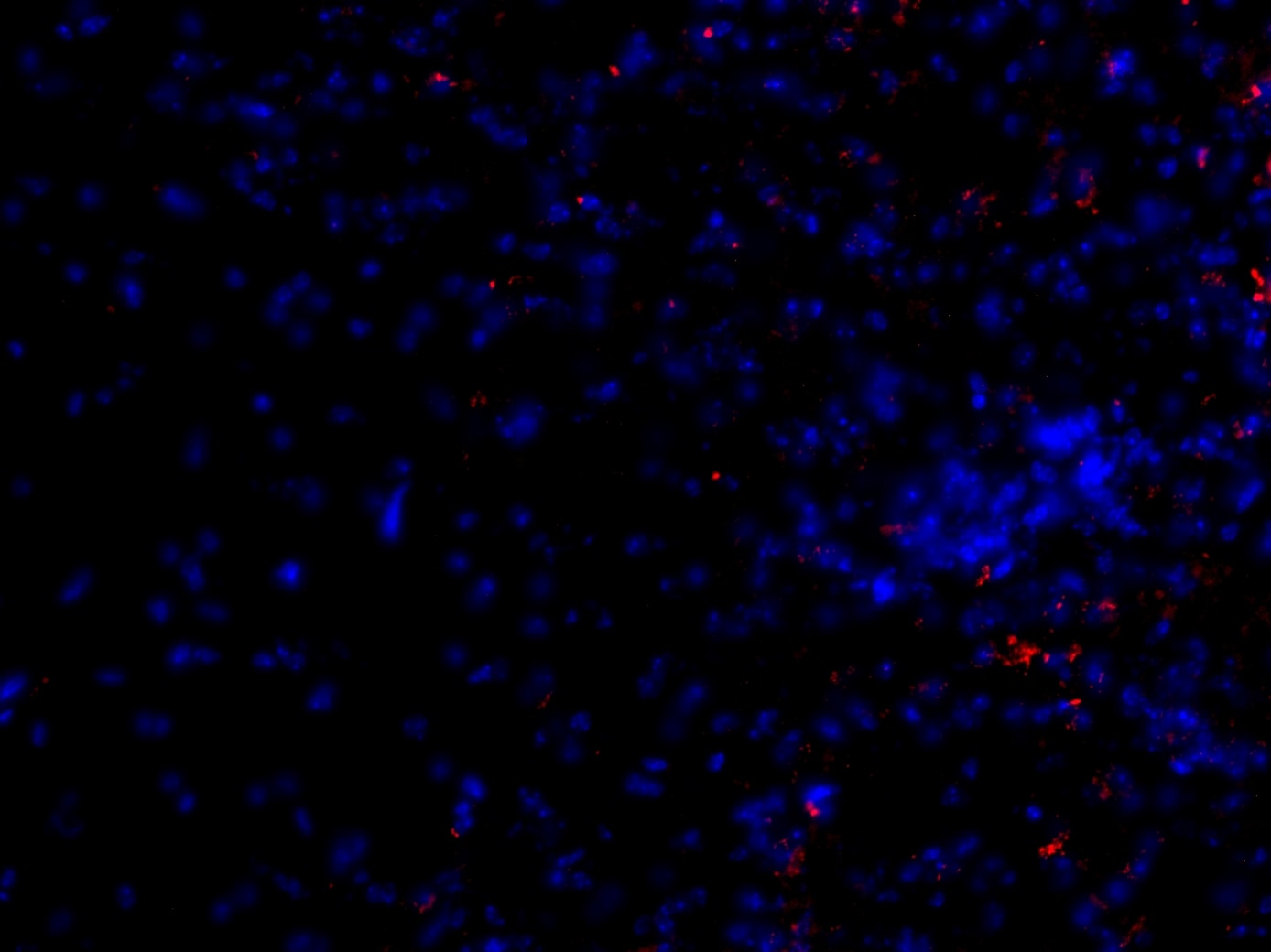
Application: Immunohistochemistry-FrozenSample Tested: Fixed frozen mouse spinal cordSpecies: MouseVerified Customer | Posted 11/08/2019
 Bio-Techne ResponseThis review was submitted through the legacy Novus Innovators Program, reflecting a new species or application tested on a primary antibody.
 -
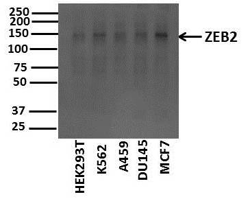
Application: Western BlotSample Tested: Cancer Cell linesSpecies: HumanVerified Customer | Posted 11/12/2017ZEB2 in various cancer cell linesSample: Whole cell lysates of various cancer cell lines (10ug) Blocking: 5% nonfat dry milk reconstituted in TBS-T for 1 hour at room temperature Primary Antibody diluted (1:500) in 5% nonfat dry milk reconstituted in TBS-T overnight at 4deg C Secondary Antibody: Goat Anti-Rabbit IgG H&L (HRP) (ab97051) at 1/10000 dilution in TBS-T
There are no reviews that match your criteria.
Protocols
Find general support by application which include: protocols, troubleshooting, illustrated assays, videos and webinars.
- Antigen Retrieval Protocol (PIER)
- Antigen Retrieval for Frozen Sections Protocol
- Appropriate Fixation of IHC/ICC Samples
- Cellular Response to Hypoxia Protocols
- Chromogenic IHC Staining of Formalin-Fixed Paraffin-Embedded (FFPE) Tissue Protocol
- Chromogenic Immunohistochemistry Staining of Frozen Tissue
- ClariTSA™ Fluorophore Kits
- Detection & Visualization of Antibody Binding
- Fluorescent IHC Staining of Frozen Tissue Protocol
- Graphic Protocol for Heat-induced Epitope Retrieval
- Graphic Protocol for the Preparation and Fluorescent IHC Staining of Frozen Tissue Sections
- Graphic Protocol for the Preparation and Fluorescent IHC Staining of Paraffin-embedded Tissue Sections
- Graphic Protocol for the Preparation of Gelatin-coated Slides for Histological Tissue Sections
- IHC Sample Preparation (Frozen sections vs Paraffin)
- Immunofluorescent IHC Staining of Formalin-Fixed Paraffin-Embedded (FFPE) Tissue Protocol
- Immunohistochemistry (IHC) and Immunocytochemistry (ICC) Protocols
- Immunohistochemistry Frozen Troubleshooting
- Immunohistochemistry Paraffin Troubleshooting
- Preparing Samples for IHC/ICC Experiments
- Preventing Non-Specific Staining (Non-Specific Binding)
- Primary Antibody Selection & Optimization
- Protocol for Heat-Induced Epitope Retrieval (HIER)
- Protocol for Making a 4% Formaldehyde Solution in PBS
- Protocol for VisUCyte™ HRP Polymer Detection Reagent
- Protocol for the Preparation & Fixation of Cells on Coverslips
- Protocol for the Preparation and Chromogenic IHC Staining of Frozen Tissue Sections
- Protocol for the Preparation and Chromogenic IHC Staining of Frozen Tissue Sections - Graphic
- Protocol for the Preparation and Chromogenic IHC Staining of Paraffin-embedded Tissue Sections
- Protocol for the Preparation and Chromogenic IHC Staining of Paraffin-embedded Tissue Sections - Graphic
- Protocol for the Preparation and Fluorescent IHC Staining of Frozen Tissue Sections
- Protocol for the Preparation and Fluorescent IHC Staining of Paraffin-embedded Tissue Sections
- Protocol for the Preparation of Gelatin-coated Slides for Histological Tissue Sections
- R&D Systems Quality Control Western Blot Protocol
- TUNEL and Active Caspase-3 Detection by IHC/ICC Protocol
- The Importance of IHC/ICC Controls
- Troubleshooting Guide: Immunohistochemistry
- Troubleshooting Guide: Western Blot Figures
- Western Blot Conditions
- Western Blot Protocol
- Western Blot Protocol for Cell Lysates
- Western Blot Troubleshooting
- Western Blot Troubleshooting Guide
- View all Protocols, Troubleshooting, Illustrated assays and Webinars
Loading...