Bromodeoxyuridine/BrdU Antibody - Azide and BSA Free
Novus Biologicals | Catalog # NB500-235
![Immunocytochemistry/ Immunofluorescence: Bromodeoxyuridine/BrdU Antibody [NB500-235]](https://resources.rndsystems.com/images/products/Bromodeoxyuridine-BrdU-Antibody-Immunocytochemistry-Immunofluorescence-NB500-235-img0006.jpg "Immunocytochemistry/ Immunofluorescence: Bromodeoxyuridine/BrdU Antibody [NB500-235]")
Key Product Details
Validated by
Species Reactivity
Validated:
Cited:
Applications
Validated:
Cited:
Label
Antibody Source
Format
Product Specifications
Immunogen
Reactivity Notes
Marker
Specificity
Clonality
Host
Isotype
Scientific Data Images for Bromodeoxyuridine/BrdU Antibody - Azide and BSA Free
![Immunoprecipitation: Bromodeoxyuridine/BrdU Antibody [NB500-235]](https://resources.rndsystems.com/images/products/Bromodeoxyuridine-BrdU-Antibody-Immunoprecipitation-NB500-235-img0005.jpg "Immunoprecipitation: Bromodeoxyuridine/BrdU Antibody [NB500-235]")
Immunoprecipitation: Bromodeoxyuridine/BrdU Antibody [NB500-235]
Immunoprecipitation: Bromodeoxyuridine/BrdU Antibody [NB500-235] - Lysate was immunoprecipitated with anti-brdu antibody (NB500-235), 5-methyl cytosine (5-Me), or a no antibody control (No A). An ELISA was then performed to measure total activity.
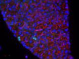
Immunocytochemistry/ Immunofluorescence: Bromodeoxyuridine/BrdU Antibody - Azide and BSA Free [NB500-235] -
Cavtratin inhibits the proliferation of vascular smooth muscle cells(A) MTT assay using human umbilical vein smooth muscle cells (HUVSMCs) cultured with 10% FBS. The cells were treated with cavtratin or AP at different concentrations for different times as indicated. A570 represented the cell proliferation. (B and C) Anti-BrdU staining of HUVSMCs 4 days after BrdU and cavtratin or AP application. *, P<0.05 versus the AP group under the same conditions. Scale bar, 50 μm. Image collected and cropped by CiteAb from the following open publication (https://pubmed.ncbi.nlm.nih.gov/29100301), licensed under a CC-BY license. Not internally tested by Novus Biologicals.
Immunocytochemistry/ Immunofluorescence: Bromodeoxyuridine/BrdU Antibody - Azide and BSA Free [NB500-235] -
Cavtratin inhibits the survival and proliferation of endothelial cells(A) Cavtratin decreased HUVEC survival in an MTT assay with 0.5% of FBS in the culture medium. Cells were treated with different concentrations (2 μM, 10 μM and 50 μM) of cavtratin or with 50 μM AP as a control. Absorbance at 570 nm (A570) indicated the cell survival. (B) HUVEC proliferation was arrested under treatment with cavtratin in an MTT assay with 10% of FBS in the culture medium. Cells were treated with 2 μM, 10 μM or 50 μM cavtratin, or with 50 μM AP as a control. A570 represented the cell proliferation. (C, D) Treatment with cavtratin significantly decreased BrdU incorporation into HUVECs. Cells were treated with 50 μM cavtratin or AP for 4 days. BrdU was also added to the medium to label the proliferating cells, which were later visualized by anti-BrdU staining. Images were captured under the same confocal setting. All BrdU+ cells were counted, including those with weak fluorescence. All data represents the mean +/- SEM. *, P < 0.05 versus the AP group under the same conditions. Scale bar, 50 μm. Image collected and cropped by CiteAb from the following open publication (https://pubmed.ncbi.nlm.nih.gov/29100301), licensed under a CC-BY license. Not internally tested by Novus Biologicals.Applications for Bromodeoxyuridine/BrdU Antibody - Azide and BSA Free
Immunocytochemistry/ Immunofluorescence
Immunohistochemistry
Immunohistochemistry-Frozen
Immunohistochemistry-Paraffin
Immunoprecipitation
Reviewed Applications
Read 2 reviews rated 5 using NB500-235 in the following applications:
Formulation, Preparation, and Storage
Purification
Formulation
Format
Preservative
Concentration
Shipping
Stability & Storage
Background: Bromodeoxyuridine/BrdU
Alternate Names
Additional Bromodeoxyuridine/BrdU Products
Product Documents for Bromodeoxyuridine/BrdU Antibody - Azide and BSA Free
Certificate of Analysis
To download a Certificate of Analysis, please enter a lot or batch number in the search box below.
Product Specific Notices for Bromodeoxyuridine/BrdU Antibody - Azide and BSA Free
This product is for research use only and is not approved for use in humans or in clinical diagnosis. Primary Antibodies are guaranteed for 1 year from date of receipt.
Related Research Areas
Citations for Bromodeoxyuridine/BrdU Antibody - Azide and BSA Free
 Powered by Bioz
Customer Reviews for Bromodeoxyuridine/BrdU Antibody - Azide and BSA Free (2)
Have you used Bromodeoxyuridine/BrdU Antibody - Azide and BSA Free?
Submit a review and receive an Amazon gift card!
$25/€18/£15/$25CAN/¥2500 Yen for a review with an image
$10/€7/£6/$10CAN/¥1110 Yen for a review without an image
Submit a review
Customer Images
-
Application: ImmunofluorescenceSample Tested: Mouse Lacrimal GlandSpecies: MouseVerified Customer | Posted 03/15/2011

-
Application: Immunohistochemistry-FrozenSample Tested: mouse brain tissue section (dorsal striatum)Species: MouseVerified Customer | Posted 05/27/2009
There are no reviews that match your criteria.
Protocols
View specific protocols for Bromodeoxyuridine/BrdU Antibody - Azide and BSA Free (NB500-235):
Immunohistochemistry protocol for Bromodeoxyuridine (BrdU) Polyclonal Antibody
Original antigenic studies performed by Dr David Stollar, Tufts University, Boston, MA
ABSTRACT
New methods for double and triple colour labeling using monoclonal antibodies to the proliferation-associated markers 5-methly-cytosine, BrdU and Ki67 are described. In order to make incorporated 5-methyl-cytosine or BrdU accessible to most antibodies, mild denaturation of the DNA is needed, and this is usually obtained by exposing the cells to acid or base. This procedure destroys most cellular antigen, including nuclear TdT and Ki67. In this study, we show that fixation in cold methanol instead of 70% ethanol for 30 minutes followed by immersion in 7 x 10⁻³ N NaOH for 10-15 seconds allows BrdU staining with the simultaneous detection of nuclear cytoplasmic and membrane assigns as well as preservation of morphological detail. This method is optimal for detection of nuclear Ki67 and TdT. These reagents, together with antibodies to membrane assigns can be included in triple colour labeling using second layers conjugated to FITC, TRITC and colloidal gold. With these methods it is now possible to characterize the phenotype of dividing cell populations such as precursors in central lymphoid tissues and germinal centre blasts in peripheral lymphoid organs.
Reference for tissue staining with anti-BrdU
Campana D, Coustan-Smith E., Janossy G., Department of Immunology, Royal Free Hospital School of Medicine, London, UK. J Immunol Methods 107(1):79-88 1988 Feb 24 Double and triple staining methods for studying the proliferative activity of human B and T lymphoid cells Abstract
For best results on unconjugated antibody, use the product at a concentration of 25 to 100 ug/mL. Nearly complete immunoprecipitation was obtained at concentrations of 100 to 500ug/mL. The product has also been tested for utility for staining of bromodeoxyuridine incorporated into DNA of replicating cells. When utilized at a purified concentration of 10 ug/mL, the product is comparable to commercially available monoclonal antibodies commonly used for the same purpose.
A key issue is that it is necessary to denature the DNA so as to expose the base to the antibodies. One protocol is: Sachiko Matsuuraa and Kazuo Suzukia Immunohistochemical Analysis of DNA Synthesis During Chronic Stimulation with Isoproterenol in Mouse Submandibular Gland. Journal of Histochemistry and Cytochemistry, Vol. 45, 1137-1146, Immunohistochemistry DNA-synthesizing cells were detected by peroxidase-anti-peroxidase (PAP) immunostaining with anti-BrdU antibodies by the method of Harms et al. 1986, which was slightly modified as follows: Sections were treated with 2 N HCl at 60C for 15 min and washed in running water for 15 min. After dehydration in an ethanol series, the sections were etched with xylene (15 min, twice). Nuclear proteins, which mask incorporated BrdU, were digested with 0.025% protease (Type V; Sigma) in PBS at 37C for 10 min, and the sections were then washed with cold PBS three times for 5 min.
Find general support by application which include: protocols, troubleshooting, illustrated assays, videos and webinars.
- Antigen Retrieval Protocol (PIER)
- Antigen Retrieval for Frozen Sections Protocol
- Appropriate Fixation of IHC/ICC Samples
- Cellular Response to Hypoxia Protocols
- Chromogenic IHC Staining of Formalin-Fixed Paraffin-Embedded (FFPE) Tissue Protocol
- Chromogenic Immunohistochemistry Staining of Frozen Tissue
- ClariTSA™ Fluorophore Kits
- Detection & Visualization of Antibody Binding
- Fluorescent IHC Staining of Frozen Tissue Protocol
- Graphic Protocol for Heat-induced Epitope Retrieval
- Graphic Protocol for the Preparation and Fluorescent IHC Staining of Frozen Tissue Sections
- Graphic Protocol for the Preparation and Fluorescent IHC Staining of Paraffin-embedded Tissue Sections
- Graphic Protocol for the Preparation of Gelatin-coated Slides for Histological Tissue Sections
- ICC Cell Smear Protocol for Suspension Cells
- ICC Immunocytochemistry Protocol Videos
- ICC for Adherent Cells
- IHC Sample Preparation (Frozen sections vs Paraffin)
- Immunocytochemistry (ICC) Protocol
- Immunocytochemistry Troubleshooting
- Immunofluorescence of Organoids Embedded in Cultrex Basement Membrane Extract
- Immunofluorescent IHC Staining of Formalin-Fixed Paraffin-Embedded (FFPE) Tissue Protocol
- Immunohistochemistry (IHC) and Immunocytochemistry (ICC) Protocols
- Immunohistochemistry Frozen Troubleshooting
- Immunohistochemistry Paraffin Troubleshooting
- Immunoprecipitation Protocol
- Preparing Samples for IHC/ICC Experiments
- Preventing Non-Specific Staining (Non-Specific Binding)
- Primary Antibody Selection & Optimization
- Protocol for Heat-Induced Epitope Retrieval (HIER)
- Protocol for Making a 4% Formaldehyde Solution in PBS
- Protocol for VisUCyte™ HRP Polymer Detection Reagent
- Protocol for the Fluorescent ICC Staining of Cell Smears - Graphic
- Protocol for the Fluorescent ICC Staining of Cultured Cells on Coverslips - Graphic
- Protocol for the Preparation & Fixation of Cells on Coverslips
- Protocol for the Preparation and Chromogenic IHC Staining of Frozen Tissue Sections
- Protocol for the Preparation and Chromogenic IHC Staining of Frozen Tissue Sections - Graphic
- Protocol for the Preparation and Chromogenic IHC Staining of Paraffin-embedded Tissue Sections
- Protocol for the Preparation and Chromogenic IHC Staining of Paraffin-embedded Tissue Sections - Graphic
- Protocol for the Preparation and Fluorescent ICC Staining of Cells on Coverslips
- Protocol for the Preparation and Fluorescent ICC Staining of Non-adherent Cells
- Protocol for the Preparation and Fluorescent ICC Staining of Stem Cells on Coverslips
- Protocol for the Preparation and Fluorescent IHC Staining of Frozen Tissue Sections
- Protocol for the Preparation and Fluorescent IHC Staining of Paraffin-embedded Tissue Sections
- Protocol for the Preparation of Gelatin-coated Slides for Histological Tissue Sections
- Protocol for the Preparation of a Cell Smear for Non-adherent Cell ICC - Graphic
- TUNEL and Active Caspase-3 Detection by IHC/ICC Protocol
- The Importance of IHC/ICC Controls
- Troubleshooting Guide: Immunohistochemistry
- View all Protocols, Troubleshooting, Illustrated assays and Webinars
FAQs for Bromodeoxyuridine/BrdU Antibody - Azide and BSA Free
-
Q: I have been using your BrdU antibody for some time now and saw that you have discontinued it. I was wondering if you had any alternative antibodies that would also work? it is antibody NB500-169 lot# 1706-5
A:
We recommend this product: NB500-235