
Dengue Virus 2 Envelope Antibody (GT643) - Azide and BSA Free
Novus Biologicals | Catalog # NBP3-13488

![Western Blot: Dengue Virus 2 Envelope Antibody (GT643) [NBP3-13488]](https://resources.rndsystems.com/images/products/Dengue-Virus-2-Envelope-Antibody-GT643-Western-Blot-NBP3-13488-img0004.jpg "Western Blot: Dengue Virus 2 Envelope Antibody (GT643) [NBP3-13488]")
Scientific images may be resized, cropped, or adjusted for brightness or contrast for presentation purposes. These updates are limited to presentation and do not affect the underlying scientific interpretation of the data. Where available, additional source data may be provided upon request.
Loading...
Key Product Details
Species Reactivity
Virus
Applications
Immunohistochemistry, Immunohistochemistry-Paraffin, Western Blot, Flow Cytometry, Immunocytochemistry/ Immunofluorescence
Label
Unconjugated
Antibody Source
Monoclonal Mouse IgG2B Clone # GT643
Format
Azide and BSA Free
Loading...
Product Specifications
Immunogen
Recombinant protein encompassing a sequence within the center region of Dengue Virus 2 Envelope (Dengue virus 2 (strain 16681 PDK 53)). The exact sequence is proprietary.
Reactivity Notes
Dengue virus
Clonality
Monoclonal
Host
Mouse
Isotype
IgG2B
Description
Centrifuge briefly prior to opening.
Scientific Data Images for Dengue Virus 2 Envelope Antibody (GT643) - Azide and BSA Free
Western Blot: Dengue Virus 2 Envelope Antibody (GT643) [NBP3-13488]
Western Blot: Dengue Virus 2 Envelope Antibody (GT643) [NBP3-13488] - Envelop protein (Dengue virus 2) antibody [GT643] detects Envelop protein (Dengue virus 2) protein by western blot analysis. A. 30 ug BHK21 whole cell lysate/extract B. 30 ug whole cell lysate/extract of Dengue virus type 2 infected BHK21 cells C. 30 ug whole cell lysate/extract of Japanese encephalitis virus infected BHK21 cells 12% SDS-PAGE Envelop protein (Dengue virus 2) antibody [GT643] (NBP3-13488) dilution: 1:1000 The HRP-conjugated anti-mouse IgG antibody (NBP2-19382) was used to detect the primary antibody.![Immunohistochemistry-Paraffin: Dengue Virus 2 Envelope Antibody (GT643) [NBP3-13488]](https://resources.rndsystems.com/images/products/Dengue-Virus-2-Envelope-Antibody-GT643-Immunohistochemistry-Paraffin-NBP3-13488-img0001.jpg "Immunohistochemistry-Paraffin: Dengue Virus 2 Envelope Antibody (GT643) [NBP3-13488]")
Immunohistochemistry-Paraffin: Dengue Virus 2 Envelope Antibody (GT643) [NBP3-13488]
Immunohistochemistry-Paraffin: Dengue Virus 2 Envelope Antibody (GT643) [NBP3-13488] - Dengue Virus 2 Envelope antibody [GT643] detects Dengue Virus 2 Envelope protein at cytoplasm by immunohistochemical analysis. Sample: Paraffin-embedded dengue virus BHK-21. Red: Dengue Virus 2 Envelope stained by Dengue Virus 2 Envelope antibody [GT643] (NBP3-13488) diluted at 1:2000. Blue: Fluoroshield with DAPI. Antigen Retrieval: Citrate buffer, pH 6.0, 15 min![Immunocytochemistry/ Immunofluorescence: Dengue Virus 2 Envelope Antibody (GT643) [NBP3-13488]](https://resources.rndsystems.com/images/products/Dengue-Virus-2-Envelope-Antibody-GT643-Immunocytochemistry-Immunofluorescence-NBP3-13488-img0002.jpg "Immunocytochemistry/ Immunofluorescence: Dengue Virus 2 Envelope Antibody (GT643) [NBP3-13488]")
Immunocytochemistry/ Immunofluorescence: Dengue Virus 2 Envelope Antibody (GT643) [NBP3-13488]
Immunocytochemistry/Immunofluorescence: Dengue Virus 2 Envelope Antibody (GT643) [NBP3-13488] - Envelope protein (Dengue virus) antibody [GT643] detects Envelope protein (Dengue virus) protein at cytoplasm by immunofluorescent analysis. Samples: BHK-21 cells mock (left) and infected with Dengue virus 2 (right) were fixed in paraformaldehyde. Green: Envelope protein (Dengue virus) protein stained by Envelope protein (Dengue virus) antibody [GT643] (NBP3-13488) diluted at 1:2000. Blue: Hoechst 33342 staining.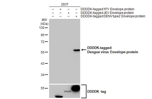
Western Blot: Dengue Virus 2 Envelope Antibody (GT643) [NBP3-13488] -
Western Blot: Dengue Virus 2 Envelope Antibody (GT643) [NBP3-13488] - Non-transfected (-) and transfected (+) 293T whole cell extracts were separated by 10% SDS-PAGE, and the membrane was blotted with Dengue Virus 2 Envelope antibody [GT643] (NBP3-13488) diluted at 1:5000. The HRP-conjugated anti-mouse IgG antibody was used to detect the primary antibody.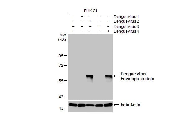
Western Blot: Dengue Virus 2 Envelope Antibody (GT643) [NBP3-13488] -
Western Blot: Dengue Virus 2 Envelope Antibody (GT643) [NBP3-13488] - Non-infected (-) and infected (+) BHK-21 whole cell extracts were separated by 10% SDS-PAGE, and the membrane was blotted with Dengue Virus 2 Envelope antibody [GT643] (NBP3-13488) diluted at 1:1000. The HRP-conjugated anti-mouse IgG antibody was used to detect the primary antibody.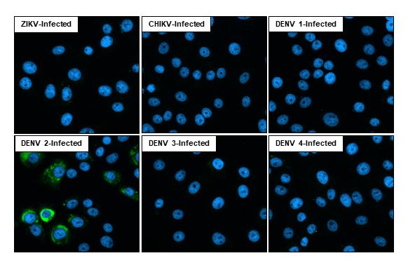
Immunocytochemistry/ Immunofluorescence: Dengue Virus 2 Envelope Antibody (GT643) [NBP3-13488] -
Immunofluorescent analysis of Dengue virus infected cells using Dengue virus Envelope protein antibody [GT643] antibody (NBP3-13488).Sample: Multiple virus infected cells slide.
Green: Dengue virus Envelope protein antibody [GT643] antibody (NBP3-13488) diluted at 1:100.
Blue: Fluoroshield with DAPI.
Applications for Dengue Virus 2 Envelope Antibody (GT643) - Azide and BSA Free
Application
Recommended Usage
Flow Cytometry
Optimal dilutions of this antibody should be experimentally determined.
Immunocytochemistry/ Immunofluorescence
Optimal dilutions of this antibody should be experimentally determined.
Immunohistochemistry
Optimal dilutions of this antibody should be experimentally determined.
Immunohistochemistry-Paraffin
Optimal dilutions of this antibody should be experimentally determined.
Western Blot
Optimal dilutions of this antibody should be experimentally determined.
Flow Cytometry Panel Builder
Bio-Techne Knows Flow Cytometry
Save time and reduce costly mistakes by quickly finding compatible reagents using the Panel Builder Tool.
Advanced Features
- Spectra Viewer - Custom analysis of spectra from multiple fluorochromes
- Spillover Popups - Visualize the spectra of individual fluorochromes
- Antigen Density Selector - Match fluorochrome brightness with antigen density

Formulation, Preparation, and Storage
Purification
Protein G purified
Formulation
PBS
Format
Azide and BSA Free
Preservative
No Preservative
Concentration
Concentrations vary lot to lot. See vial label for concentration. If unlisted please contact technical services.
Shipping
The product is shipped with polar packs. Upon receipt, store it immediately at the temperature recommended below.
Stability & Storage
Store at 4C short term. Aliquot and store at -20C long term. Avoid freeze-thaw cycles.
Background: Dengue Virus 2 Envelope
Dengue virus entry into host cells occurs via receptor-mediated endocytosis by receptor molecules including the mannose receptor, heparan sulfate, glycosaminoglycans, and DC-SIGN (1,3). Following attachment, the virus is endocytosed in clathrin-coated vesicles (1,3). Following internalization, clathrin disassembles and endosomal processing occurs, allowing viral fusion, disassembly, and release of viral RNA (1,3). This release results in viral translation and replication, virus assembly and maturation, and eventual exocytosis of the mature virus (1,3). Infection can result in a wide range of clinical symptoms including mild disease such as Dengue fever which is characterized by fever, headache, joint pain, rash, and retro-orbital pain, or severe, life-threatening conditions like Dengue hemorrhagic fever or Dengue shock syndrome which involves vascular permeability and leakage (1-3). Host immune response against infection includes innate immune response via interferon secretion and pro-inflammatory cytokine production, as well as adaptive immune response involving cellular and humoral components like T cell activation and B-cell mediated antibody production (1-3). As far as treatment for Dengue virus infection, there no commercial antiviral agents, though some anti-pyretics and certain phenolic compounds do show promise in treating infection (1-3). However, Resveratol, an antiviral for other Flavivirus, has been shown to directly attack the Dengue virus genome (1). While more work needs to be done, there are some live-attenuated tetravalent Dengue virus vaccine candidates in clinal trials including DENVax and TV003/TV005 (1-3).
References
1. Nanaware N, Banerjee A, Mullick Bagchi S, Bagchi P, Mukherjee A. Dengue Virus Infection: A Tale of Viral Exploitations and Host Responses. Viruses. 2021;13(10):1967. Published 2021 Sep 30. https://doi.org/10.3390/v13101967
2. Harapan H, Michie A, Sasmono RT, Imrie A. Dengue: A Minireview. Viruses. 2020;12(8):829. Published 2020 Jul 30. https://doi.org/10.3390/v12080829
3. Roy SK, Bhattacharjee S. Dengue virus: epidemiology, biology, and disease aetiology. Can J Microbiol. 2021;67(10):687-702. https://doi.org/10.1139/cjm-2020-0572
Alternate Names
Dengue 2 envelope;Dengue envelope;envelope protein, Envelope protein E
Additional Dengue Virus 2 Envelope Products
Product Documents for Dengue Virus 2 Envelope Antibody (GT643) - Azide and BSA Free
Certificate of Analysis
To download a Certificate of Analysis, please enter a lot or batch number in the search box below.
Product Specific Notices for Dengue Virus 2 Envelope Antibody (GT643) - Azide and BSA Free
This product is for research use only and is not approved for use in humans or in clinical diagnosis. Primary Antibodies are guaranteed for 1 year from date of receipt.
Customer Reviews for Dengue Virus 2 Envelope Antibody (GT643) - Azide and BSA Free
There are currently no reviews for this product. Be the first to review Dengue Virus 2 Envelope Antibody (GT643) - Azide and BSA Free and earn rewards!
Have you used Dengue Virus 2 Envelope Antibody (GT643) - Azide and BSA Free?
Submit a review and receive an Amazon gift card!
$25/€18/£15/$25CAN/¥2500 for a review with an image
$10/€7/£6/$10CAN/¥1110 for a review without an image
Submit a review
Protocols
Find general support by application which include: protocols, troubleshooting, illustrated assays, videos and webinars.
- 7-Amino Actinomycin D (7-AAD) Cell Viability Flow Cytometry Protocol
- Antigen Retrieval Protocol (PIER)
- Antigen Retrieval for Frozen Sections Protocol
- Appropriate Fixation of IHC/ICC Samples
- Cellular Response to Hypoxia Protocols
- Chromogenic IHC Staining of Formalin-Fixed Paraffin-Embedded (FFPE) Tissue Protocol
- Chromogenic Immunohistochemistry Staining of Frozen Tissue
- ClariTSA™ Fluorophore Kits
- Detection & Visualization of Antibody Binding
- Extracellular Membrane Flow Cytometry Protocol
- Flow Cytometry Protocol for Cell Surface Markers
- Flow Cytometry Protocol for Staining Membrane Associated Proteins
- Flow Cytometry Staining Protocols
- Flow Cytometry Troubleshooting Guide
- Fluorescent IHC Staining of Frozen Tissue Protocol
- Graphic Protocol for Heat-induced Epitope Retrieval
- Graphic Protocol for the Preparation and Fluorescent IHC Staining of Frozen Tissue Sections
- Graphic Protocol for the Preparation and Fluorescent IHC Staining of Paraffin-embedded Tissue Sections
- Graphic Protocol for the Preparation of Gelatin-coated Slides for Histological Tissue Sections
- ICC Cell Smear Protocol for Suspension Cells
- ICC Immunocytochemistry Protocol Videos
- ICC for Adherent Cells
- IHC Sample Preparation (Frozen sections vs Paraffin)
- Immunocytochemistry (ICC) Protocol
- Immunocytochemistry Troubleshooting
- Immunofluorescence of Organoids Embedded in Cultrex Basement Membrane Extract
- Immunofluorescent IHC Staining of Formalin-Fixed Paraffin-Embedded (FFPE) Tissue Protocol
- Immunohistochemistry (IHC) and Immunocytochemistry (ICC) Protocols
- Immunohistochemistry Frozen Troubleshooting
- Immunohistochemistry Paraffin Troubleshooting
- Intracellular Flow Cytometry Protocol Using Alcohol (Methanol)
- Intracellular Flow Cytometry Protocol Using Detergents
- Intracellular Nuclear Staining Flow Cytometry Protocol Using Detergents
- Intracellular Staining Flow Cytometry Protocol Using Alcohol Permeabilization
- Intracellular Staining Flow Cytometry Protocol Using Detergents to Permeabilize Cells
- Preparing Samples for IHC/ICC Experiments
- Preventing Non-Specific Staining (Non-Specific Binding)
- Primary Antibody Selection & Optimization
- Propidium Iodide Cell Viability Flow Cytometry Protocol
- Protocol for Heat-Induced Epitope Retrieval (HIER)
- Protocol for Liperfluo
- Protocol for Making a 4% Formaldehyde Solution in PBS
- Protocol for VisUCyte™ HRP Polymer Detection Reagent
- Protocol for the Characterization of Human Th22 Cells
- Protocol for the Characterization of Human Th9 Cells
- Protocol for the Fluorescent ICC Staining of Cell Smears - Graphic
- Protocol for the Fluorescent ICC Staining of Cultured Cells on Coverslips - Graphic
- Protocol for the Preparation & Fixation of Cells on Coverslips
- Protocol for the Preparation and Chromogenic IHC Staining of Frozen Tissue Sections
- Protocol for the Preparation and Chromogenic IHC Staining of Frozen Tissue Sections - Graphic
- Protocol for the Preparation and Chromogenic IHC Staining of Paraffin-embedded Tissue Sections
- Protocol for the Preparation and Chromogenic IHC Staining of Paraffin-embedded Tissue Sections - Graphic
- Protocol for the Preparation and Fluorescent ICC Staining of Cells on Coverslips
- Protocol for the Preparation and Fluorescent ICC Staining of Non-adherent Cells
- Protocol for the Preparation and Fluorescent ICC Staining of Stem Cells on Coverslips
- Protocol for the Preparation and Fluorescent IHC Staining of Frozen Tissue Sections
- Protocol for the Preparation and Fluorescent IHC Staining of Paraffin-embedded Tissue Sections
- Protocol for the Preparation of Gelatin-coated Slides for Histological Tissue Sections
- Protocol for the Preparation of a Cell Smear for Non-adherent Cell ICC - Graphic
- Protocol: Annexin V and PI Staining by Flow Cytometry
- Protocol: Annexin V and PI Staining for Apoptosis by Flow Cytometry
- R&D Systems Quality Control Western Blot Protocol
- TUNEL and Active Caspase-3 Detection by IHC/ICC Protocol
- The Importance of IHC/ICC Controls
- Troubleshooting Guide: Fluorokine Flow Cytometry Kits
- Troubleshooting Guide: Immunohistochemistry
- Troubleshooting Guide: Western Blot Figures
- Western Blot Conditions
- Western Blot Protocol
- Western Blot Protocol for Cell Lysates
- Western Blot Troubleshooting
- Western Blot Troubleshooting Guide
- View all Protocols, Troubleshooting, Illustrated assays and Webinars
Loading...