
Key Product Details
Validated by
Knockout/Knockdown
Species Reactivity
Human
Applications
Knockout Validated, Immunohistochemistry, Western Blot, Immunocytochemistry, COMET
Label
Unconjugated
Antibody Source
Monoclonal Mouse IgG2B Clone # 1036020
Loading...
Product Specifications
Immunogen
Human embryonic kidney cell HEK293-derived human EMMPRIN/CD147 protein
Glu138-Ala323
Accession # P35613.2
Glu138-Ala323
Accession # P35613.2
Specificity
Detects human EMMPRIN/CD147 in direct ELISAs and Western blots.
Clonality
Monoclonal
Host
Mouse
Isotype
IgG2B
Scientific Data Images for Human EMMPRIN/CD147 Antibody (1036020)
Detection of EMMPRIN/CD147 in Colon via seqIF™ staining on COMET™
EMMPRIN/CD147 was detected in immersion fixed paraffin-embedded sections of human colon using Mouse Anti-Human EMMPRIN/CD147 Monoclonal Antibody (Catalog # MAB9721) at 1 µg/mL at 37 ° Celsius for 4 minutes. Before incubation with the primary antibody, tissue underwent an all-in-one dewaxing and antigen retrieval preprocessing using PreTreatment Module (PT Module) and Dewax and HIER Buffer H (pH 9; Epredia Catalog # TA-999-DHBH). Tissue was stained using the Alexa Fluor™ 647 Goat anti-Mouse IgG Secondary Antibody at 1:200 at 37 ° Celsius for 2 minutes. (Yellow; Lunaphore Catalog # DR647MS) and counterstained with DAPI (blue; Lunaphore Catalog # DR100). Specific staining was localized to the membrane. Protocol available in COMET™ Panel Builder.
Detection of EMMPRIN/CD147 in Human Liver Cancer via seqIF™ staining on COMET™
EMMPRIN/CD147 was detected in immersion fixed paraffin-embedded sections of human liver cancer using Mouse Anti-Human EMMPRIN/CD147 Monoclonal Antibody (Catalog # MAB9721) at 1 µg/mL at 37 ° Celsius for 4 minutes. Before incubation with the primary antibody, tissue underwent an all-in-one dewaxing and antigen retrieval preprocessing using PreTreatment Module (PT Module) and Dewax and HIER Buffer H (pH 9; Epredia Catalog # TA-999-DHBH). Tissue was stained using the Alexa Fluor™ 647 Goat anti-Mouse IgG Secondary Antibody at 1:200 at 37 ° Celsius for 2 minutes. (Yellow; Lunaphore Catalog # DR647MS) and counterstained with DAPI (blue; Lunaphore Catalog # DR100). Specific staining was localized to the membrane. Protocol available in COMET™ Panel Builder.
Detection of EMMPRIN/CD147 in Human Pancreatic Cancer via seqIF™ staining on COMET™
EMMPRIN/CD147 was detected in immersion fixed paraffin-embedded sections of human pancreatic cancer using Mouse Anti-Human EMMPRIN/CD147 Monoclonal Antibody (Catalog # MAB9721) at 1 µg/mL at 37 ° Celsius for 4 minutes. Before incubation with the primary antibody, tissue underwent an all-in-one dewaxing and antigen retrieval preprocessing using PreTreatment Module (PT Module) and Dewax and HIER Buffer H (pH 9; Epredia Catalog # TA-999-DHBH). Tissue was stained using the Alexa Fluor™ 647 Goat anti-Mouse IgG Secondary Antibody at 1:200 at 37 ° Celsius for 2 minutes. (Yellow; Lunaphore Catalog # DR647MS) and counterstained with DAPI (blue; Lunaphore Catalog # DR100). Specific staining was localized to the membrane. Protocol available in COMET™ Panel Builder.
Detection of Human EMMPRIN/CD147 by Western Blot.
Western blot shows lysates of human brain (cerebellum), human heart (ventricle), and human liver. PVDF membrane was probed with 2 µg/mL of Mouse Anti-Human EMMPRIN/CD147 Monoclonal Antibody (Catalog # MAB9721) followed by HRP-conjugated Anti-Mouse IgG Secondary Antibody (HAF018). A specific band was detected for EMMPRIN/CD147 at approximately 45-60 kDa (as indicated). This experiment was conducted under reducing conditions and using Western Blot Buffer Group 1.
EMMPRIN/CD147 in 789-O Human Cell Line.
EMMPRIN/CD147 was detected in immersion fixed 789-O Human renal cell carcinoma cell line using Mouse Anti-Human EMMPRIN/CD147 Monoclonal Antibody (Catalog # MAB9721) at 8 µg/mL for 3 hours at room temperature. Cells were stained using the NorthernLights™ 557-conjugated Anti-Mouse IgG Secondary Antibody (red; NL007) and counterstained with DAPI (blue). Specific staining was localized to cell membrane. Staining was performed using our protocol for Fluorescent ICC Staining of Non-adherent Cells.
EMMPRIN/CD147 in Human Heart.
EMMPRIN/CD147 was detected in immersion fixed paraffin-embedded sections of human heart using Mouse Anti-Human EMMPRIN/CD147 Monoclonal Antibody (Catalog # MAB9721) at 5 µg/mL for 1 hour at room temperature followed by incubation with the Anti-Mouse IgG VisUCyte™ HRP Polymer Antibody (VC001). Before incubation with the primary antibody, tissue was subjected to heat-induced epitope retrieval using Antigen Retrieval Reagent-Basic (CTS013). Tissue was stained using DAB (brown) and counterstained with hematoxylin (blue). Specific staining was localized to cell membrane in capillaries. Staining was performed using our protocol for IHC Staining with VisUCyte HRP Polymer Detection Reagents.
Western Blot Shows Human EMMPRIN/CD147 Specificity by Using Knockout Cell Line.
Western blot shows lysates of HEK293T human embryonic kidney cell line and human EMMPRIN knockout HEK293T human embryonic kidney cell line (KO). PVDF membrane was probed with 2 µg/mL of Mouse Anti-Human EMMPRIN/CD147 Monoclonal Antibody (Catalog # MAB9721) followed by HRP-conjugated Anti-Mouse IgG Secondary Antibody (HAF018). A specific band was detected for EMMPRIN/CD147 at approximately 45-60 kDa (as indicated) in the parental cell line, but is not detectable in knockout cell line. GAPDH (MAB5718) is shown as a loading control. This experiment was conducted under reducing conditions and using Western Blot Buffer Group 1.Applications for Human EMMPRIN/CD147 Antibody (1036020)
Application
Recommended Usage
COMET
1 µg/mL
Sample: Immersion fixed paraffin-embedded sections of human colon, liver cancer and pancreatic cancer.
Sample: Immersion fixed paraffin-embedded sections of human colon, liver cancer and pancreatic cancer.
Immunocytochemistry
8-25 µg/mL
Sample: Immersion fixed 789-O Human renal cell carcinoma cell line
Sample: Immersion fixed 789-O Human renal cell carcinoma cell line
Immunohistochemistry
5-25 µg/mL
Sample: Immersion fixed paraffin-embedded sections of human heart
Sample: Immersion fixed paraffin-embedded sections of human heart
Knockout Validated
2 µg/mL
Sample: EMMPRIN/CD147 is specifically detected in HEK293T human embryonic kidney parental cell line but is not detectable in EMMPRIN/CD147 knockout HEK293T human embryonic kidney cell line.
Sample: EMMPRIN/CD147 is specifically detected in HEK293T human embryonic kidney parental cell line but is not detectable in EMMPRIN/CD147 knockout HEK293T human embryonic kidney cell line.
Western Blot
2 µg/mL
Sample: Human brain (cerebellum), human heart (ventricle), and human liver
Sample: Human brain (cerebellum), human heart (ventricle), and human liver
Reviewed Applications
Read 1 review rated 5 using MAB9721 in the following applications:
Formulation, Preparation, and Storage
Purification
Protein A or G purified from hybridoma culture supernatant
Reconstitution
Reconstitute at 0.5 mg/mL in sterile PBS. For liquid material, refer to CoA for concentration.
Loading...
Formulation
Lyophilized from a 0.2 μm filtered solution in PBS with Trehalose. See Certificate of Analysis for details.
*Small pack size (-SP) is supplied either lyophilized or as a 0.2 µm filtered solution in PBS.
*Small pack size (-SP) is supplied either lyophilized or as a 0.2 µm filtered solution in PBS.
Shipping
Lyophilized product is shipped at ambient temperature. Liquid small pack size (-SP) is shipped with polar packs. Upon receipt, store immediately at the temperature recommended below.
Stability & Storage
Use a manual defrost freezer and avoid repeated freeze-thaw cycles.
- 12 months from date of receipt, -20 to -70 °C as supplied.
- 1 month, 2 to 8 °C under sterile conditions after reconstitution.
- 6 months, -20 to -70 °C under sterile conditions after reconstitution.
Calculators
Background: EMMPRIN/CD147
References
- Gabison, E. E. et al. (2005) Biochimie 87:361.
- Yurchenko, V. et al. (2006) Immunology 117:301.
- Kasinrerk, W. et al. (1992) J. Immunol. 149:847.
- Iacono, K.T. et al. (2007) Exp. Mol. Pathol. 83:283.
- Hanna, S. M. et al. (2003) BMC Biochem. 4:17.
- Liao, C-G. et al. (2011) Mol. Cell. Biol. 31:2591.
- Riethdorf, S. et al. (2006) Int. J. Cancer 119:1800.
- Braundmeier, A. G. et al. (2006) J. Clin. Endocrinol. Metab. 91:2358.
- Tang, Y. et al. (2006) Mol. Cancer Res. 4:371.
- Quemener, C. et al. (2007) Cancer Res. 67:9.
- Wilson, M. C. et al. (2005) J. Biol. Chem. 280:27213.
- Xu, D. and M. E. Hemler, (2005) Mol. Cell. Proteomics 4:1061.
- Tang, W. et al. (2004) Mol. Biol. Cell 15:4043.
- Zhao, P. et al. (2010) Cancer Sci. 101:387.
- Dai, J. et al. (2009) BMC Cancer 9:337.
- Li, Y. et al. (2012) J. Biol. Chem. 287:4759.
- Hibino, T. et al. (2013) Cancer Res. 73:172.
- Arora, K. et al. (2005) J. Immunol. 175:517.
- Pushkarsky, T. et al. (2005) J. Biol. Chem. 280:27866.
- Egawa, N. et al. (2006) J. Biol. Chem. 281:37576.
- Sidhu, S. S. et al. (2004) Oncogene 23:956.
- Ulrich, H. et al. (2020) Stem Cell Rev. and Rep., https://doi.org/10.1007/s12015-020-09976-7.
Long Name
Extracellular Matrix Metalloproteinase Inducer
Alternate Names
Basigin, BSG, CD147
Gene Symbol
BSG
UniProt
Additional EMMPRIN/CD147 Products
Product Documents for Human EMMPRIN/CD147 Antibody (1036020)
Certificate of Analysis
To download a Certificate of Analysis, please enter a lot or batch number in the search box below.
Note: Certificate of Analysis not available for kit components.
Product Specific Notices for Human EMMPRIN/CD147 Antibody (1036020)
For research use only
Customer Reviews for Human EMMPRIN/CD147 Antibody (1036020) (1)
5 out of 5
1 Customer Rating
Have you used Human EMMPRIN/CD147 Antibody (1036020)?
Submit a review and receive an Amazon gift card!
$25/€18/£15/$25CAN/¥2500 Yen for a review with an image
$10/€7/£6/$10CAN/¥1110 Yen for a review without an image
Submit a review
Customer Images
Showing
1
-
1 的
1 review
Showing All
Filter By:
-
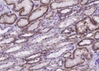
Application: ImmunohistochemistrySample Tested: Kidney tissueSpecies: HumanVerified Customer | Posted 01/06/2022

There are no reviews that match your criteria.
Protocols
Find general support by application which include: protocols, troubleshooting, illustrated assays, videos and webinars.
- Antigen Retrieval Protocol (PIER)
- Antigen Retrieval for Frozen Sections Protocol
- Appropriate Fixation of IHC/ICC Samples
- Cellular Response to Hypoxia Protocols
- Chromogenic IHC Staining of Formalin-Fixed Paraffin-Embedded (FFPE) Tissue Protocol
- Chromogenic Immunohistochemistry Staining of Frozen Tissue
- ClariTSA™ Fluorophore Kits
- Detection & Visualization of Antibody Binding
- Fluorescent IHC Staining of Frozen Tissue Protocol
- Graphic Protocol for Heat-induced Epitope Retrieval
- Graphic Protocol for the Preparation and Fluorescent IHC Staining of Frozen Tissue Sections
- Graphic Protocol for the Preparation and Fluorescent IHC Staining of Paraffin-embedded Tissue Sections
- Graphic Protocol for the Preparation of Gelatin-coated Slides for Histological Tissue Sections
- ICC Cell Smear Protocol for Suspension Cells
- ICC Immunocytochemistry Protocol Videos
- ICC for Adherent Cells
- IHC Sample Preparation (Frozen sections vs Paraffin)
- Immunocytochemistry (ICC) Protocol
- Immunocytochemistry Troubleshooting
- Immunofluorescence of Organoids Embedded in Cultrex Basement Membrane Extract
- Immunofluorescent IHC Staining of Formalin-Fixed Paraffin-Embedded (FFPE) Tissue Protocol
- Immunohistochemistry (IHC) and Immunocytochemistry (ICC) Protocols
- Immunohistochemistry Frozen Troubleshooting
- Immunohistochemistry Paraffin Troubleshooting
- Preparing Samples for IHC/ICC Experiments
- Preventing Non-Specific Staining (Non-Specific Binding)
- Primary Antibody Selection & Optimization
- Protocol for Heat-Induced Epitope Retrieval (HIER)
- Protocol for Making a 4% Formaldehyde Solution in PBS
- Protocol for VisUCyte™ HRP Polymer Detection Reagent
- Protocol for the Fluorescent ICC Staining of Cell Smears - Graphic
- Protocol for the Fluorescent ICC Staining of Cultured Cells on Coverslips - Graphic
- Protocol for the Preparation & Fixation of Cells on Coverslips
- Protocol for the Preparation and Chromogenic IHC Staining of Frozen Tissue Sections
- Protocol for the Preparation and Chromogenic IHC Staining of Frozen Tissue Sections - Graphic
- Protocol for the Preparation and Chromogenic IHC Staining of Paraffin-embedded Tissue Sections
- Protocol for the Preparation and Chromogenic IHC Staining of Paraffin-embedded Tissue Sections - Graphic
- Protocol for the Preparation and Fluorescent ICC Staining of Cells on Coverslips
- Protocol for the Preparation and Fluorescent ICC Staining of Non-adherent Cells
- Protocol for the Preparation and Fluorescent ICC Staining of Stem Cells on Coverslips
- Protocol for the Preparation and Fluorescent IHC Staining of Frozen Tissue Sections
- Protocol for the Preparation and Fluorescent IHC Staining of Paraffin-embedded Tissue Sections
- Protocol for the Preparation of Gelatin-coated Slides for Histological Tissue Sections
- Protocol for the Preparation of a Cell Smear for Non-adherent Cell ICC - Graphic
- R&D Systems Quality Control Western Blot Protocol
- TUNEL and Active Caspase-3 Detection by IHC/ICC Protocol
- The Importance of IHC/ICC Controls
- Troubleshooting Guide: Immunohistochemistry
- Troubleshooting Guide: Western Blot Figures
- Western Blot Conditions
- Western Blot Protocol
- Western Blot Protocol for Cell Lysates
- Western Blot Troubleshooting
- Western Blot Troubleshooting Guide
- View all Protocols, Troubleshooting, Illustrated assays and Webinars
Loading...