
Estrogen-related receptor alpha (ERR-alpha, ERR1, ESRL1; NR3B1) is a member of the orphan nuclear receptor family. ERR-alpha is expressed in the liver, kidney, brain, heart, uterus, vagina and cervix. ERR-alpha takes part in the cell cycle and the fatty acid metabolizing of the musculus skeleti. ERR-alpha was shown to interact with ER-alpha. ERR-beta (ERR2; NR3B2) is also expressed in the liver, brain, uterus, vagina, and cervix. ERR-beta has a role in early placental development. ERR-gamma (ESRRG; NR3B3) is expressed in the brain, kidney, testis, lung, adrenal gland, pancreas, placenta and bone marrow. ERR-gamma was shown to be located in the critical region for Usher's syndrome.
Human ERR gamma/NR3B3 Antibody (H6812)
R&D Systems | Catalog # PP-H6812-00

Scientific images may be resized, cropped, or adjusted for brightness or contrast for presentation purposes. These updates are limited to presentation and do not affect the underlying scientific interpretation of the data. Where available, additional source data may be provided upon request.
Loading...
Key Product Details
Species Reactivity
Validated:
Human
Cited:
Human, Mouse, Avian - Chicken, Transgenic Mouse, Xenopus
Applications
Validated:
Immunohistochemistry, Western Blot, Direct ELISA, Immunoprecipitation
Cited:
Immunohistochemistry, Western Blot, Immunoprecipitation, Chromatin Immunoprecipitation (ChIP)
Label
Unconjugated
Antibody Source
Monoclonal Mouse IgG2A Clone # H6812
Loading...
Product Specifications
Clonality
Monoclonal
Host
Mouse
Isotype
IgG2A
Scientific Data Images for Human ERR gamma/NR3B3 Antibody (H6812)
Detection of ERR gamma/NR3B3 by Immunohistochemistry
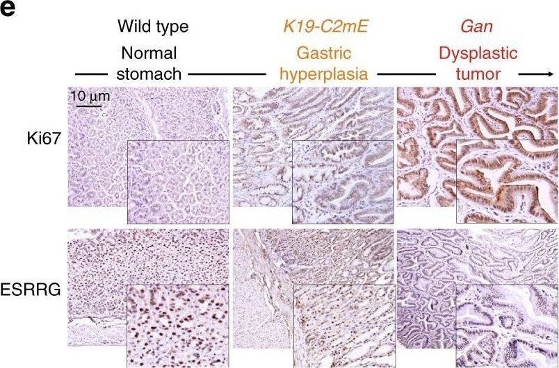
ESRRG expression in GC patients and in a mouse model. a Venn diagram of genes showing significant differential expression between normal and cancer tissue in the three different GC patient cohorts. A univariate test (two-sample t-test) with a multivariate permutation test (10,000 random permutations) was employed. In each comparison, a cut-off p-value of less than 0.001 was applied to retain genes with an expression level that differed significantly between the two groups of tissues examined. b Expression patterns of selected genes shared in the three GC patient cohorts. The expression of 521 genes was commonly up- or downregulated in all three cohorts. Colored bars at the top of the heat map represent samples as indicated. Genes involved in transcription are highlighted in blue or red text. c ESRRG expression from TCGA data. d Western blot analysis from normal gastric and tumor tissues. e, f Gastritis in K19-C2mE mice and gastric tumors in K19-Wnt1/C2mE mice. e Immunohistochemistry (IHC) staining of Ki67 and ESRRG in wild type (left), K19-C2mE (middle), and Gan (right) mouse stomachs. f ESRRG mRNA levels in the GC mouse model. Original magnification, ×200. Data represent the mean ± s.d. (error bars) from the indicated samples (n = 4 per group). Student’s t-test was used to examine statistical significance (* p < 0.05, *** p < 0.005) Image collected and cropped by CiteAb from the following open publication (https://pubmed.ncbi.nlm.nih.gov/29765046), licensed under a CC-BY license. Not internally tested by R&D Systems.Applications for Human ERR gamma/NR3B3 Antibody (H6812)
Application
Recommended Usage
Direct ELISA
Optimal dilutions of this antibody should be experimentally determined.
Immunohistochemistry
5-15 µg/mL
Sample: Perfusion fixed paraffin-embedded sections of rat retina (inner nuclear layer)
Sample: Perfusion fixed paraffin-embedded sections of rat retina (inner nuclear layer)
Immunoprecipitation
Optimal dilutions of this antibody should be experimentally determined.
Western Blot
Optimal dilutions of this antibody should be experimentally determined.
Formulation, Preparation, and Storage
Formulation
A liquid formulation in physiologic saline with 0.1% NaN<sub>3</sub>.
Shipping
The product is shipped with dry ice or equivalent. Upon receipt, store it immediately at the temperature recommended below.
Background: ERR gamma/NR3B3
Long Name
Estrogen-related Receptor gamma
Alternate Names
ERR3, ESRRG, NR3B3
Entrez Gene IDs
2104 (Human)
Gene Symbol
ESRRG
Additional ERR gamma/NR3B3 Products
Product Documents for Human ERR gamma/NR3B3 Antibody (H6812)
Certificate of Analysis
To download a Certificate of Analysis, please enter a lot or batch number in the search box below.
Note: Certificate of Analysis not available for kit components.
Product Specific Notices for Human ERR gamma/NR3B3 Antibody (H6812)
For research use only
Related Research Areas
Citations for Human ERR gamma/NR3B3 Antibody (H6812)
 Powered by Bioz
Customer Reviews for Human ERR gamma/NR3B3 Antibody (H6812)
There are currently no reviews for this product. Be the first to review Human ERR gamma/NR3B3 Antibody (H6812) and earn rewards!
Have you used Human ERR gamma/NR3B3 Antibody (H6812)?
Submit a review and receive an Amazon gift card!
$25/€18/£15/$25CAN/¥2500 for a review with an image
$10/€7/£6/$10CAN/¥1110 for a review without an image
Submit a review
Protocols
Find general support by application which include: protocols, troubleshooting, illustrated assays, videos and webinars.
- Antigen Retrieval Protocol (PIER)
- Antigen Retrieval for Frozen Sections Protocol
- Appropriate Fixation of IHC/ICC Samples
- Cellular Response to Hypoxia Protocols
- Chromogenic IHC Staining of Formalin-Fixed Paraffin-Embedded (FFPE) Tissue Protocol
- Chromogenic Immunohistochemistry Staining of Frozen Tissue
- ClariTSA™ Fluorophore Kits
- Detection & Visualization of Antibody Binding
- ELISA Sample Preparation & Collection Guide
- ELISA Troubleshooting Guide
- Fluorescent IHC Staining of Frozen Tissue Protocol
- Graphic Protocol for Heat-induced Epitope Retrieval
- Graphic Protocol for the Preparation and Fluorescent IHC Staining of Frozen Tissue Sections
- Graphic Protocol for the Preparation and Fluorescent IHC Staining of Paraffin-embedded Tissue Sections
- Graphic Protocol for the Preparation of Gelatin-coated Slides for Histological Tissue Sections
- How to Run an R&D Systems DuoSet ELISA
- How to Run an R&D Systems Quantikine ELISA
- How to Run an R&D Systems Quantikine™ QuicKit™ ELISA
- IHC Sample Preparation (Frozen sections vs Paraffin)
- Immunofluorescent IHC Staining of Formalin-Fixed Paraffin-Embedded (FFPE) Tissue Protocol
- Immunohistochemistry (IHC) and Immunocytochemistry (ICC) Protocols
- Immunohistochemistry Frozen Troubleshooting
- Immunohistochemistry Paraffin Troubleshooting
- Immunoprecipitation Protocol
- Preparing Samples for IHC/ICC Experiments
- Preventing Non-Specific Staining (Non-Specific Binding)
- Primary Antibody Selection & Optimization
- Protocol for Heat-Induced Epitope Retrieval (HIER)
- Protocol for Making a 4% Formaldehyde Solution in PBS
- Protocol for VisUCyte™ HRP Polymer Detection Reagent
- Protocol for the Preparation & Fixation of Cells on Coverslips
- Protocol for the Preparation and Chromogenic IHC Staining of Frozen Tissue Sections
- Protocol for the Preparation and Chromogenic IHC Staining of Frozen Tissue Sections - Graphic
- Protocol for the Preparation and Chromogenic IHC Staining of Paraffin-embedded Tissue Sections
- Protocol for the Preparation and Chromogenic IHC Staining of Paraffin-embedded Tissue Sections - Graphic
- Protocol for the Preparation and Fluorescent IHC Staining of Frozen Tissue Sections
- Protocol for the Preparation and Fluorescent IHC Staining of Paraffin-embedded Tissue Sections
- Protocol for the Preparation of Gelatin-coated Slides for Histological Tissue Sections
- Quantikine HS ELISA Kit Assay Principle, Alkaline Phosphatase
- Quantikine HS ELISA Kit Principle, Streptavidin-HRP Polymer
- R&D Systems Quality Control Western Blot Protocol
- Sandwich ELISA (Colorimetric) – Biotin/Streptavidin Detection Protocol
- Sandwich ELISA (Colorimetric) – Direct Detection Protocol
- TUNEL and Active Caspase-3 Detection by IHC/ICC Protocol
- The Importance of IHC/ICC Controls
- Troubleshooting Guide: ELISA
- Troubleshooting Guide: Immunohistochemistry
- Troubleshooting Guide: Western Blot Figures
- Western Blot Conditions
- Western Blot Protocol
- Western Blot Protocol for Cell Lysates
- Western Blot Troubleshooting
- Western Blot Troubleshooting Guide
- View all Protocols, Troubleshooting, Illustrated assays and Webinars
Loading...