FAP (also known as seprase) is a Type II transmembrane serine protease that is structurally related to dipeptidyl peptidase IV (1). FAP has substrate specificity similar to dipeptidyl peptidase IV, which is specific for N-terminal Xaa-Pro sequences, but FAP is also an endopeptidase able to degrade gelatin and Type I collagen (2). The enzymatically active form of FAP is a dimer (3). FAP has a restricted tissue distribution. It is not detectable in normal tissues or resting fibroblasts, but is highly expressed on reactive stromal fibroblasts in epithelial cancers (4), in granulation tissue during wound healing, and in bone and soft tissue sarcomas (5). Because of its expression patterns and enzymatic activities, FAP is believed to play roles in tumor invasion, tissue remodeling, and wound repair.
Human Fibroblast Activation Protein alpha/FAP Antibody
R&D Systems | Catalog # AF3715

Key Product Details
Validated by
Knockout/Knockdown
Species Reactivity
Validated:
Human
Cited:
Human, Mouse, Transgenic Mouse, Xenograft
Applications
Validated:
Knockout Validated, Immunohistochemistry, Western Blot, Dual RNAscope ISH-IHC Compatible, Simple Western, Immunoprecipitation
Cited:
Immunohistochemistry, Immunohistochemistry-Paraffin, Western Blot, Flow Cytometry, Immunocytochemistry, Immunoprecipitation, Photoimmunotherapy, Westen Blot
Label
Unconjugated
Antibody Source
Polyclonal Sheep IgG
Loading...
Product Specifications
Immunogen
S. frugiperda insect ovarian cell line Sf 21-derived recombinant human Fibroblast Activation Protein alpha /FAP
Leu26-Asp760
Accession # Q12884
Leu26-Asp760
Accession # Q12884
Specificity
Detects human Fibroblast Activation Protein alpha /FAP in direct ELISAs and Western blots.
Clonality
Polyclonal
Host
Sheep
Isotype
IgG
Scientific Data Images for Human Fibroblast Activation Protein alpha/FAP Antibody
Detection of Human Fibroblast Activation Protein alpha /FAP by Western Blot.
Western blot shows lysates of WI-38 human lung fibroblast cell line. PVDF membrane was probed with 0.5 µg/mL of Sheep Anti-Human Fibroblast Activation Protein a/FAP Antigen Affinity-purified Polyclonal Antibody (Catalog # AF3715) followed by HRP-conjugated Anti-Sheep IgG Secondary Antibody (Catalog # HAF016). A specific band was detected for Fibroblast Activation Protein a/FAP at approximately 97 kDa (as indicated). This experiment was conducted under reducing conditions and using Immunoblot Buffer Group 1.
Fibroblast Activation Protein alpha /FAP in Human Squamous Cell Carcinoma.
Fibroblast Activation Protein a/FAP was detected in immersion fixed paraffin-embedded sections of human squamous cell carcinoma using Sheep Anti-Human Fibroblast Activation Protein a/FAP Antigen Affinity-purified Polyclonal Antibody (Catalog # AF3715) at 15 µg/mL overnight at 4 °C. Tissue was stained using the Anti-Sheep HRP-DAB Cell & Tissue Staining Kit (brown; Catalog # CTS019) and counterstained with hematoxylin (blue). Specific staining was localized to connective tissue. View our protocol for Chromogenic IHC Staining of Paraffin-embedded Tissue Sections.
Fibroblast Activation Protein alpha /FAP in Human Basal Cell Carcinoma.
Fibroblast Activation Protein alpha /FAP was detected in immersion fixed paraffin-embedded sections of human basal cell carcinoma using Sheep Anti-Human Fibroblast Activation Protein alpha /FAP Antigen Affinity-purified Polyclonal Antibody (Catalog # AF3715) at 10 µg/mL overnight at 4 °C. Tissue was stained using the Anti-Sheep HRP-DAB Cell & Tissue Staining Kit (brown; Catalog # CTS019) and counterstained with hematoxylin (blue). Specific staining was localized to cytoplasm. View our protocol for Chromogenic IHC Staining of Paraffin-embedded Tissue Sections.
Detection of Human Fibroblast Activation Protein alpha /FAP by Simple WesternTM.
Simple Western lane view shows lysates of IMR‑90 human lung fibroblast cell line and WI‑38 human lung fibroblast cell line, loaded at 0.2 mg/mL. A specific band was detected for Fibroblast Activation Protein alpha /FAP at approximately 130 kDa (as indicated) using 10 µg/mL of Sheep Anti-Human Fibroblast Activation Protein alpha /FAP Antigen Affinity-purified Polyclonal Antibody (Catalog # AF3715) followed by 1:50 dilution of HRP-conjugated Anti-Sheep IgG Secondary Antibody (HAF016). This experiment was conducted under reducing conditions and using the 12-230 kDa separation system.
Western Blot Shows Human Fibroblast Activation Protein alpha /FAP Specificity by Using Knockout Cell Line.
Western blot shows lysates of WI‑38 human lung fibroblast cell line and human FAP knockout WI‑38 human lung fibroblast cell line. PVDF membrane was probed with 0.5 µg/mL of Sheep Anti-Human Fibroblast Activation Protein alpha /FAP Antigen Affinity-purified Polyclonal Antibody (Catalog # AF3715) followed by HRP-conjugated Anti-Sheep IgG Secondary Antibody (HAF016). A specific band was detected for Fibroblast Activation Protein alpha /FAP at approximately 97 kDa (as indicated) in the parental WI‑38 human lung fibroblast cell line, but is not detectable in knockout WI‑38 human lung fibroblast cell line. GAPDH (AF5718) is shown as a loading control. This experiment was conducted under reducing conditions and using Western Blot Buffer Group 1.
Detection of Fibroblast Activation Protein alpha /FAP in Human Esophagus.
Formalin-fixed paraffin-embedded tissue sections of human squamous cell carcinoma in the esophagus were probed for FAP mRNA (ACD RNAScope Probe, catalog # 411971; Fast Red chromogen, ACD catalog # 322360). Adjacent tissue section was processed for immunohistochemistry using sheep anti-human FAP polyclonal antibody (R&D Systems catalog # AF3715) at 5ug/mL with overnight incubation at 4 degrees Celsius followed by incubation with anti-sheep IgG VisUCyte HRP Polymer Antibody (Catalog # VC006) and DAB chromogen (yellow-brown). Tissue was counterstained with hematoxylin (blue). Specific staining was localized to connective tissue.
Detection of Fibroblast Activation Protein alpha /FAP by Western Blot
FGFR2 drives the process of liver fibrosis. (A) The expression of liver fibrosis markers (ACTA2, fibroblast activation protein alpha (FAP), alpha-1 type I collagen (COL1A1)) was measured by qPCR. The samples were divided into four groups according to whether cells were induced with or without TGF-beta and whether cells were overexpressed by FGFR2. (B) The expression of markers in wild-type and FGFR2-OE cell lines under TGF-beta induction was analyzed by Western blot analysis. (C) The expression levels of alpha -SMA and (D) collagen secretion in LX-2 and Huh-7 were evaluated, when FGFR2 was overexpressed or knocked down upon equal TGF-beta induction. (E) Cells were collected after 48 h of co-culture, and the expression of alpha -SMA and (F) type I collagen in the lower layer cells was determined by ELISA. The results are marked as significant “*” when p < 0.05, “**” when p < 0.01, and not significant (ns) if p ≥ 0.05. Image collected and cropped by CiteAb from the following open publication (https://pubmed.ncbi.nlm.nih.gov/37111305), licensed under a CC-BY license. Not internally tested by R&D Systems.
Detection of Fibroblast Activation Protein alpha /FAP by Western Blot
CYN blocks the activation and development of liver fibrosis in vitro. (A) Evaluation of the inhibitory effect of CYN on the fibrosis-promoting effect of FGFR2. The fibrotic transformation of wild-type and FGFR2-OE LX-2 cells and Huh-7 cells was induced through TGF-beta activation, followed by an intervention with CYN for the relevant groups. The expression changes of the fibrosis markers ACTA2 and COL1A1 were analyzed by qPCR. (B) Wild-type cell lines were employed in the aforementioned experiments, and the activation of FGFR2 was triggered by supplementation with the exogenous basic fibroblast growth factor (bFGF) factor. (C) Analysis of the extent of antagonism of CYN towards TGF-beta Signaling. Activation induction models were established by adding or not adding TGF-beta to the cell culture environment with or without the CYN intervention, and the expression of liver fibrosis markers was determined by qPCR, (D) Western blot, and (E,F) ELISA analyses. (G) A co-culture model was used to evaluate the blocking effect of CYN on liver fibrosis activation of signaling transmission. The activation intensity of lower-layer wild-type cells was measured and compared using alpha -SMA expression and collagen secretion. The results are marked as significant “*” when p < 0.05, “**” when p < 0.01, and not significant (ns) if p ≥ 0.05. Image collected and cropped by CiteAb from the following open publication (https://pubmed.ncbi.nlm.nih.gov/37111305), licensed under a CC-BY license. Not internally tested by R&D Systems.
Detection of Fibroblast Activation Protein alpha /FAP by Western Blot
High expression of fibroblast growth factor receptor 2 (FGFR2) coincides with liver fibrosis. (A) The expression of FGFR2 in fibrotic liver and normal liver tissues was evaluated through data mining of human samples affected by Hepatitis B infection (GSE38941), alcohol abuse (GSE28619), and nonalcoholic steatohepatitis (GSE48452), as well as mouse samples induced with carbon tetrachloride (CCl4) (GSE152329, GSE98577). The differential fold expression was determined by comparing the expression of FGFR2 in the fibrotic liver and normal liver tissues. (B) A comparison of changes in FGFR2 expression in liver fibrosis patients in remission (improved) or not in remission (not improved) with mining data of GSE175448. (C) The correlation between FGFR2 expression and the liver fibrosis score (Ishak score) was analyzed by comparing FGFR2 expression in three pairs of liver fibrotic tissues and normal liver tissues through immunohistochemistry. (D) FGFR2 expression in liver fibrosis tissues and normal liver tissues was verified by Western blot and (E) q-PCR analyses. (E) Liver tissues from mice treated with CCl4 for different durations were used to determine the expression of the liver fibrosis markers Actin Alpha 2 (ACTA2) and FGFR2 by q-PCR and the trends of their expression with increasing days of induction. (F) The expression of ACTA2 and (G) FGFR2 was determined by q-PCR in liver tissues from mice treated with CCl4 for varying durations. The statistical significance was determined based on the p-value. The results are marked as significant “*” when p < 0.05, “**” when p < 0.01, “***” when p < 0.001, and not significant (ns) if p ≥ 0.05. Image collected and cropped by CiteAb from the following open publication (https://pubmed.ncbi.nlm.nih.gov/37111305), licensed under a CC-BY license. Not internally tested by R&D Systems.
Detection of Fibroblast Activation Protein alpha /FAP by Western Blot
Expression analysis of Fap in OA synovium. a Immunostaining of human FAP in the synovium of control and OA patients. DAPI staining indicates the nucleus. Scale bars: 100 μm. b qPCR analysis of human FAP mRNA levels in the synovium of control and OA patients (n = 8 samples per group). c Western blot analysis of human FAP protein levels in the synovium of control and OA patients (n = 3 samples per group). d Immunostaining of mouse Fap in the knee joint of sham and DMM-treated mice. Sham or DMM surgery was performed in 8-week-old FapLacZ/+ mice, which were sacrificed 8 weeks later, and LacZ immunostaining was used to detect Fap expression at the posterior horn of the medial meniscus (F: femur; T: tibia; M: meniscus; S: synovium). DAPI staining indicates the nucleus. Scale bars: 100 μm. The statistical significance was assessed using two-tailed Student’s unpaired t tests. Data are presented as the mean ± SD (**P < 0.01) Image collected and cropped by CiteAb from the following open publication (https://pubmed.ncbi.nlm.nih.gov/36588124), licensed under a CC-BY license. Not internally tested by R&D Systems.
Detection of Fibroblast Activation Protein alpha /FAP by Western Blot
FGFR2 drives the process of liver fibrosis. (A) The expression of liver fibrosis markers (ACTA2, fibroblast activation protein alpha (FAP), alpha-1 type I collagen (COL1A1)) was measured by qPCR. The samples were divided into four groups according to whether cells were induced with or without TGF-beta and whether cells were overexpressed by FGFR2. (B) The expression of markers in wild-type and FGFR2-OE cell lines under TGF-beta induction was analyzed by Western blot analysis. (C) The expression levels of alpha -SMA and (D) collagen secretion in LX-2 and Huh-7 were evaluated, when FGFR2 was overexpressed or knocked down upon equal TGF-beta induction. (E) Cells were collected after 48 h of co-culture, and the expression of alpha -SMA and (F) type I collagen in the lower layer cells was determined by ELISA. The results are marked as significant “*” when p < 0.05, “**” when p < 0.01, and not significant (ns) if p ≥ 0.05. Image collected and cropped by CiteAb from the following open publication (https://pubmed.ncbi.nlm.nih.gov/37111305), licensed under a CC-BY license. Not internally tested by R&D Systems.
Detection of Fibroblast Activation Protein alpha /FAP by Western Blot
Expression analysis of Fap in OA synovium. a Immunostaining of human FAP in the synovium of control and OA patients. DAPI staining indicates the nucleus. Scale bars: 100 μm. b qPCR analysis of human FAP mRNA levels in the synovium of control and OA patients (n = 8 samples per group). c Western blot analysis of human FAP protein levels in the synovium of control and OA patients (n = 3 samples per group). d Immunostaining of mouse Fap in the knee joint of sham and DMM-treated mice. Sham or DMM surgery was performed in 8-week-old FapLacZ/+ mice, which were sacrificed 8 weeks later, and LacZ immunostaining was used to detect Fap expression at the posterior horn of the medial meniscus (F: femur; T: tibia; M: meniscus; S: synovium). DAPI staining indicates the nucleus. Scale bars: 100 μm. The statistical significance was assessed using two-tailed Student’s unpaired t tests. Data are presented as the mean ± SD (**P < 0.01) Image collected and cropped by CiteAb from the following open publication (https://pubmed.ncbi.nlm.nih.gov/36588124), licensed under a CC-BY license. Not internally tested by R&D Systems.
Detection of Fibroblast Activation Protein alpha /FAP by Western Blot
AREG promotes migration and CAF-like differentiation of MSCs: (A,B) To investigate the differentiation of MSCs into CAFs upon treatment with rhAREG (100 ng/mL), the mRNA and protein expression levels of FAP, IL-6, and alpha SMA were compared using qRT-PCR (A) and Western blotting (B). beta -actin was used as a loading control (B). (C,D) The effects of rhAREG (100 ng/mL) on the survival (C) and growth (D) of MSCs were evaluated using the MTS assay. (E) The effect of rhAREG (100 ng/mL) on the migration of MSCs was evaluated using the transwell migration assay. MSCs were seeded in the upper chamber, and migrated cells were counted in five fields of view after 48 h. Representative images for each condition are presented below the graph (E). The data are presented as the mean ± SEM of three independent experiments (A,C–E). N.S., not significant; ** p < 0.01, *** p < 0.001. Scale bars: 100 μm (E). Image collected and cropped by CiteAb from the following open publication (https://pubmed.ncbi.nlm.nih.gov/39451251), licensed under a CC-BY license. Not internally tested by R&D Systems.Applications for Human Fibroblast Activation Protein alpha/FAP Antibody
Application
Recommended Usage
Dual RNAscope ISH-IHC Compatible
5-15 µg/mL
Sample: Immersion fixed paraffin-embedded sections of human esophagus
Sample: Immersion fixed paraffin-embedded sections of human esophagus
Immunohistochemistry
5-15 µg/mL
Sample: Immersion fixed paraffin-embedded sections of human squamous cell carcinoma and human basal cell carcinoma
Sample: Immersion fixed paraffin-embedded sections of human squamous cell carcinoma and human basal cell carcinoma
Immunoprecipitation
25 µg/mL
Sample: Conditioned cell culture medium spiked with Recombinant Human Fibroblast Activation Protein alpha /FAP (Catalog # 3715-SE), see our available Western blot detection antibodies
Sample: Conditioned cell culture medium spiked with Recombinant Human Fibroblast Activation Protein alpha /FAP (Catalog # 3715-SE), see our available Western blot detection antibodies
Knockout Validated
Fibroblast
Activation Protein alpha /FAP is specifically detected in
WI‑38 human lung fibroblast parental cell line but is not
detectable in Fibroblast Activation Protein alpha /FAP knockout
WI‑38 human lung fibroblast cell line.
Simple Western
10 µg/mL
Sample: IMR‑90 human lung fibroblast cell line and WI‑38 human lung fibroblast cell line
Sample: IMR‑90 human lung fibroblast cell line and WI‑38 human lung fibroblast cell line
Western Blot
0.5 µg/mL
Sample: WI‑38 human lung fibroblast cell line
Sample: WI‑38 human lung fibroblast cell line
Reviewed Applications
Read 4 reviews rated 4.5 using AF3715 in the following applications:
Formulation, Preparation, and Storage
Purification
Antigen Affinity-purified
Reconstitution
Reconstitute at 0.2 mg/mL in sterile PBS. For liquid material, refer to CoA for concentration.
Loading...
Formulation
Lyophilized from a 0.2 μm filtered solution in PBS with Trehalose. See Certificate of Analysis for details.
*Small pack size (-SP) is supplied either lyophilized or as a 0.2 µm filtered solution in PBS.
*Small pack size (-SP) is supplied either lyophilized or as a 0.2 µm filtered solution in PBS.
Shipping
Lyophilized product is shipped at ambient temperature. Liquid small pack size (-SP) is shipped with polar packs. Upon receipt, store immediately at the temperature recommended below.
Stability & Storage
Use a manual defrost freezer and avoid repeated freeze-thaw cycles.
- 12 months from date of receipt, -20 to -70 °C as supplied.
- 1 month, 2 to 8 °C under sterile conditions after reconstitution.
- 6 months, -20 to -70 °C under sterile conditions after reconstitution.
Calculators
Background: Fibroblast Activation Protein alpha/FAP
References
- Scanlan, M.J. et al. (1994) Proc. Natl. Acad. Sci. USA 91:5657.
- Park, J.E. et al. (1999) J. Biol. Chem. 274:36505.
- Pineiro-Sanchez, M.L. et al. (1997) J. Biol. Chem. 272:7595.
- Garin-Chesa, P. et al. (1990) Proc. Natl. Acad. Sci. USA 87:7235.
- Rettig, W.J. et al. (1988) Proc. Natl. Acad. Sci. USA 85:3110.
Alternate Names
FAP, Seprase
Gene Symbol
FAP
UniProt
Additional Fibroblast Activation Protein alpha/FAP Products
- All Products for Fibroblast Activation Protein alpha/FAP
- Fibroblast Activation Protein alpha/FAP cDNA Clones
- Fibroblast Activation Protein alpha/FAP ELISA Kits
- Fibroblast Activation Protein alpha/FAP Lysates
- Fibroblast Activation Protein alpha/FAP Primary Antibodies
- Fibroblast Activation Protein alpha/FAP Proteins and Enzymes
- Fibroblast Activation Protein alpha/FAP Simple Plex
Product Documents for Human Fibroblast Activation Protein alpha/FAP Antibody
Certificate of Analysis
To download a Certificate of Analysis, please enter a lot or batch number in the search box below.
Note: Certificate of Analysis not available for kit components.
Product Specific Notices for Human Fibroblast Activation Protein alpha/FAP Antibody
For research use only
Related Research Areas
Citations for Human Fibroblast Activation Protein alpha/FAP Antibody
 Powered by Bioz
Customer Reviews for Human Fibroblast Activation Protein alpha/FAP Antibody (4)
4.5 out of 5
4 Customer Ratings
Have you used Human Fibroblast Activation Protein alpha/FAP Antibody?
Submit a review and receive an Amazon gift card!
$25/€18/£15/$25CAN/¥2500 Yen for a review with an image
$10/€7/£6/$10CAN/¥1110 Yen for a review without an image
Submit a review
Customer Images
Showing
1
-
4 的
4 reviews
Showing All
Filter By:
-
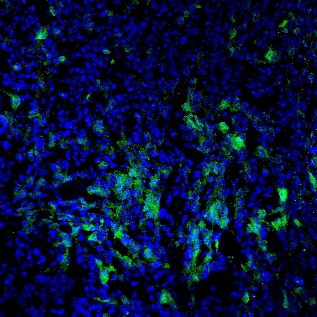
Application: Immunocytochemistry/ImmunofluorescenceSample Tested: A375 human melanoma cell lineSpecies: MouseVerified Customer | Posted 09/27/2023

-
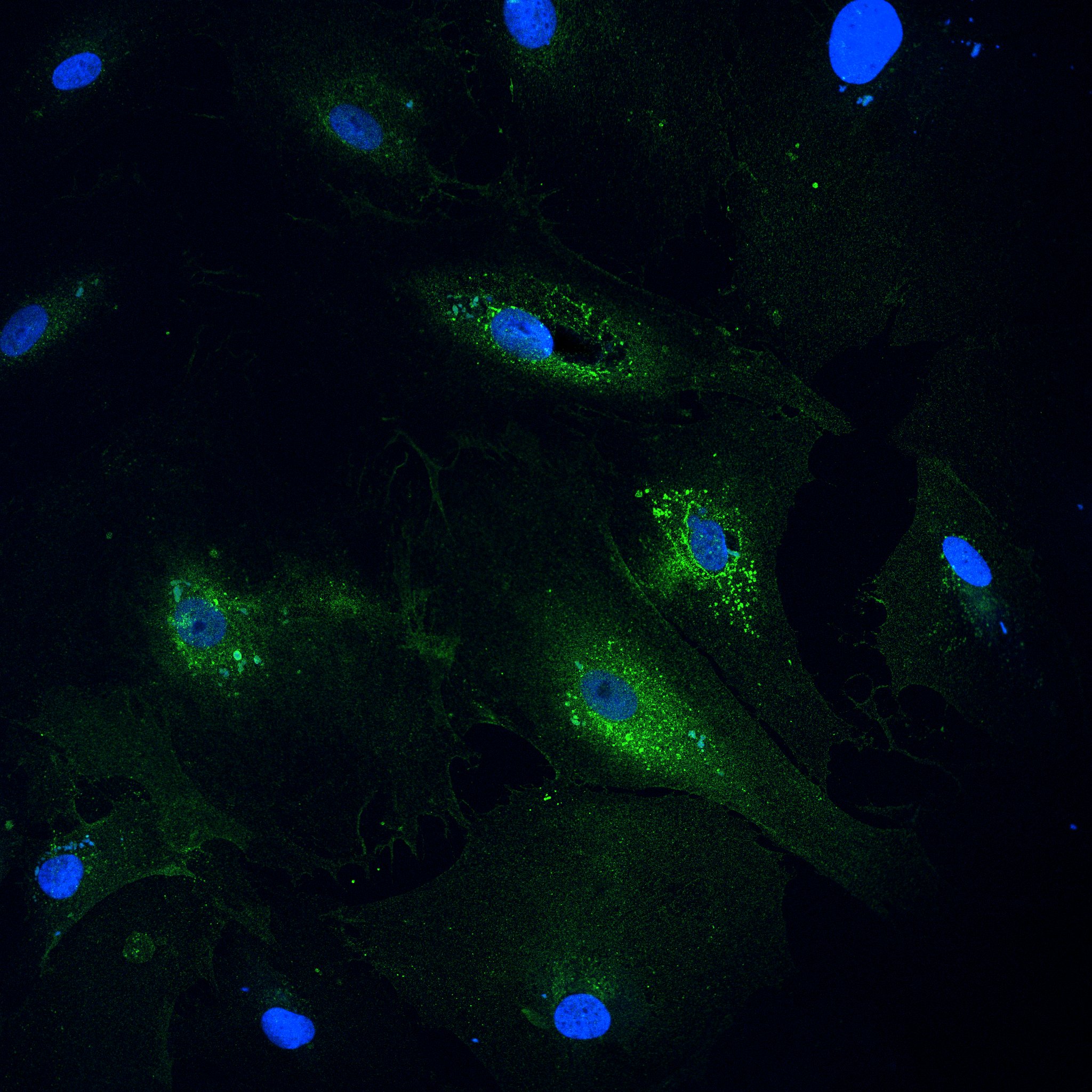
Application: ImmunocytochemistrySample Tested: primary human cardiac fibroblastsSpecies: HumanVerified Customer | Posted 08/01/2023Primary human cardiac fibroblasts stained with FAP(green, 1:100) and DAPI (blue)
Bio-Techne ResponseThis review was submitted through the legacy Novus Innovators Program, reflecting a new species or application tested on a primary antibody.
-
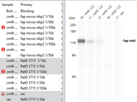
Application: Simple WesternSample Tested: Cellules musculaire lisses mésentériquesSpecies: HumanVerified Customer | Posted 08/12/2021Human Fibroblast Activation Protein alpha FAP Antibody ref AF3715 RetD Systems Polyclonal Sheep. 95kd mais peut présenter Des glycosylations d’où la taille aux alentours de 120kd! La prochaine fois: - Utiliser des concentrations de protéines >0,6mg/ml - Diluer l’anticorps secondaire sheep 1/25è au lieu du 1/10è pour éviter un peu plus le bruit de fond. - Reconstituer l’anticorps à 0,5mg/ml Au lieu de 0,2mg/ml recommandé par R et D. Efficacité validée dans conditions suivantes: Dilution anticorps FAP= 1/10è (0,2mg/ml solution stock) [CMLh]= 0,7mg/ml Dilution anticorps 2aire sheep = 1/10è
-
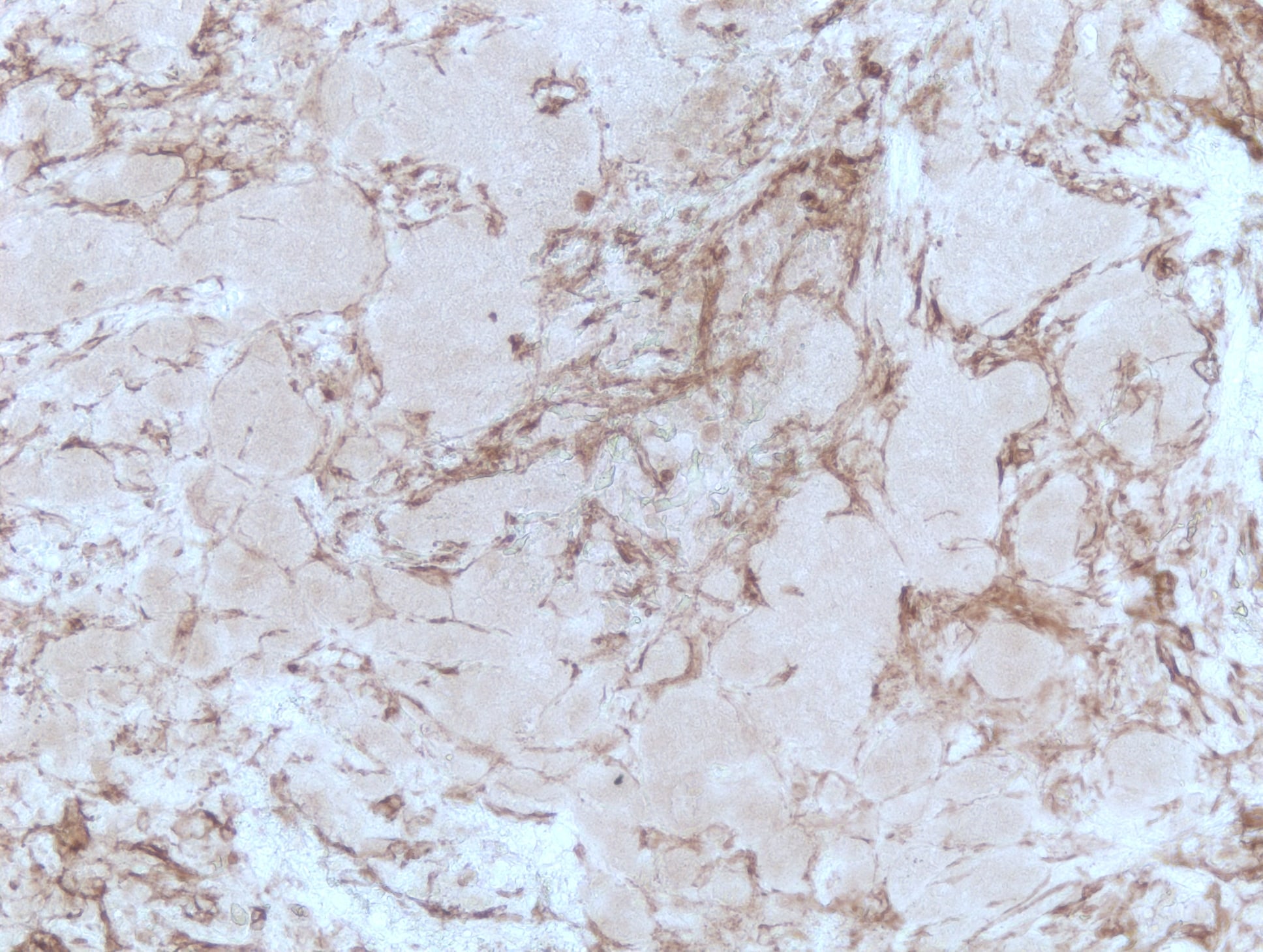
Application: Immunohistochemistry-ParaffinSample Tested: Breast tumorSpecies: HumanVerified Customer | Posted 04/15/2019FAP staining on human breast tumor FFPE tissue following HIER in Tris/EDTA buffer (pH 9)
There are no reviews that match your criteria.
Protocols
Find general support by application which include: protocols, troubleshooting, illustrated assays, videos and webinars.
- Antigen Retrieval Protocol (PIER)
- Antigen Retrieval for Frozen Sections Protocol
- Appropriate Fixation of IHC/ICC Samples
- Cellular Response to Hypoxia Protocols
- Chromogenic IHC Staining of Formalin-Fixed Paraffin-Embedded (FFPE) Tissue Protocol
- Chromogenic Immunohistochemistry Staining of Frozen Tissue
- ClariTSA™ Fluorophore Kits
- Detection & Visualization of Antibody Binding
- Fluorescent IHC Staining of Frozen Tissue Protocol
- Graphic Protocol for Heat-induced Epitope Retrieval
- Graphic Protocol for the Preparation and Fluorescent IHC Staining of Frozen Tissue Sections
- Graphic Protocol for the Preparation and Fluorescent IHC Staining of Paraffin-embedded Tissue Sections
- Graphic Protocol for the Preparation of Gelatin-coated Slides for Histological Tissue Sections
- IHC Sample Preparation (Frozen sections vs Paraffin)
- ISH-IHC Protocol for Chromogenic Detection on Formalin Fixed Paraffin Embedded (FFPE) Tissue
- Immunofluorescent IHC Staining of Formalin-Fixed Paraffin-Embedded (FFPE) Tissue Protocol
- Immunohistochemistry (IHC) and Immunocytochemistry (ICC) Protocols
- Immunohistochemistry Frozen Troubleshooting
- Immunohistochemistry Paraffin Troubleshooting
- Immunoprecipitation Protocol
- Preparing Samples for IHC/ICC Experiments
- Preventing Non-Specific Staining (Non-Specific Binding)
- Primary Antibody Selection & Optimization
- Protocol for Heat-Induced Epitope Retrieval (HIER)
- Protocol for Making a 4% Formaldehyde Solution in PBS
- Protocol for VisUCyte™ HRP Polymer Detection Reagent
- Protocol for the Preparation & Fixation of Cells on Coverslips
- Protocol for the Preparation and Chromogenic IHC Staining of Frozen Tissue Sections
- Protocol for the Preparation and Chromogenic IHC Staining of Frozen Tissue Sections - Graphic
- Protocol for the Preparation and Chromogenic IHC Staining of Paraffin-embedded Tissue Sections
- Protocol for the Preparation and Chromogenic IHC Staining of Paraffin-embedded Tissue Sections - Graphic
- Protocol for the Preparation and Fluorescent IHC Staining of Frozen Tissue Sections
- Protocol for the Preparation and Fluorescent IHC Staining of Paraffin-embedded Tissue Sections
- Protocol for the Preparation of Gelatin-coated Slides for Histological Tissue Sections
- R&D Systems Quality Control Western Blot Protocol
- TUNEL and Active Caspase-3 Detection by IHC/ICC Protocol
- The Importance of IHC/ICC Controls
- Troubleshooting Guide: Immunohistochemistry
- Troubleshooting Guide: Western Blot Figures
- Western Blot Conditions
- Western Blot Protocol
- Western Blot Protocol for Cell Lysates
- Western Blot Troubleshooting
- Western Blot Troubleshooting Guide
- View all Protocols, Troubleshooting, Illustrated assays and Webinars
Loading...