SPARC, an acronym for “secreted protein, acidic and rich in cysteine”, is also known as osteonectin or BM-40 (1-5). It is the founding member of a family of secreted matricellular proteins with similar domain structure. The 286 amino acid (aa), 43 kDa protein contains an N-terminal acidic region that binds calcium, a follistatin domain that contains Kazal-like sequences, and a C-terminal extracellular calcium (EC) binding domain with two EF-hand motifs (1-5). Crystal structure modeling shows that residues implicated in cell binding, inhibition of cell spreading, and disassembly of focal adhesions cluster on one face of SPARC, while a collagen binding epitope and an N-glycosylation site are opposite this face (6). SPARC is produced by fibroblasts, capillary endothelial cells, platelets and macrophages, especially in areas of tissue morphogenesis and remodeling (3, 7). SPARC shows context-specific effects, but generally inhibits adhesion, spreading and proliferation, and promotes collagen matrix formation (3-5). For endothelial cells, SPARC disrupts focal adhesions and binds and sequesters PDGF and VEGF (3-5). SPARC is abundantly expressed in bone, where it promotes osteoblast differentiation and inhibits adipogenesis (5, 8). SPARC is potentially cleaved by metalloproteinases, producing an angiogenic peptide that includes the copper-binding sequence KGHK (7). Paradoxically, SPARC is highly expressed in many tumor types undergoing an endothelial to mesenchymal transistion; its expression, however, mainly decreases the likelihood of metastasis and confers sensitivity to chemotherapy and radiation (4, 9-11). Stabilin-1, which is expressed on alternately activated macrophages, is the first SPARC receptor to be identified. It binds the SPARC EC domain and mediates endocytosis for degradation (12). Mature human SPARC shows 92%, 92%, 97%, 99%, 96%, and 85% aa identity with mouse, rat, canine, bovine, porcine, and chick SPARC, respectively.

Key Product Details
Validated by
Species Reactivity
Validated:
Cited:
Applications
Validated:
Cited:
Label
Antibody Source
Product Specifications
Immunogen
Ala18-Ile303
Accession # P09486
Specificity
Clonality
Host
Isotype
Scientific Data Images for Human SPARC Antibody
Detection of SPARC in MG-63 cells by Flow Cytometry
MG-63 cells were stained with Goat Anti-Human SPARC Antigen Affinity-purified Polyclonal Antibody (Catalog # AF941, filled histogram) or isotype control antibody (Catalog # AB-108-C, open histogram) followed by Allophycocyanin-conjugated Anti-Goat IgG Secondary Antibody (Catalog # F0108). To facilitate intracellular staining, cells were fixed with Flow Cytometry Fixation Buffer (Catalog # FC004) and permeabilized with Flow Cytometry Permeabilization/Wash Buffer I (Catalog # FC005). View our protocol for Staining Intracellular Molecules.
Detection of Human SPARC by Western Blot.
Western blot shows lysates of MG-63 human osteosarcoma cell line. PVDF membrane was probed with 2 µg/mL of Goat Anti-Human SPARC Antigen Affinity-purified Polyclonal Antibody (Catalog # AF941) followed by HRP-conjugated Anti-Goat IgG Secondary Antibody (HAF019). A specific band was detected for SPARC at approximately 43 kDa (as indicated). This experiment was conducted under reducing conditions and using Immunoblot Buffer Group 1.
SPARC/Osteonectin in Human Ovary.
SPARC/Osteonectin was detected in immersion fixed paraffin-embedded sections of human ovary using Goat Anti-Human SPARC/Osteonectin Antigen Affinity-purified Polyclonal Antibody (Catalog # AF941) at 3 µg/mL overnight at 4 °C. Tissue was stained using the Anti-Goat HRP-DAB Cell & Tissue Staining Kit (brown; CTS008) and counterstained with hematoxylin (blue). View our protocol for Chromogenic IHC Staining of Paraffin-embedded Tissue Sections.
Detection of Human SPARC by Simple WesternTM.
Simple Western lane view shows lysates of MG-63 human osteosarcoma cell line, loaded at 0.2 mg/mL. A specific band was detected for SPARC at approximately 57 kDa (as indicated) using 20 µg/mL of Goat Anti-Human SPARC Antigen Affinity-purified Polyclonal Antibody (Catalog # AF941) followed by 1:50 dilution of HRP-conjugated Anti-Goat IgG Secondary Antibody (HAF109). This experiment was conducted under reducing conditions and using the 12-230 kDa separation system.
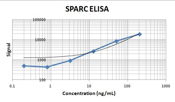
Human SPARC ELISA Standard Curve.
Recombinant Human SPARC protein was serially diluted 2-fold and captured by Mouse Anti-Human SPARC Monoclonal Antibody (Catalog # MAB941) coated on a Clear Polystyrene Microplate (DY990). Goat Anti-Human SPARC Antigen Affinity-purified Polyclonal Antibody (Catalog # AF941) was biotinylated and incubated with the protein captured on the plate. Detection of the standard curve was achieved by incubating Streptavidin-HRP (DY998) followed by Substrate Solution (DY999) and stopping the enzymatic reaction with Stop Solution (DY994).
Detection of Human SPARC by Immunocytochemistry/ Immunofluorescence
Expression of SPARC in human ovarian tissues using antibody AF941 and bs-1133R.(A) Normal human ovarian tissue using antibody AF941, (B) Benign ovarian tumor using antibody AF941, (C) High differentiation of ovarian carcinoma using antibody AF941, (D) Medium differentiation of ovarian carcinoma using antibody AF941, (E) Low differentiation of ovarian carcinoma using antibody AF941, (F) Normal human ovarian tissue using antibody bs-1133R, (G) Benign ovarian tumor using antibody bs-1133R, (H) High differentiation of ovarian carcinoma using antibody bs-1133R, (I) Medium differentiation of ovarian carcinoma using antibody bs-1133R, (J) Low differentiation of ovarian carcinoma using antibody bs-1133R (Magnification ×200). Black arrows indicate cell cytoplasm stained, red arrows indicate stroma stained. Image collected and cropped by CiteAb from the following open publication (https://pubmed.ncbi.nlm.nih.gov/22879971), licensed under a CC-BY license. Not internally tested by R&D Systems.
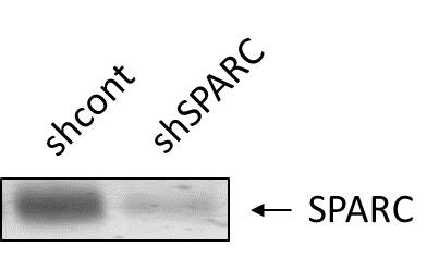
Detection of Human SPARC by Western Blot
Verification of knockdown of SPARC expression by lentivirus-mediated RNA interference. (A) GFP expression images showed shRNA delivery efficiency. (Magnification × 200). (B) SPARC protein expressions of SPARC shRNA infected cells, control shRNA infected cells and non-infected cells as measured by Western blot. (C) SPARC mRNA expressions of SPARC shRNA infected cells, control shRNA infected cells and non-infected cells as measured by q-RT-PCR. (D) SPARC protein expressions of SPARC shRNA infected cells, control shRNA infected cells and non-infected cells as measured by ICC staining (Magnification ×200). *P<0.05 versus control. Image collected and cropped by CiteAb from the following open publication (https://bmccancer.biomedcentral.com/articles/10.1186/1471-2407-12-464), licensed under a CC-BY license. Not internally tested by R&D Systems.
Detection of Human SPARC by Immunocytochemistry/ Immunofluorescence
Overview of Multi-dimensional Microscopic Molecular Profiling (MMMP).The overall MMMP approach is depicted using an example tissue section from normal human duodenum (sample #1.9.7). (a) Slides were subjected to repeated cycles of staining and imaging with fluorescent primary antibodies and DAPI. At the end of each cycle, fluorescent signal was removed by a chemical bleaching process, and slides were again imaged, before proceeding to the next round of this iterative procedure. After the final antibody stain (#15 Sma), slides were analyzed with a series of histochemical stains. (b) A set of tiling images spanning each tissue section was initially generated by the microscope system. The tiling images were then computationally ‘stitched’ together to produce a single image per staining cycle for each sample. (c) Image registration was performed to align images from the same tissue section across cycles. Mean intensities of the DAPI signal from all immuno-fluorescence images are shown from before (Unregistered) and after (Registered) the image registration procedure was completed. (d) Following registration, signal intensities from the relevant channels for each image (columns) in the MMMP series were extracted for each pixel (rows) within the tissue section and compiled into a large data matrix of in situ molecular profiles. Image collected and cropped by CiteAb from the following open publication (https://dx.plos.org/10.1371/journal.pone.0128975), licensed under a CC-BY license. Not internally tested by R&D Systems.
Detection of Human SPARC by Immunocytochemistry/ Immunofluorescence
Expression of SPARC in human ovarian tissues using antibody AF941 and bs-1133R.(A) Normal human ovarian tissue using antibody AF941, (B) Benign ovarian tumor using antibody AF941, (C) High differentiation of ovarian carcinoma using antibody AF941, (D) Medium differentiation of ovarian carcinoma using antibody AF941, (E) Low differentiation of ovarian carcinoma using antibody AF941, (F) Normal human ovarian tissue using antibody bs-1133R, (G) Benign ovarian tumor using antibody bs-1133R, (H) High differentiation of ovarian carcinoma using antibody bs-1133R, (I) Medium differentiation of ovarian carcinoma using antibody bs-1133R, (J) Low differentiation of ovarian carcinoma using antibody bs-1133R (Magnification ×200). Black arrows indicate cell cytoplasm stained, red arrows indicate stroma stained. Image collected and cropped by CiteAb from the following open publication (https://pubmed.ncbi.nlm.nih.gov/22879971), licensed under a CC-BY license. Not internally tested by R&D Systems.Applications for Human SPARC Antibody
CyTOF-ready
ELISA
This antibody functions as an ELISA detection antibody when paired with Mouse Anti-Human SPARC Monoclonal Antibody (Catalog # MAB941).
This product is intended for assay development on various assay platforms requiring antibody pairs. We recommend the Human SPARC DuoSet ELISA Kit (Catalog # DY941-05) for convenient development of a sandwich ELISA or the Human SPARC Quantikine ELISA Kit (Catalog # DSP00) for a complete optimized ELISA.
Immunohistochemistry
Sample: Immersion fixed paraffin-embedded sections of human ovary, normal human liver, and human liver cancer tissues
Intracellular Staining by Flow Cytometry
Sample: MG‑63 human osteosarcoma cell line fixed with paraformaldehyde and permeabilized with saponin
Simple Western
Sample: MG‑63 human osteosarcoma cell line
Western Blot
Sample: MG‑63 human osteosarcoma cell line
Reviewed Applications
Read 4 reviews rated 4.8 using AF941 in the following applications:
Flow Cytometry Panel Builder
Bio-Techne Knows Flow Cytometry
Save time and reduce costly mistakes by quickly finding compatible reagents using the Panel Builder Tool.
Advanced Features
- Spectra Viewer - Custom analysis of spectra from multiple fluorochromes
- Spillover Popups - Visualize the spectra of individual fluorochromes
- Antigen Density Selector - Match fluorochrome brightness with antigen density

Formulation, Preparation, and Storage
Purification
Reconstitution
Reconstitute at 0.2 mg/mL in sterile PBS. For liquid material, refer to CoA for concentration.
Formulation
*Small pack size (-SP) is supplied either lyophilized or as a 0.2 µm filtered solution in PBS.
Shipping
Stability & Storage
- 12 months from date of receipt, -20 to -70 °C as supplied.
- 1 month, 2 to 8 °C under sterile conditions after reconstitution.
- 6 months, -20 to -70 °C under sterile conditions after reconstitution.
Calculators
Background: SPARC
References
- Lankat-Buttgereit, B. et al. (1988) FEBS Lett. 236:352.
- Sweetwyne, M.T. et al. (2004) J. Histochem. Cytochem. 52:723.
- Sage, H. et al. (1989) J. Cell Biol. 109:341.
- Framson, P.E. and E.H. Sage (2004) J. Cell. Biochem. 92:679.
- Alford, A.I. and K. D. Hankenson (2006) Bone 38:749.
- Hohenester, E et al. (1997) EMBO J. 16:3778.
- Sage, E.H. et al. (2003) J. Biol. Chem. 278:37849.
- Delany, A.M. et al. (2003) Endocrinology 144:2588.
- Robert, G. et al. (2006) Cancer Res. 66:7516.
- Koblinski, J.E. et al. (2005) Cancer Res. 65:7370.
- Tai, I.T. et al. (2005) J. Clin. Invest. 115:1492.
- Kzhyshkowska, J. et al. (2006) J. Immunol. 176:5825.
Long Name
Alternate Names
Gene Symbol
UniProt
Additional SPARC Products
Product Documents for Human SPARC Antibody
Certificate of Analysis
To download a Certificate of Analysis, please enter a lot or batch number in the search box below.
Note: Certificate of Analysis not available for kit components.
Product Specific Notices for Human SPARC Antibody
For research use only
Citations for Human SPARC Antibody
 Powered by Bioz
Customer Reviews for Human SPARC Antibody (4)
Have you used Human SPARC Antibody?
Submit a review and receive an Amazon gift card!
$25/€18/£15/$25CAN/¥2500 Yen for a review with an image
$10/€7/£6/$10CAN/¥1110 Yen for a review without an image
Submit a review
Customer Images
_Figure-1(1)_fae021f5-6a37-4023-9a52-cf1445712c48.png)
-
Application: Simple WesternSample Tested: endothelial cellsSpecies: MouseVerified Customer | Posted 09/05/2024basal SPARC expression in mEC1:10 in milk-free antibody diluent

-
Application: ELISASample Tested: Serum and PlasmaSpecies: HumanVerified Customer | Posted 11/08/2019This antibody was used as detection along with the capture MAB941. It worked great as an ELISA to detect human serum and plasma samples.
-
Application: LuminexSample Tested: SerumSpecies: HumanVerified Customer | Posted 11/08/2018
-
Application: Western BlotSample Tested: Trabecular meshworkSpecies: HumanVerified Customer | Posted 01/12/2018
There are no reviews that match your criteria.
Protocols
Find general support by application which include: protocols, troubleshooting, illustrated assays, videos and webinars.
- 7-Amino Actinomycin D (7-AAD) Cell Viability Flow Cytometry Protocol
- Antigen Retrieval Protocol (PIER)
- Antigen Retrieval for Frozen Sections Protocol
- Appropriate Fixation of IHC/ICC Samples
- Cellular Response to Hypoxia Protocols
- Chromogenic IHC Staining of Formalin-Fixed Paraffin-Embedded (FFPE) Tissue Protocol
- Chromogenic Immunohistochemistry Staining of Frozen Tissue
- ClariTSA™ Fluorophore Kits
- Detection & Visualization of Antibody Binding
- ELISA Sample Preparation & Collection Guide
- ELISA Troubleshooting Guide
- Extracellular Membrane Flow Cytometry Protocol
- Flow Cytometry Protocol for Cell Surface Markers
- Flow Cytometry Protocol for Staining Membrane Associated Proteins
- Flow Cytometry Staining Protocols
- Flow Cytometry Troubleshooting Guide
- Fluorescent IHC Staining of Frozen Tissue Protocol
- Graphic Protocol for Heat-induced Epitope Retrieval
- Graphic Protocol for the Preparation and Fluorescent IHC Staining of Frozen Tissue Sections
- Graphic Protocol for the Preparation and Fluorescent IHC Staining of Paraffin-embedded Tissue Sections
- Graphic Protocol for the Preparation of Gelatin-coated Slides for Histological Tissue Sections
- How to Run an R&D Systems DuoSet ELISA
- How to Run an R&D Systems Quantikine ELISA
- How to Run an R&D Systems Quantikine™ QuicKit™ ELISA
- IHC Sample Preparation (Frozen sections vs Paraffin)
- Immunofluorescent IHC Staining of Formalin-Fixed Paraffin-Embedded (FFPE) Tissue Protocol
- Immunohistochemistry (IHC) and Immunocytochemistry (ICC) Protocols
- Immunohistochemistry Frozen Troubleshooting
- Immunohistochemistry Paraffin Troubleshooting
- Intracellular Flow Cytometry Protocol Using Alcohol (Methanol)
- Intracellular Flow Cytometry Protocol Using Detergents
- Intracellular Nuclear Staining Flow Cytometry Protocol Using Detergents
- Intracellular Staining Flow Cytometry Protocol Using Alcohol Permeabilization
- Intracellular Staining Flow Cytometry Protocol Using Detergents to Permeabilize Cells
- Preparing Samples for IHC/ICC Experiments
- Preventing Non-Specific Staining (Non-Specific Binding)
- Primary Antibody Selection & Optimization
- Propidium Iodide Cell Viability Flow Cytometry Protocol
- Protocol for Heat-Induced Epitope Retrieval (HIER)
- Protocol for Liperfluo
- Protocol for Making a 4% Formaldehyde Solution in PBS
- Protocol for VisUCyte™ HRP Polymer Detection Reagent
- Protocol for the Characterization of Human Th22 Cells
- Protocol for the Characterization of Human Th9 Cells
- Protocol for the Preparation & Fixation of Cells on Coverslips
- Protocol for the Preparation and Chromogenic IHC Staining of Frozen Tissue Sections
- Protocol for the Preparation and Chromogenic IHC Staining of Frozen Tissue Sections - Graphic
- Protocol for the Preparation and Chromogenic IHC Staining of Paraffin-embedded Tissue Sections
- Protocol for the Preparation and Chromogenic IHC Staining of Paraffin-embedded Tissue Sections - Graphic
- Protocol for the Preparation and Fluorescent IHC Staining of Frozen Tissue Sections
- Protocol for the Preparation and Fluorescent IHC Staining of Paraffin-embedded Tissue Sections
- Protocol for the Preparation of Gelatin-coated Slides for Histological Tissue Sections
- Protocol: Annexin V and PI Staining by Flow Cytometry
- Protocol: Annexin V and PI Staining for Apoptosis by Flow Cytometry
- Quantikine HS ELISA Kit Assay Principle, Alkaline Phosphatase
- Quantikine HS ELISA Kit Principle, Streptavidin-HRP Polymer
- R&D Systems Quality Control Western Blot Protocol
- Sandwich ELISA (Colorimetric) – Biotin/Streptavidin Detection Protocol
- Sandwich ELISA (Colorimetric) – Direct Detection Protocol
- TUNEL and Active Caspase-3 Detection by IHC/ICC Protocol
- The Importance of IHC/ICC Controls
- Troubleshooting Guide: ELISA
- Troubleshooting Guide: Fluorokine Flow Cytometry Kits
- Troubleshooting Guide: Immunohistochemistry
- Troubleshooting Guide: Western Blot Figures
- Western Blot Conditions
- Western Blot Protocol
- Western Blot Protocol for Cell Lysates
- Western Blot Troubleshooting
- Western Blot Troubleshooting Guide
- View all Protocols, Troubleshooting, Illustrated assays and Webinars
Associated Pathways
