
Key Product Details
Species Reactivity
Human
Applications
Western Blot, Flow Cytometry, CyTOF-ready
Label
Unconjugated
Antibody Source
Monoclonal Mouse IgG2A Clone # 528903
Loading...
Product Specifications
Immunogen
Mouse myeloma cell line NS0-derived human VSIG4 protein
Arg20-Pro283
Accession # Q9Y279
Arg20-Pro283
Accession # Q9Y279
Specificity
Detects human VSIG4 in direct ELISAs.
Clonality
Monoclonal
Host
Mouse
Isotype
IgG2A
Scientific Data Images for Human VSIG4 Antibody (528903)
Detection of Human VSIG4 by Western Blot.
Western blot shows lysates of HEK293T human embryonic kidney cell line either mock transfected or transfected with human VSIG4. PVDF membrane was probed with 2 µg/mL of Mouse Anti-Human VSIG4 Monoclonal Antibody (Catalog # MAB4646) followed by HRP-conjugated Anti-Mouse IgG Secondary Antibody (HAF018). A specific band was detected for VSIG4 at approximately 75 kDa (as indicated). This experiment was conducted under reducing conditions and using Western Blot Buffer Group 1.
Detection of VSIG4 in Human Macrophages by Flow Cytometry
Human CD14+ monocytes were positively selected from PBMC (MAGH105) and cultured for 7 days with 10% Human AB serum, 50 ng/ml M-CSF (216-MC), and 10μM hydrocortisone (4093). Cells were harvested and stained with either (A) Mouse anti-Human VSIG4 (Catalog # MAB4646) or (B) Isotype control antibody (MAB003) followed by Allophycocyanin-conjugated anti-Mouse IgG Secondary Antibody (F0101B) and Mouse anti-Human CD11b Alexa Fluor® 488-conjugated Monoclonal Antibody (FAB16991G). Staining was performed using our Staining Membrane-Associated Proteins protocol.Applications for Human VSIG4 Antibody (528903)
Application
Recommended Usage
CyTOF-ready
Ready to be labeled using established conjugation methods. No BSA or other carrier proteins that could interfere with conjugation.
Flow Cytometry
0.25 µg/106 cells
Sample: Human PBMC-derived macrophages
Sample: Human PBMC-derived macrophages
Western Blot
2 µg/mL
Sample: HEK293T human embryonic kidney cell line transfected with human VSIG4
Sample: HEK293T human embryonic kidney cell line transfected with human VSIG4
Reviewed Applications
Read 1 review rated 5 using MAB4646 in the following applications:
Flow Cytometry Panel Builder
Bio-Techne Knows Flow Cytometry
Save time and reduce costly mistakes by quickly finding compatible reagents using the Panel Builder Tool.
Advanced Features
- Spectra Viewer - Custom analysis of spectra from multiple fluorochromes
- Spillover Popups - Visualize the spectra of individual fluorochromes
- Antigen Density Selector - Match fluorochrome brightness with antigen density

Formulation, Preparation, and Storage
Purification
Protein A or G purified from hybridoma culture supernatant
Reconstitution
Reconstitute at 0.5 mg/mL in sterile PBS. For liquid material, refer to CoA for concentration.
Loading...
Formulation
Lyophilized from a 0.2 μm filtered solution in PBS with Trehalose. *Small pack size (SP) is supplied either lyophilized or as a 0.2 µm filtered solution in PBS.
Shipping
Lyophilized product is shipped at ambient temperature. Liquid small pack size (-SP) is shipped with polar packs. Upon receipt, store immediately at the temperature recommended below.
Stability & Storage
Use a manual defrost freezer and avoid repeated freeze-thaw cycles.
- 12 months from date of receipt, -20 to -70 °C as supplied.
- 1 month, 2 to 8 °C under sterile conditions after reconstitution.
- 6 months, -20 to -70 °C under sterile conditions after reconstitution.
Calculators
Background: VSIG4
References
- He, J.Q. et al. (2008) Mol. Immunol. 4041.
- Vogt, L. et al. (2006) J. Clin. Invest. 116:2817.
- Langnaese, K. et al. (2000) Biochim. Biophys. Acta 1492:522.
- Helmy, K. et al. (2006) Cell 124:915.
- Entrez protein Accession # EAX05393, NP_001093901, CAI42052, CAI4205, EAX05394.
- Tanaka, M. et al. (2008) Clin. Exp. Immunol. 154:38.
- Lee, M.Y. et al. (2006) J. Leukoc. Biol. 80:922.
- Gorgani, N.N. et al. (2008) J. Immunol. 181:7902.
- Walker, M.G. (2002) Biochim. Biophys. Acta 1574:387.
- Wiesmann, C. et al. (2006) Nature 444:217.
- Katschke, K.J. et al. (2007) J. Exp. Med. 204:1319.
- Small, A.G. et al. (2016) Swiss Med Wkly. 5:146.
- Roh J. et al. (2017) Oncotarget. 8:58122.
- Kim K.H. et al. (2016) Autophagy. 12:1647.
Long Name
V-Set and Immunoglobulin Domain Containing 4
Alternate Names
Z39IG
Gene Symbol
VSIG4
UniProt
Additional VSIG4 Products
Product Documents for Human VSIG4 Antibody (528903)
Certificate of Analysis
To download a Certificate of Analysis, please enter a lot or batch number in the search box below.
Note: Certificate of Analysis not available for kit components.
Product Specific Notices for Human VSIG4 Antibody (528903)
For research use only
Related Research Areas
Customer Reviews for Human VSIG4 Antibody (528903) (1)
5 out of 5
1 Customer Rating
Have you used Human VSIG4 Antibody (528903)?
Submit a review and receive an Amazon gift card!
$25/€18/£15/$25CAN/¥2500 Yen for a review with an image
$10/€7/£6/$10CAN/¥1110 Yen for a review without an image
Submit a review
Customer Images
Showing
1
-
1 的
1 review
Showing All
Filter By:
-
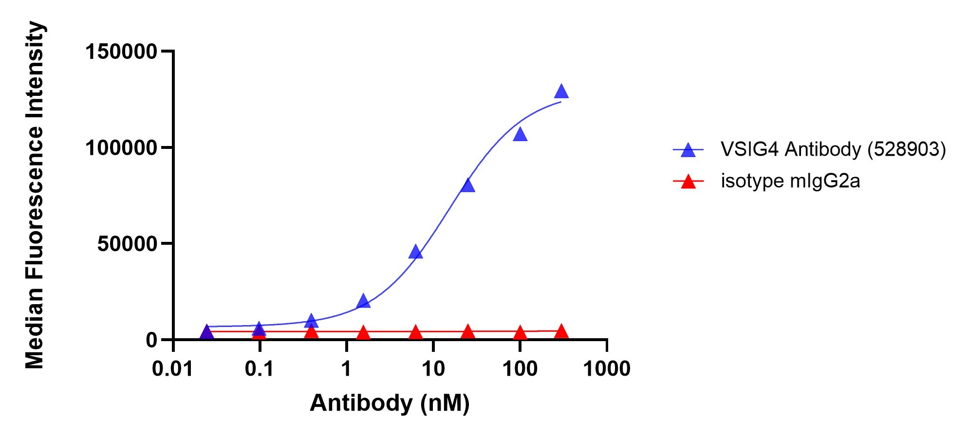
Application: Flow CytometrySample Tested: macrophagesSpecies: HumanVerified Customer | Posted 06/20/2026Binding of VSIG4 in human monocyte-derived macrophages. The cells were incubated with a titration of the human VSIG4 antibody or isotype control for 30 minutes on ice, followed by a secondary a-mouse IgG APCThe antibody was tested for binding to VSIG4 expressed in human monocyte-derived macrophages. The antibody was titrated starting at 100 nM and detected with a secondary a-mouse IgG APC

There are no reviews that match your criteria.
Protocols
Find general support by application which include: protocols, troubleshooting, illustrated assays, videos and webinars.
- 7-Amino Actinomycin D (7-AAD) Cell Viability Flow Cytometry Protocol
- Cellular Response to Hypoxia Protocols
- Extracellular Membrane Flow Cytometry Protocol
- Flow Cytometry Protocol for Cell Surface Markers
- Flow Cytometry Protocol for Staining Membrane Associated Proteins
- Flow Cytometry Staining Protocols
- Flow Cytometry Troubleshooting Guide
- Intracellular Flow Cytometry Protocol Using Alcohol (Methanol)
- Intracellular Flow Cytometry Protocol Using Detergents
- Intracellular Nuclear Staining Flow Cytometry Protocol Using Detergents
- Intracellular Staining Flow Cytometry Protocol Using Alcohol Permeabilization
- Intracellular Staining Flow Cytometry Protocol Using Detergents to Permeabilize Cells
- Propidium Iodide Cell Viability Flow Cytometry Protocol
- Protocol for Liperfluo
- Protocol for the Characterization of Human Th22 Cells
- Protocol for the Characterization of Human Th9 Cells
- Protocol: Annexin V and PI Staining by Flow Cytometry
- Protocol: Annexin V and PI Staining for Apoptosis by Flow Cytometry
- R&D Systems Quality Control Western Blot Protocol
- Troubleshooting Guide: Fluorokine Flow Cytometry Kits
- Troubleshooting Guide: Western Blot Figures
- Western Blot Conditions
- Western Blot Protocol
- Western Blot Protocol for Cell Lysates
- Western Blot Troubleshooting
- Western Blot Troubleshooting Guide
- View all Protocols, Troubleshooting, Illustrated assays and Webinars
Loading...