IKK beta Antibody (10A9B6) - BSA Free
Novus Biologicals | Catalog # NB100-56513
Key Product Details
Species Reactivity
Validated:
Human, Mouse
Cited:
Human, Mouse
Applications
Validated:
Immunohistochemistry, Immunohistochemistry-Frozen, Western Blot, Flow Cytometry, Flow (Intracellular), Immunocytochemistry/ Immunofluorescence, Simple Western, Immunoprecipitation, Gel Super Shift Assays
Cited:
Western Blot, Immunocytochemistry/ Immunofluorescence, EMSA
Label
Unconjugated
Antibody Source
Monoclonal Mouse IgG1 kappa Clone # 10A9B6
Format
BSA Free
Loading...
Product Specifications
Immunogen
Full-length human IKKb/IKK2 recombinant protein was used as immunogen (NP_001547).
Clonality
Monoclonal
Host
Mouse
Isotype
IgG1 kappa
Scientific Data Images for IKK beta Antibody (10A9B6) - BSA Free
![Western Blot: IKK beta Antibody (10A9B6)BSA Free [NB100-56513]](https://resources.rndsystems.com/images/products/IKK-beta-Antibody-10A9B6-Western-Blot-NB100-56513-img0005.jpg "Western Blot: IKK beta Antibody (10A9B6)BSA Free [NB100-56513]")
Western Blot: IKK beta Antibody (10A9B6)BSA Free [NB100-56513]
Western Blot: IKK beta Antibody (10A9B6) [NB100-56513] - IKK beta in Daudi cell lysate using IKK beta antibody at 5 ug/ml.![Immunohistochemistry-Frozen: IKK beta Antibody (10A9B6) - BSA Free [NB100-56513]](https://resources.rndsystems.com/images/products/IKK-beta-Antibody-10A9B6-Immunohistochemistry-Frozen-NB100-56513-img0013.jpg "Immunohistochemistry-Frozen: IKK beta Antibody (10A9B6) - BSA Free [NB100-56513]")
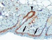
Immunohistochemistry-Frozen: IKK beta Antibody (10A9B6) - BSA Free [NB100-56513]
Immunohistochemistry-Frozen: IKK beta Antibody (10A9B6) [NB100-56513] - Mouse skin tissue section stained with IKK beta antibody. IHC-Fr image submitted by a verified customer review.![Flow (Intracellular): IKK beta Antibody (10A9B6) - BSA Free [NB100-56513]](https://resources.rndsystems.com/images/products/IKK-beta-Antibody-10A9B6-Flow-Intracellular-NB100-56513-img0011.jpg "Flow (Intracellular): IKK beta Antibody (10A9B6) - BSA Free [NB100-56513]")
Flow (Intracellular): IKK beta Antibody (10A9B6) - BSA Free [NB100-56513]
Flow (Intracellular): IKK beta Antibody (10A9B6) [NB100-56513] - Analysis using the Alexa Fluor (R) 488 conjugate. Staining of IKK in HEK 293 cells using 0.1 ug of this antibody. Green histogram represents the isotype control, red represents the IKK antibody. Image using the Azide Free format of this antibody.![Simple Western: IKK beta Antibody (10A9B6)BSA Free [NB100-56513]](https://resources.rndsystems.com/images/products/IKK-beta-Antibody-10A9B6-Simple-Western-NB100-56513-img0010.jpg "Simple Western: IKK beta Antibody (10A9B6)BSA Free [NB100-56513]")
Simple Western: IKK beta Antibody (10A9B6)BSA Free [NB100-56513]
Simple Western: IKK beta Antibody (10A9B6) [NB100-56513] - Analysis using Azide Free version of NB100-56513. Lane view shows a specific band for IKK beta in 1.0 mg/ml of HeLa lysate. This experiment was performed under reducing conditions using the 12-230 kDa separation system. *Non-specific interaction with the![Immunoprecipitation: IKK beta Antibody (10A9B6) - BSA Free [NB100-56513]](https://resources.rndsystems.com/images/products/IKK-beta-Antibody-10A9B6-Western-Blot-NB100-56513-img0012.jpg "Immunoprecipitation: IKK beta Antibody (10A9B6) - BSA Free [NB100-56513]")
Immunoprecipitation: IKK beta Antibody (10A9B6) - BSA Free [NB100-56513]
Immunoprecipitation: IKK beta Antibody (10A9B6) [NB100-56513] - Antibody was used to immunoprecipitate IKKb from 1x10^6 Daudi cells and the immuno-precipitated protein was detected by western blotting using NB100-56509. IKKb was detected as a protein of ~87 kD. Image using the Azide Free format of this antibody.Applications for IKK beta Antibody (10A9B6) - BSA Free
Application
Recommended Usage
Flow Cytometry
1:10 - 1:1000
Gel Super Shift Assays
reported in scientific literature (PMID 19662631)
Immunocytochemistry/ Immunofluorescence
reported in scientific literature (PMID 24825920)
Immunohistochemistry-Frozen
reported by customer review
Immunoprecipitation
2 - 5 ug / 10^6 cells
Simple Western
1:12.5
Western Blot
2 - 5 ug/mL
Application Notes
An approx. 87 kDa band should be observed. In Simple Western only 10 - 15 uL of the recommended dilution is used per data point.
See Simple Western Antibody Database for Simple Western validation: Tested in HeLa lysate 1.0 mg/mL, separated by Size, antibody dilution of 1:12.5, apparent MW was 89 kDa. Separated by Size-Wes, Sally Sue/Peggy Sue.
See Simple Western Antibody Database for Simple Western validation: Tested in HeLa lysate 1.0 mg/mL, separated by Size, antibody dilution of 1:12.5, apparent MW was 89 kDa. Separated by Size-Wes, Sally Sue/Peggy Sue.
Reviewed Applications
Read 1 review rated 5 using NB100-56513 in the following applications:
Flow Cytometry Panel Builder
Bio-Techne Knows Flow Cytometry
Save time and reduce costly mistakes by quickly finding compatible reagents using the Panel Builder Tool.
Advanced Features
- Spectra Viewer - Custom analysis of spectra from multiple fluorochromes
- Spillover Popups - Visualize the spectra of individual fluorochromes
- Antigen Density Selector - Match fluorochrome brightness with antigen density

Formulation, Preparation, and Storage
Purification
Protein G purified
Formulation
PBS
Format
BSA Free
Preservative
0.05% Sodium Azide
Concentration
1.0 mg/ml
Shipping
The product is shipped with polar packs. Upon receipt, store it immediately at the temperature recommended below.
Stability & Storage
Store at 4C short term. Aliquot and store at -20C long term. Avoid freeze-thaw cycles.
Background: IKK beta
Long Name
IkB Kinase beta
Alternate Names
IkBKB, IKK2, NFKBIKB
Gene Symbol
IKBKB
UniProt
Additional IKK beta Products
Product Documents for IKK beta Antibody (10A9B6) - BSA Free
Certificate of Analysis
To download a Certificate of Analysis, please enter a lot or batch number in the search box below.
Product Specific Notices for IKK beta Antibody (10A9B6) - BSA Free
This product is for research use only and is not approved for use in humans or in clinical diagnosis. Primary Antibodies are guaranteed for 1 year from date of receipt.
Citations for IKK beta Antibody (10A9B6) - BSA Free
 Powered by Bioz
Customer Reviews for IKK beta Antibody (10A9B6) - BSA Free (1)
5 out of 5
1 Customer Rating
Have you used IKK beta Antibody (10A9B6) - BSA Free?
Submit a review and receive an Amazon gift card!
$25/€18/£15/$25CAN/¥2500 Yen for a review with an image
$10/€7/£6/$10CAN/¥1110 Yen for a review without an image
Submit a review
Customer Images
Showing
1
-
1 的
1 review
Showing All
Filter By:
-
Application: Immunohistochemistry-FrozenSample Tested: mouse skinSpecies: MouseVerified Customer | Posted 08/17/2021IKK beta Antibody, mouse skin
 Bio-Techne ResponseThis review was submitted through the legacy Novus Innovators Program, reflecting a new species or application tested on a primary antibody.
There are no reviews that match your criteria.
Protocols
Find general support by application which include: protocols, troubleshooting, illustrated assays, videos and webinars.
- 7-Amino Actinomycin D (7-AAD) Cell Viability Flow Cytometry Protocol
- Antigen Retrieval Protocol (PIER)
- Antigen Retrieval for Frozen Sections Protocol
- Appropriate Fixation of IHC/ICC Samples
- Cellular Response to Hypoxia Protocols
- Chromogenic IHC Staining of Formalin-Fixed Paraffin-Embedded (FFPE) Tissue Protocol
- Chromogenic Immunohistochemistry Staining of Frozen Tissue
- ClariTSA™ Fluorophore Kits
- Detection & Visualization of Antibody Binding
- Extracellular Membrane Flow Cytometry Protocol
- Flow Cytometry Protocol for Cell Surface Markers
- Flow Cytometry Protocol for Staining Membrane Associated Proteins
- Flow Cytometry Staining Protocols
- Flow Cytometry Troubleshooting Guide
- Fluorescent IHC Staining of Frozen Tissue Protocol
- Graphic Protocol for Heat-induced Epitope Retrieval
- Graphic Protocol for the Preparation and Fluorescent IHC Staining of Frozen Tissue Sections
- Graphic Protocol for the Preparation and Fluorescent IHC Staining of Paraffin-embedded Tissue Sections
- Graphic Protocol for the Preparation of Gelatin-coated Slides for Histological Tissue Sections
- ICC Cell Smear Protocol for Suspension Cells
- ICC Immunocytochemistry Protocol Videos
- ICC for Adherent Cells
- IHC Sample Preparation (Frozen sections vs Paraffin)
- Immunocytochemistry (ICC) Protocol
- Immunocytochemistry Troubleshooting
- Immunofluorescence of Organoids Embedded in Cultrex Basement Membrane Extract
- Immunofluorescent IHC Staining of Formalin-Fixed Paraffin-Embedded (FFPE) Tissue Protocol
- Immunohistochemistry (IHC) and Immunocytochemistry (ICC) Protocols
- Immunohistochemistry Frozen Troubleshooting
- Immunohistochemistry Paraffin Troubleshooting
- Immunoprecipitation Protocol
- Intracellular Flow Cytometry Protocol Using Alcohol (Methanol)
- Intracellular Flow Cytometry Protocol Using Detergents
- Intracellular Nuclear Staining Flow Cytometry Protocol Using Detergents
- Intracellular Staining Flow Cytometry Protocol Using Alcohol Permeabilization
- Intracellular Staining Flow Cytometry Protocol Using Detergents to Permeabilize Cells
- Preparing Samples for IHC/ICC Experiments
- Preventing Non-Specific Staining (Non-Specific Binding)
- Primary Antibody Selection & Optimization
- Propidium Iodide Cell Viability Flow Cytometry Protocol
- Protocol for Heat-Induced Epitope Retrieval (HIER)
- Protocol for Liperfluo
- Protocol for Making a 4% Formaldehyde Solution in PBS
- Protocol for VisUCyte™ HRP Polymer Detection Reagent
- Protocol for the Characterization of Human Th22 Cells
- Protocol for the Characterization of Human Th9 Cells
- Protocol for the Fluorescent ICC Staining of Cell Smears - Graphic
- Protocol for the Fluorescent ICC Staining of Cultured Cells on Coverslips - Graphic
- Protocol for the Preparation & Fixation of Cells on Coverslips
- Protocol for the Preparation and Chromogenic IHC Staining of Frozen Tissue Sections
- Protocol for the Preparation and Chromogenic IHC Staining of Frozen Tissue Sections - Graphic
- Protocol for the Preparation and Chromogenic IHC Staining of Paraffin-embedded Tissue Sections
- Protocol for the Preparation and Chromogenic IHC Staining of Paraffin-embedded Tissue Sections - Graphic
- Protocol for the Preparation and Fluorescent ICC Staining of Cells on Coverslips
- Protocol for the Preparation and Fluorescent ICC Staining of Non-adherent Cells
- Protocol for the Preparation and Fluorescent ICC Staining of Stem Cells on Coverslips
- Protocol for the Preparation and Fluorescent IHC Staining of Frozen Tissue Sections
- Protocol for the Preparation and Fluorescent IHC Staining of Paraffin-embedded Tissue Sections
- Protocol for the Preparation of Gelatin-coated Slides for Histological Tissue Sections
- Protocol for the Preparation of a Cell Smear for Non-adherent Cell ICC - Graphic
- Protocol: Annexin V and PI Staining by Flow Cytometry
- Protocol: Annexin V and PI Staining for Apoptosis by Flow Cytometry
- R&D Systems Quality Control Western Blot Protocol
- TUNEL and Active Caspase-3 Detection by IHC/ICC Protocol
- The Importance of IHC/ICC Controls
- Troubleshooting Guide: Fluorokine Flow Cytometry Kits
- Troubleshooting Guide: Immunohistochemistry
- Troubleshooting Guide: Western Blot Figures
- Western Blot Conditions
- Western Blot Protocol
- Western Blot Protocol for Cell Lysates
- Western Blot Troubleshooting
- Western Blot Troubleshooting Guide
- View all Protocols, Troubleshooting, Illustrated assays and Webinars
Loading...
Associated Pathways


Notch Signaling Pathways
 Pathogen or Damage-activated C-Type Lectin Receptor Signaling Pathways
 Toll-Like Receptor Signaling Pathways
