Ki67/MKI67 Antibody (8D5) - BSA Free
Novus Biologicals | Catalog # NBP2-22112
Key Product Details
Validated by
Knockout/Knockdown
Species Reactivity
Validated:
Human, Mouse, Rabbit
Cited:
Human, Mouse, Rabbit
Applications
Validated:
Knockout Validated, Immunohistochemistry, Immunohistochemistry-Paraffin, Immunohistochemistry-Frozen, Western Blot, ELISA, Immunocytochemistry/ Immunofluorescence
Cited:
Immunohistochemistry, Immunohistochemistry-Paraffin, Immunohistochemistry-Frozen, Western Blot, Immunocytochemistry/ Immunofluorescence, IF/IHC
Label
Unconjugated
Antibody Source
Monoclonal Mouse IgG1 Clone # 8D5
Format
BSA Free
Loading...
Product Specifications
Immunogen
The immunogen for this KI67/MKI67 Antibody (8D5) was made using a synthetic peptide from the internal region of Human KI67/MKI67 (amino acids: 1100-1113, 1222-1235 and 3 more spots); sequence: CEDLAGFKELFQTPG [Uniprot: P46013].
Reactivity Notes
Use in Rabbit reported in scientific literature (PMID:33691202). Human reactivity reported in the scientific literature (PMID: 23777661). Rat reactivity reported in scientific literature (PMID: 23447644). Porcine reactivity reported in scientific literature (PMID: 27046485).
Localization
Nuclear
Marker
Proliferation Marker
Clonality
Monoclonal
Host
Mouse
Isotype
IgG1
Theoretical MW
359 kDa.
Disclaimer note: The observed molecular weight of the protein may vary from the listed predicted molecular weight due to post translational modifications, post translation cleavages, relative charges, and other experimental factors.
Disclaimer note: The observed molecular weight of the protein may vary from the listed predicted molecular weight due to post translational modifications, post translation cleavages, relative charges, and other experimental factors.
Scientific Data Images for Ki67/MKI67 Antibody (8D5) - BSA Free
![Immunohistochemistry-Paraffin: Ki67/MKI67 Antibody (8D5) - BSA Free [NBP2-22112]](https://resources.rndsystems.com/images/products/Ki-67-MKI67-Antibody-8D5-Immunohistochemistry-Paraffin-NBP2-22112-img0002.jpg "Immunohistochemistry-Paraffin: Ki67/MKI67 Antibody (8D5) - BSA Free [NBP2-22112]")
Immunohistochemistry-Paraffin: Ki67/MKI67 Antibody (8D5) - BSA Free [NBP2-22112]
Immunohistochemistry-Paraffin: Ki67/MKI67 Antibody (8D5) [NBP2-22112] - Analysis of paraffin-embedded lung cancer (left) and rectal cancer (right) using Ki67 mouse mAb with DAB staining.![Immunocytochemistry/ Immunofluorescence: Ki67/MKI67 Antibody (8D5) - BSA Free [NBP2-22112]](https://resources.rndsystems.com/images/products/Ki-67-MKI67-Antibody-8D5-Immunocytochemistry-Immunofluorescence-NBP2-22112-img0006.jpg "Immunocytochemistry/ Immunofluorescence: Ki67/MKI67 Antibody (8D5) - BSA Free [NBP2-22112]")
Immunocytochemistry/ Immunofluorescence: Ki67/MKI67 Antibody (8D5) - BSA Free [NBP2-22112]
Immunocytochemistry/Immunofluorescence: Ki67/MKI67 Antibody (8D5) [NBP2-22112] - A549 human alveolar adenocarcinoma cell line, fixation with PFA 4%, blocking (PBS, 1% BSA, 0.1% Tween), primary antibody: 1:100 in blocking buffer, O/N. Secondary antibody: AlexaFluor 594, 1 hour, RT. Counterstained with DAPI. Imaged with a fluorescence microscope. This image was submitted via customer review.![Knockout Validated: Ki67/MKI67 Antibody (8D5) - BSA Free [NBP2-22112]](https://resources.rndsystems.com/images/products/Ki67-MKI67-Antibody-8D5-Knockout-Validated-NBP2-22112-img0009.jpg "Simple Western: Ki67/MKI67 Antibody (8D5) - BSA Free [NBP2-22112]")
Simple Western: Ki67/MKI67 Antibody (8D5) - BSA Free [NBP2-22112]
Simple Western: Ki67/MKI67 Antibody (8D5) [NBP2-22112] - Detection of Ki67/MKI67 by Simple WesternTM. Simple Western lane view shows lysates of HeLa parental cell line and Ki67 knockout (KO) HeLa cell line. A specific band was detected for Ki67/MKI67 at approximately 317 kDa (as indicated) in the parental cell line, but is not detectable in the knockout HeLa cell line using 1:100 of Mouse Anti-Ki67/MKI67 Monoclonal Antibody (Catalog # NBP2-22112). GAPDH is shown as a loading control. This experiment was conducted under reducing conditions and using the 12-230 kDa separation system.![Immunocytochemistry/ Immunofluorescence: Ki67/MKI67 Antibody (8D5) - BSA Free [NBP2-22112]](https://resources.rndsystems.com/images/products/Ki67-MKI67-Antibody-8D5-Immunocytochemistry-Immunofluorescence-NBP2-22112-img0008.jpg "Immunocytochemistry/ Immunofluorescence: Ki67/MKI67 Antibody (8D5) - BSA Free [NBP2-22112]")
Immunocytochemistry/ Immunofluorescence: Ki67/MKI67 Antibody (8D5) - BSA Free [NBP2-22112]
Immunocytochemistry/Immunofluorescence: Ki67/MKI67 Antibody (8D5) [NBP2-22112] - NIH3T3 cells were fixed in 4% paraformaldehyde for 10 minutes and permeabilized in 0.5% Triton X-100 in PBS for 5 minutes. The cells were incubated with anti- NBP2-22112 at 2 ug/ml overnight at 4C and detected with an anti-mouse Dylight 488 (Green) at a 1:1000 dilution for 60 minutes. Nuclei were counterstained with DAPI (Blue). Cells were imaged using a 100X objective and digitally deconvolved.![Immunocytochemistry/ Immunofluorescence: Ki67/MKI67 Antibody (8D5) - BSA Free [NBP2-22112]](https://resources.rndsystems.com/images/products/Ki67-MKI67-Antibody-8D5-Immunocytochemistry-Immunofluorescence-NBP2-22112-img0007.jpg "Immunocytochemistry/ Immunofluorescence: Ki67/MKI67 Antibody (8D5) - BSA Free [NBP2-22112]")
Immunocytochemistry/ Immunofluorescence: Ki67/MKI67 Antibody (8D5) - BSA Free [NBP2-22112]
Immunocytochemistry/Immunofluorescence: Ki67/MKI67 Antibody (8D5) [NBP2-22112] - A431 cells were fixed in 4% paraformaldehyde for 10 minutes and permeabilized in 0.5% Triton X-100 in PBS for 5 minutes. The cells were incubated with anti- NBP2-22112 at 2 ug/ml overnight at 4C and detected with an anti-mouse Dylight 488 (Green) at a 1:1000 dilution for 60 minutes. Nuclei were counterstained with DAPI (Blue). Cells were imaged using a 100X objective and digitally deconvolved.![Western Blot: Ki67/MKI67 Antibody (8D5)BSA Free [NBP2-22112]](https://resources.rndsystems.com/images/products/Ki-67-MKI67-Antibody-8D5-Western-Blot-NBP2-22112-img0001.jpg "Western Blot: Ki67/MKI67 Antibody (8D5)BSA Free [NBP2-22112]")
Western Blot: Ki67/MKI67 Antibody (8D5)BSA Free [NBP2-22112]
Western Blot: Ki67/MKI67 Antibody (8D5) [NBP2-22112] - Analysis using Ki67 mouse mAb against Hela (1), MCF-7 (2) and Raji (3) cell lysate.![Immunocytochemistry/ Immunofluorescence: Ki67/MKI67 Antibody (8D5) - BSA Free [NBP2-22112]](https://resources.rndsystems.com/images/products/Ki67-MKI67-Antibody-8D5-Immunocytochemistry-Immunofluorescence-NBP2-22112-img0011.jpg "Immunocytochemistry/ Immunofluorescence: Ki67/MKI67 Antibody (8D5) - BSA Free [NBP2-22112]")
Immunocytochemistry/ Immunofluorescence: Ki67/MKI67 Antibody (8D5) - BSA Free [NBP2-22112]
Immunocytochemistry/Immunofluorescence: Ki67/MKI67 Antibody (8D5) [NBP2-22112] - U-251 MG cells were fixed in 4% paraformaldehyde for 10 minutes and permeabilized in 0.5% Triton X-100 in PBS for 5 minutes. The cells were incubated with anti-Ki67/MKI67 Antibody [8D5] conjugated to FITC (NBP2-22112F) at 5 ug/ml for 1 hour at room temperature. Nuclei were counterstained with DAPI (Blue). Cells were imaged using a 100X objective and digitally deconvolved.![Immunocytochemistry/ Immunofluorescence: Ki67/MKI67 Antibody (8D5) - BSA Free [NBP2-22112]](https://resources.rndsystems.com/images/products/Ki-67-MKI67-Antibody-8D5-Immunocytochemistry-Immunofluorescence-NBP2-22112-img0005.jpg "Immunocytochemistry/ Immunofluorescence: Ki67/MKI67 Antibody (8D5) - BSA Free [NBP2-22112]")
Immunocytochemistry/ Immunofluorescence: Ki67/MKI67 Antibody (8D5) - BSA Free [NBP2-22112]
Immunocytochemistry/Immunofluorescence: Ki67/MKI67 Antibody (8D5) [NBP2-22112] - HeLa cells were fixed for 10 minutes using 10% formalin and then permeabilized for 5 minutes using 1X PBS + 0.5% Triton X-100. The cells were incubated with anti-Ki-67 (8D5) conjugated to Alexa Fluor 488 [NBP2-22112AF488] at 10ug/ml for 1 hour at room temperature. Nuclei were counterstained with DAPI (Blue). Cells were imaged using a 40X objective.![Immunocytochemistry/ Immunofluorescence: Ki67/MKI67 Antibody (8D5) - BSA Free [NBP2-22112]](https://resources.rndsystems.com/images/products/Ki-67-MKI67-Antibody-8D5-Immunocytochemistry-Immunofluorescence-NBP2-22112-img0004.jpg "Immunocytochemistry/ Immunofluorescence: Ki67/MKI67 Antibody (8D5) - BSA Free [NBP2-22112]")
Immunocytochemistry/ Immunofluorescence: Ki67/MKI67 Antibody (8D5) - BSA Free [NBP2-22112]
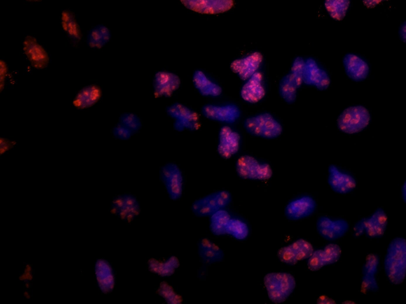
Immunocytochemistry/Immunofluorescence: Ki67/MKI67 Antibody (8D5) [NBP2-22112] - Ki67 antibody was tested at 1:100 in HeLa cells with DyLight 488 (green). Nuclei and alpha-tubulin were counterstained with DAPI (blue) and DyLight 550 (red). Image objective 40x. in A431 Human Cell Line -")
Ki67/MKI67 (8D5) in A431 Human Cell Line -
Ki67/MKI67 (8D5) was detected in immersion fixed A431 human skin carcinoma cell line using Mouse anti-Ki67/MKI67 (8D5) Protein-G purified Monoclonal Antibody conjugated to Alexa Fluor® 488 (Catalog # NBP2-22112AF488) (green) at 5 µg/mL overnight at 4C. Cells were counterstained with DAPI (blue). Cells were imaged using a 100X objective and digitally deconvolved. in A431 Human Cell Line -")
Ki67/MKI67 (8D5) in A431 Human Cell Line -
Ki67/MKI67 (8D5) was detected in immersion fixed A431 human skin carcinoma cell line using Mouse anti-Ki67/MKI67 (8D5) Protein-G purified Monoclonal Antibody conjugated to FITC (Catalog # NBP2-22112F) (green) at 5 µg/mL overnight at 4C. Cells were counterstained with DAPI (blue). Cells were imaged using a 100X objective and digitally deconvolved. in A431 Human Cell Line.")
Ki67/MKI67 (8D5) in A431 Human Cell Line.
Ki67/MKI67 (8D5) was detected in immersion fixed A431 human skin carcinoma cell line using Mouse anti-Ki67/MKI67 (8D5) Protein-G purified Monoclonal Antibody conjugated to Alexa Fluor® 647 (Catalog # NBP2-22112AF647) (light blue) at 2 µg/mL overnight at 4C. Cells were counterstained with DAPI (blue). Cells were imaged using a 100X objective and digitally deconvolved. in A431 Human Cell Line.")
Ki67/MKI67 (8D5) in A431 Human Cell Line.
Ki67/MKI67 (8D5) was detected in immersion fixed A431 human skin carcinoma cell line using Mouse anti-Ki67/MKI67 (8D5) Protein-G purified Monoclonal Antibody conjugated to DyLight 488 (Catalog # NBP2-22112G) (green) at 2 µg/mL overnight at 4C. Cells were counterstained with DAPI (blue). Cells were imaged using a 100X objective and digitally deconvolved. - BSA Free [NBP2-22112] -")
Western Blot: Ki67/MKI67 Antibody (8D5) - BSA Free [NBP2-22112] -
RPTECs trigger CD4+ T-cells to proliferate, anoxia-reoxygenation increases proliferation further, whereas CD4+ T-cell differentiation to regulatory T-cells does not occur. CD4+ T-cells were cultured alone or co-cultured with RPTECs for 7 days. RPTECs were previously subjected or not to 90 min of anoxia. Four such experiments were performed. Compared to isolated CD4+ T-cell culture, proliferation index was higher in RPTEC CD4+ T-cell co-culture and increased further in the case of previous RPTEC culture under anoxia. * indicates a p < 0.05 compared to isolated CD4+ T-cell culture, # a p < 0.05 compared to RPTEC CD4+ T-cell co-culture, and ^ a p < 0.05 compared to RPTEC (A). CD4+ T-cells were cultured alone or co-cultured with RPTECs. RPTECs were previously subjected or not to anoxia. After 3 days of co-culture, the CD4+ T-cells were collected. Four experiments were performed. Compared to isolated CD4+ T-cell culture, in CD4+ T-cell derived from RPTEC CD4+ T-cell co-culture Ki-67 (B,C) increased. Previous culture of RPTECs under anoxia induced further Ki-67 in CD4+ T-cells (B,C). FOXP3 expression in CD4+ T-cells remained unaffected under all conditions (B,D). The Western blotting results were normalized for beta -actin and then depicted after normalization for the control group. Error bars correspond to SEM. * indicates a p < 0.05 compared to isolated CD4+ T-cell culture, # a p < 0.05 compared to CD4+ harvested from RPTEC CD4+ T-cell co-culture, and ^ a p < 0.05 compared to CD4+ collected from RPTEC CD4+ T-cell co-culture in which RPTECs were previously subjected to anoxia. Image collected and cropped by CiteAb from the following open publication (https://pubmed.ncbi.nlm.nih.gov/33572206), licensed under a CC-BY license. Not internally tested by Novus Biologicals. - BSA Free [NBP2-22112] -")
Western Blot: Ki67/MKI67 Antibody (8D5) - BSA Free [NBP2-22112] -
Albumin overload induces cellular senescence and EMT. Panel (A) depicts the results of one out of three performed experiments. Albumin overload increased the expression of the cell cycle arrest inducers p21 (B) and p16 (C). In addition, it decreased the level of the cell proliferation marker Ki-67 (D) and enhanced the cellular senescence marker GLB-1 (E). RPTECs exposed to high albumin concentration overproduced IL-1 beta (F) and TGF-beta 1 (G). Exposure of RPTECs to high albumin concentration resulted in EMT as the level of alpha -SMA increased (H). TUDCA and 4-PBA ameliorated but did not eliminate all the changes above. * p < 0.05 vs. control; # p < 0.05 vs. RPTECs treated with 4-PBA; ^ p < 0.05 vs. RPTECs treated with TUDCA; + p < 0.05 vs. RPTECs exposed to BSA; & p < 0.05 vs. RPTECs exposed to BSA and 4-PBA; ! p < 0.05 vs. RPTECs exposed to BSA and TUDCA. 4-PBA, 4-Phenylbutyric acid; alpha -smooth muscle actin; BSA, bovine serum albumin; GLB-1, beta -galactosidase; IL-1 beta, interleukin-1 beta ; Ki-67, marker of proliferation Ki-67; p16, p16 INK4A; p21, p21 Waf1/Cip1; TGF-beta 1, transforming growth factor-beta 1; TUDCA, tauroursodeoxycholic acid. Image collected and cropped by CiteAb from the following open publication (https://pubmed.ncbi.nlm.nih.gov/37298591), licensed under a CC-BY license. Not internally tested by Novus Biologicals. - BSA Free [NBP2-22112] -")
Western Blot: Ki67/MKI67 Antibody (8D5) - BSA Free [NBP2-22112] -
The effect of high glucose and dapagliflozin on cell cycle inhibitors p21 and p16, cell proliferation, and senescence. High glucose upregulated p21, while dapagliflozin normalized the p21 levels (A,B). Dapagliflozin downregulated p16 expression under normal and high-glucose conditions (A,C). High glucose decreased Ki-67, whereas dapagliflozin restored Ki-67 levels (A,D). High glucose increased GLB-1 expression, whereas dapagliflozin decreased GLB-1 under normal and high-glucose conditions (A,E). * p < 0.05 vs. control; # p < 0.05 vs. RPTECs treated with 15 ng/mL dapagliflozin; ^ p < 0.05 vs. RPTECs in high glucose; + p < 0.05 vs. RPTECs in high glucose and treated with 15 ng/mL dapagliflozin. GLB-1, beta-galactosidase; Ki-67, marker of proliferation Ki-67; p16, p16 INK4A, p21, p21 Waf1/Cip1. Image collected and cropped by CiteAb from the following open publication (https://pubmed.ncbi.nlm.nih.gov/36555751), licensed under a CC-BY license. Not internally tested by Novus Biologicals. - BSA Free [NBP2-22112] -")
Western Blot: Ki67/MKI67 Antibody (8D5) - BSA Free [NBP2-22112] -
Anoxia-reoxygenation induces senescence phenotype, while LW6 prevents it. Anoxia-reoxygenation increases the p21 level, whereas the MDH-2 inhibitor LW6 prevents reoxygenation-induced p21 upregulation (A,B). Reoxygenation downregulated Ki-67, while LW6 prevented reoxygenation-induced Ki-67 reduction (A,C). Under reoxygenation, GLB-1 increased, but LW6 reversed the above change (A,D). RPTECs under reoxygenation produced more IL-1 beta, whereas LW6 downregulated reoxygenation-induced IL-1 beta overproduction (E). * p < 0.05 vs. control; # p < 0.05 vs. LW6-treated RPTECs under normoxia; + p < 0.05 vs. RPTECs under reoxygenation, ^ p < 0.05 vs. LW6-treated RPTECs under reoxygenation. GLB-1, beta-galactosidase; IL-1 beta, interleukin-1 beta. Image collected and cropped by CiteAb from the following open publication (https://pubmed.ncbi.nlm.nih.gov/36291624), licensed under a CC-BY license. Not internally tested by Novus Biologicals. - BSA Free [NBP2-22112] -")
Immunohistochemistry: Ki67/MKI67 Antibody (8D5) - BSA Free [NBP2-22112] -
Immunofluorescence of KIF22 and KI67 proteins in the cortex. (a) KIF22 and KI67 at 12 PCW, low magnification scale bars = 4 mm, high magnification scale bars = 100 μm. (b) KIF22 and KI67 at 14 PCW, low magnification scale bars = 4 mm, high magnification scale bars = 100 μm. c) high magnification of KI67/KIF22 expressing cells. Scale bars = 10 μm. (d) Percentage of cells expressing KIF22, KI67, or both at 12 PCW. (e) Percentage of cells expressing KIF22, KI67, or both at 14 PCW. (f) Combined data of percentage of cells expressing KIF22, KI67, or both. Image collected and cropped by CiteAb from the following open publication (https://pubmed.ncbi.nlm.nih.gov/33825894), licensed under a CC-BY license. Not internally tested by Novus Biologicals. - BSA Free [NBP2-22112] -")
Immunohistochemistry: Ki67/MKI67 Antibody (8D5) - BSA Free [NBP2-22112] -
Immunofluorescence of KIF22 and KI67 proteins in the cortex. (a) KIF22 and KI67 at 12 PCW, low magnification scale bars = 4 mm, high magnification scale bars = 100 μm. (b) KIF22 and KI67 at 14 PCW, low magnification scale bars = 4 mm, high magnification scale bars = 100 μm. c) high magnification of KI67/KIF22 expressing cells. Scale bars = 10 μm. (d) Percentage of cells expressing KIF22, KI67, or both at 12 PCW. (e) Percentage of cells expressing KIF22, KI67, or both at 14 PCW. (f) Combined data of percentage of cells expressing KIF22, KI67, or both. Image collected and cropped by CiteAb from the following open publication (https://pubmed.ncbi.nlm.nih.gov/33825894), licensed under a CC-BY license. Not internally tested by Novus Biologicals.Applications for Ki67/MKI67 Antibody (8D5) - BSA Free
Application
Recommended Usage
ELISA
1:10000
Immunocytochemistry/ Immunofluorescence
1:100. Use reported by customer review
Immunohistochemistry-Frozen
reported in scientific literature (PMID 33131911)
Immunohistochemistry-Paraffin
1:200-1:1000
Western Blot
1:500-1:2000
Reviewed Applications
Read 1 review rated 5 using NBP2-22112 in the following applications:
Formulation, Preparation, and Storage
Purification
Ammonium sulfate precipitation
Formulation
PBS
Format
BSA Free
Preservative
0.03% Sodium Azide
Concentration
1.0 mg/ml
Shipping
The product is shipped with polar packs. Upon receipt, store it immediately at the temperature recommended below.
Stability & Storage
Store at 4C short term. Aliquot and store at -20C long term. Avoid freeze-thaw cycles.
Background: Ki67/MKI67
Detection of Ki67 by immunostaining is commonly used as a proliferation marker in solid tumors, as well as certain hematological malignancies (3-5). The Ki67 index, which reports on positive Ki67 stained tumor cell nuclei, has been extensively studied as a prognostic biomarker in cancers such as breast cancer and cervical cancer.
References
1. Gerdes J, Schwab U, Lemke H, Stein H. (1983) Production of a mouse monoclonal antibody reactive with a human nuclear antigen associated with cell proliferation. Int J Cancer. 31:13-20. PMID: 6339421
2. Starborg M, Gell K, Brundell E and Hoog C. (1996) The murine Ki-67 cell proliferation antigen accumulates in the nucleolar and heterochromatic regions of interphase cells and at the periphery of the mitotic chromosomes in a process essential for cell cycle progression. J Cell Sci. 109:143-153. 1996
3. Karamitopoulou E, Perentes E, Tolnay M, Probst A. (1998) Prognostic significance of MIB-1, p53, and bcl-2 immunoreactivity in meningiomas. Hum Pathol. 29(2):140-5. PMID: 9490273
4. Geyer FC, Rodrigues DN, Weigelt B and Reis-Filho JS. (2012) Molecular classification of estrogen receptor-positive/luminal breast cancers. Adv Anat Pathol. 19(1):39-53. PMID: 22156833
5. Ikenberg H, Bergeron C, Schmidt D, Griesser H, Alameda F, Angeloni C, Bogers J, Dachez R, Denton K, Hariri J, Keller T, von Knebel Doeberitz M, Neumann HH, Puig-Tintore LM, Sideri M, Rehm S, Ridder R; PALMS Study Group. (2013) Screening for cervical cancer precursors with p16/Ki-67 dual-stained cytology: results of the PALMS study. J Natl Cancer Inst. 105(20):1550-7. PMID: 24096620
Long Name
Antigen Identified by Monoclonal Antibody Ki67
Alternate Names
Ki-67, KIA, MIB-1, MKI67, PPP1R105, TSG126
Entrez Gene IDs
4288 (Human)
Gene Symbol
MKI67
UniProt
Additional Ki67/MKI67 Products
Product Documents for Ki67/MKI67 Antibody (8D5) - BSA Free
Certificate of Analysis
To download a Certificate of Analysis, please enter a lot or batch number in the search box below.
Product Specific Notices for Ki67/MKI67 Antibody (8D5) - BSA Free
This product is for research use only and is not approved for use in humans or in clinical diagnosis. Primary Antibodies are guaranteed for 1 year from date of receipt.
Related Research Areas
Citations for Ki67/MKI67 Antibody (8D5) - BSA Free
 Powered by Bioz
Customer Reviews for Ki67/MKI67 Antibody (8D5) - BSA Free (1)
5 out of 5
1 Customer Rating
Have you used Ki67/MKI67 Antibody (8D5) - BSA Free?
Submit a review and receive an Amazon gift card!
$25/€18/£15/$25CAN/¥2500 Yen for a review with an image
$10/€7/£6/$10CAN/¥1110 Yen for a review without an image
Submit a review
Customer Images
Showing
1
-
1 的
1 review
Showing All
Filter By:
-
Application: ImmunocytochemistrySample Tested: A549 human alveolar adenocarcinoma cell lineSpecies: HumanVerified Customer | Posted 03/27/2018Ki-67/MKI67 staining of A549 human alveolar adenocarcinoma cellsA549 human alveolar adenocarcinoma cell line, fixation with PFA 4%, blocking with PBS 1X + BSA 1% + 0.1% Tween, primary antibody used at 1:100 in blocking buffer, O/N. Secondary antibody used AlexaFluor 594, 1 hour, RT and imaged with a fluorescence microscope and counterstained with DAPI.

There are no reviews that match your criteria.
Protocols
Find general support by application which include: protocols, troubleshooting, illustrated assays, videos and webinars.
- Antigen Retrieval Protocol (PIER)
- Antigen Retrieval for Frozen Sections Protocol
- Appropriate Fixation of IHC/ICC Samples
- Cellular Response to Hypoxia Protocols
- Chromogenic IHC Staining of Formalin-Fixed Paraffin-Embedded (FFPE) Tissue Protocol
- Chromogenic Immunohistochemistry Staining of Frozen Tissue
- ClariTSA™ Fluorophore Kits
- Detection & Visualization of Antibody Binding
- ELISA Sample Preparation & Collection Guide
- ELISA Troubleshooting Guide
- Fluorescent IHC Staining of Frozen Tissue Protocol
- Graphic Protocol for Heat-induced Epitope Retrieval
- Graphic Protocol for the Preparation and Fluorescent IHC Staining of Frozen Tissue Sections
- Graphic Protocol for the Preparation and Fluorescent IHC Staining of Paraffin-embedded Tissue Sections
- Graphic Protocol for the Preparation of Gelatin-coated Slides for Histological Tissue Sections
- How to Run an R&D Systems DuoSet ELISA
- How to Run an R&D Systems Quantikine ELISA
- How to Run an R&D Systems Quantikine™ QuicKit™ ELISA
- ICC Cell Smear Protocol for Suspension Cells
- ICC Immunocytochemistry Protocol Videos
- ICC for Adherent Cells
- IHC Sample Preparation (Frozen sections vs Paraffin)
- Immunocytochemistry (ICC) Protocol
- Immunocytochemistry Troubleshooting
- Immunofluorescence of Organoids Embedded in Cultrex Basement Membrane Extract
- Immunofluorescent IHC Staining of Formalin-Fixed Paraffin-Embedded (FFPE) Tissue Protocol
- Immunohistochemistry (IHC) and Immunocytochemistry (ICC) Protocols
- Immunohistochemistry Frozen Troubleshooting
- Immunohistochemistry Paraffin Troubleshooting
- Preparing Samples for IHC/ICC Experiments
- Preventing Non-Specific Staining (Non-Specific Binding)
- Primary Antibody Selection & Optimization
- Protocol for Heat-Induced Epitope Retrieval (HIER)
- Protocol for Making a 4% Formaldehyde Solution in PBS
- Protocol for VisUCyte™ HRP Polymer Detection Reagent
- Protocol for the Fluorescent ICC Staining of Cell Smears - Graphic
- Protocol for the Fluorescent ICC Staining of Cultured Cells on Coverslips - Graphic
- Protocol for the Preparation & Fixation of Cells on Coverslips
- Protocol for the Preparation and Chromogenic IHC Staining of Frozen Tissue Sections
- Protocol for the Preparation and Chromogenic IHC Staining of Frozen Tissue Sections - Graphic
- Protocol for the Preparation and Chromogenic IHC Staining of Paraffin-embedded Tissue Sections
- Protocol for the Preparation and Chromogenic IHC Staining of Paraffin-embedded Tissue Sections - Graphic
- Protocol for the Preparation and Fluorescent ICC Staining of Cells on Coverslips
- Protocol for the Preparation and Fluorescent ICC Staining of Non-adherent Cells
- Protocol for the Preparation and Fluorescent ICC Staining of Stem Cells on Coverslips
- Protocol for the Preparation and Fluorescent IHC Staining of Frozen Tissue Sections
- Protocol for the Preparation and Fluorescent IHC Staining of Paraffin-embedded Tissue Sections
- Protocol for the Preparation of Gelatin-coated Slides for Histological Tissue Sections
- Protocol for the Preparation of a Cell Smear for Non-adherent Cell ICC - Graphic
- Quantikine HS ELISA Kit Assay Principle, Alkaline Phosphatase
- Quantikine HS ELISA Kit Principle, Streptavidin-HRP Polymer
- R&D Systems Quality Control Western Blot Protocol
- Sandwich ELISA (Colorimetric) – Biotin/Streptavidin Detection Protocol
- Sandwich ELISA (Colorimetric) – Direct Detection Protocol
- TUNEL and Active Caspase-3 Detection by IHC/ICC Protocol
- The Importance of IHC/ICC Controls
- Troubleshooting Guide: ELISA
- Troubleshooting Guide: Immunohistochemistry
- Troubleshooting Guide: Western Blot Figures
- Western Blot Conditions
- Western Blot Protocol
- Western Blot Protocol for Cell Lysates
- Western Blot Troubleshooting
- Western Blot Troubleshooting Guide
- View all Protocols, Troubleshooting, Illustrated assays and Webinars
FAQs for Ki67/MKI67 Antibody (8D5) - BSA Free
Showing
1
-
2 的
2 FAQs
Showing All
-
Q: Whats the concentration of this Ki67/MKI67 Antibody?
A: The concentration is lot specific available upon request.
-
Q: Whats the concentration of this Ki67/MKI67 Antibody?
A: Optimal concentrations and conditions for each application should be determined by the user.
-
Q: Whats the concentration of this Ki67/MKI67 Antibody?
A: The concentration is lot specific available upon request.
-
Q: Whats the concentration of this Ki67/MKI67 Antibody?
A: Optimal concentrations and conditions for each application should be determined by the user.
Loading...