
Lysine (K)-specific Demethylase 5B/KDM5B/JARID1B Antibody - BSA Free
Novus Biologicals | Catalog # NB100-97821


Scientific images may be resized, cropped, or adjusted for brightness or contrast for presentation purposes. These updates are limited to presentation and do not affect the underlying scientific interpretation of the data. Where available, additional source data may be provided upon request.
Key Product Details
Species Reactivity
Validated:
Cited:
Applications
Validated:
Cited:
Label
Antibody Source
Format
Product Specifications
Immunogen
Clonality
Host
Isotype
Scientific Data Images for Lysine (K)-specific Demethylase 5B/KDM5B/JARID1B Antibody - BSA Free
Western Blot: Lysine (K)-specific Demethylase 5B/KDM5B/JARID1B Antibody [NB100-97821]
Western Blot: Lysine (K)-specific Demethylase 5B/KDM5B/JARID1B Antibody [NB100-97821] - Whole cell lysate (50 ug) from HeLa, 293T, and Jurkat cells prepared using NETN lysis buffer. Antibody: Affinity purified rabbit anti-JARID1B antibody used for WB at 0.4 ug/ml. Detection: Chemiluminescence with an exposure time of 3 minutes.
Immunohistochemistry-Paraffin: Lysine (K)-specific Demethylase 5B/KDM5B/JARID1B Antibody [NB100-97821]
Immunohistochemistry-Paraffin: Lysine (K)-specific Demethylase 5B/KDM5B/JARID1B Antibody [NB100-97821] - MCF-7 cells stained with Lysine (K)-specific Demethylase 5B antibody. Image from verified customer review.
Immunohistochemistry-Paraffin: Lysine (K)-specific Demethylase 5B/KDM5B/JARID1B Antibody [NB100-97821]
Immunohistochemistry-Paraffin: Lysine (K)-specific Demethylase 5B/KDM5B/JARID1B Antibody [NB100-97821] - Section of human prostate carcinoma. Antibody: Rabbit anti-JARID1B antibody used at 1:500 (2ug/ml). Secondary: HRP-conjugated goat anti-rabbit IgG (A120-501P). Substrate: DAB.
Immunoprecipitation: Lysine (K)-specific Demethylase 5B/KDM5B/JARID1B Antibody [NB100-97821]
Immunoprecipitation: Lysine (K)-specific Demethylase 5B/KDM5B/JARID1B Antibody [NB100-97821] - Detection of human JARID1B by western blot of immunoprecipitates. Samples: Whole cell lysate (1.0 mg per IP reaction; 20% of IP loaded) from 293T cells prepared using NETN lysis buffer. Antibodies: Affinity purified rabbit anti-JARID1B antibody NB100-97821 (lot NB100-97821-3) used for IP at 6 ug per reaction. JARID1B was also immunoprecipitated by a previous lot of this antibody (lot NB100-97821-2). For blotting immunoprecipitated Jarid1B, NB100-97821 was used at 1 ug/ml. Detection: Chemiluminescence with an exposure time of 75 seconds.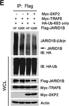
Western Blot: Lysine (K)-specific Demethylase 5B/KDM5B/JARID1B Antibody - BSA Free [NB100-97821] -
SKP2 regulates the ubiquitination of JARID1B through TRAF6(A) Immunofluorescence images show a co-localization of endogenous JARID1B and TRAF6 in PC3 cells. Scale bars represent 10 μm. (B) and (C) Co-immunoprecipitation analysis shows that endogenous TRAF6 physically interacts with JARID1B in PC3 cells, as shown by reciprocal co-immunoprecipitation between the two proteins (Also see Supplementary Figure S5). (D)In vivo ubiquitination assay shows that TRAF6 increases K63-linked ubiquitination of JARID1B and SKP2 inhibits TRAF6-mediated JARID1B ubiquitination. Cells were transfected with Flag-JARID1B, HA-Ub-K63-only, Myc-TRAF6 and Myc-SKP2 constructs as indicated. WCL indicates the whole cell lysates. (E) TRAF6 mediates JARID1B ubiquitination through lysine residue 242. HEK293T cells were transfected with Flag-JARID1B WT or Flag-JARID1B-K242R, HA-Ub-K63-only, Myc-TRAF6 and Myc-SKP2 plasmids as indicated. In vivo ubiquitination assay was performed in a standard procedure. WCL indicates the whole cell lysates. Image collected and cropped by CiteAb from the following open publication (https://pubmed.ncbi.nlm.nih.gov/25596733), licensed under a CC-BY license. Not internally tested by Novus Biologicals.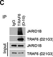
Western Blot: Lysine (K)-specific Demethylase 5B/KDM5B/JARID1B Antibody - BSA Free [NB100-97821] -
SKP2 regulates the ubiquitination of JARID1B through TRAF6(A) Immunofluorescence images show a co-localization of endogenous JARID1B and TRAF6 in PC3 cells. Scale bars represent 10 μm. (B) and (C) Co-immunoprecipitation analysis shows that endogenous TRAF6 physically interacts with JARID1B in PC3 cells, as shown by reciprocal co-immunoprecipitation between the two proteins (Also see Supplementary Figure S5). (D)In vivo ubiquitination assay shows that TRAF6 increases K63-linked ubiquitination of JARID1B and SKP2 inhibits TRAF6-mediated JARID1B ubiquitination. Cells were transfected with Flag-JARID1B, HA-Ub-K63-only, Myc-TRAF6 and Myc-SKP2 constructs as indicated. WCL indicates the whole cell lysates. (E) TRAF6 mediates JARID1B ubiquitination through lysine residue 242. HEK293T cells were transfected with Flag-JARID1B WT or Flag-JARID1B-K242R, HA-Ub-K63-only, Myc-TRAF6 and Myc-SKP2 plasmids as indicated. In vivo ubiquitination assay was performed in a standard procedure. WCL indicates the whole cell lysates. Image collected and cropped by CiteAb from the following open publication (https://pubmed.ncbi.nlm.nih.gov/25596733), licensed under a CC-BY license. Not internally tested by Novus Biologicals.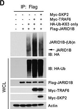
Western Blot: Lysine (K)-specific Demethylase 5B/KDM5B/JARID1B Antibody - BSA Free [NB100-97821] -
SKP2 regulates the ubiquitination of JARID1B through TRAF6(A) Immunofluorescence images show a co-localization of endogenous JARID1B and TRAF6 in PC3 cells. Scale bars represent 10 μm. (B) and (C) Co-immunoprecipitation analysis shows that endogenous TRAF6 physically interacts with JARID1B in PC3 cells, as shown by reciprocal co-immunoprecipitation between the two proteins (Also see Supplementary Figure S5). (D)In vivo ubiquitination assay shows that TRAF6 increases K63-linked ubiquitination of JARID1B and SKP2 inhibits TRAF6-mediated JARID1B ubiquitination. Cells were transfected with Flag-JARID1B, HA-Ub-K63-only, Myc-TRAF6 and Myc-SKP2 constructs as indicated. WCL indicates the whole cell lysates. (E) TRAF6 mediates JARID1B ubiquitination through lysine residue 242. HEK293T cells were transfected with Flag-JARID1B WT or Flag-JARID1B-K242R, HA-Ub-K63-only, Myc-TRAF6 and Myc-SKP2 plasmids as indicated. In vivo ubiquitination assay was performed in a standard procedure. WCL indicates the whole cell lysates. Image collected and cropped by CiteAb from the following open publication (https://pubmed.ncbi.nlm.nih.gov/25596733), licensed under a CC-BY license. Not internally tested by Novus Biologicals.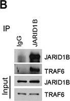
Western Blot: Lysine (K)-specific Demethylase 5B/KDM5B/JARID1B Antibody - BSA Free [NB100-97821] -
SKP2 regulates the ubiquitination of JARID1B through TRAF6(A) Immunofluorescence images show a co-localization of endogenous JARID1B and TRAF6 in PC3 cells. Scale bars represent 10 μm. (B) and (C) Co-immunoprecipitation analysis shows that endogenous TRAF6 physically interacts with JARID1B in PC3 cells, as shown by reciprocal co-immunoprecipitation between the two proteins (Also see Supplementary Figure S5). (D)In vivo ubiquitination assay shows that TRAF6 increases K63-linked ubiquitination of JARID1B and SKP2 inhibits TRAF6-mediated JARID1B ubiquitination. Cells were transfected with Flag-JARID1B, HA-Ub-K63-only, Myc-TRAF6 and Myc-SKP2 constructs as indicated. WCL indicates the whole cell lysates. (E) TRAF6 mediates JARID1B ubiquitination through lysine residue 242. HEK293T cells were transfected with Flag-JARID1B WT or Flag-JARID1B-K242R, HA-Ub-K63-only, Myc-TRAF6 and Myc-SKP2 plasmids as indicated. In vivo ubiquitination assay was performed in a standard procedure. WCL indicates the whole cell lysates. Image collected and cropped by CiteAb from the following open publication (https://pubmed.ncbi.nlm.nih.gov/25596733), licensed under a CC-BY license. Not internally tested by Novus Biologicals.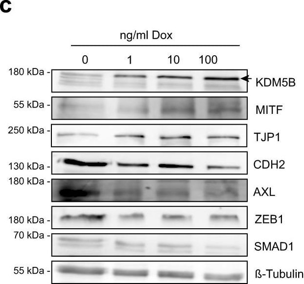
Western Blot: Lysine (K)-specific Demethylase 5B/KDM5B/JARID1B Antibody - BSA Free [NB100-97821] -
Enforced KDM5B expression facilitates melanocytic lineage-directed elimination by TMECG.a Quantitation of mRNA after 24 h, 48 h, 72 h and 7 days of Cpd1 treatment of MaMel63a cells as assessed by qPCR. Mean +/- SD. Shown is one representative example. b Regulation of differentiation, cytokinesis, and mitotic spindle assembly genes as detected by cDNA microarray analysis after KDM5B shRNA knockdown in WM3734 cells (n = 1). c, d Immunoblotting of melanocytic lineage and (de-)differentiation markers after 24 h of KDM5B induction in WM3734Tet3G-KDM5B cells (c) and after 72 h of Cpd1 treatment in MaMel63a cells (d). Shown are representative data (n = 2). e Anti-MITF immunostaining (upper panel) and Fontana-Masson staining (lower panels) of CM melanoma tumor grafts from Cpd1-treated vs. control mice. f MTT cell viability assay of WM3734 cells. Representative example is shown left (mean +/- SD, n = 2) and corresponding IC50 values on the right. TMECG was either concurrently given together with Cpd1 (“con") or added 3 days after Cpd1 pre-treatment (“pre"). Readout was performed after 72 h of TMECG treatment. g Persister-state-directed therapy model in vivo. Left: schematic representation of treatment dosing and timing in immunodeficient NMRI-(nu/nu)-nude mice. Right: tumor volumes of WM3734 xenografts (endpoint at day 30). TMECG was either concurrently given together with Cpd1 (“con") or added one week after Cpd1 pre-treatment (“pre"). Mean +/-SEM (6 mice in TMECG and Cpd1 control group, five mice in “con" and seven mice in “pre" group). Significance was determined by two-sided Mann–Whitney test. Source data are provided as a Source Data file. Image collected and cropped by CiteAb from the following open publication (https://pubmed.ncbi.nlm.nih.gov/35650266), licensed under a CC-BY license. Not internally tested by Novus Biologicals.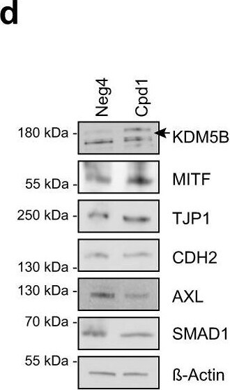
Western Blot: Lysine (K)-specific Demethylase 5B/KDM5B/JARID1B Antibody - BSA Free [NB100-97821] -
Enforced KDM5B expression facilitates melanocytic lineage-directed elimination by TMECG.a Quantitation of mRNA after 24 h, 48 h, 72 h and 7 days of Cpd1 treatment of MaMel63a cells as assessed by qPCR. Mean +/- SD. Shown is one representative example. b Regulation of differentiation, cytokinesis, and mitotic spindle assembly genes as detected by cDNA microarray analysis after KDM5B shRNA knockdown in WM3734 cells (n = 1). c, d Immunoblotting of melanocytic lineage and (de-)differentiation markers after 24 h of KDM5B induction in WM3734Tet3G-KDM5B cells (c) and after 72 h of Cpd1 treatment in MaMel63a cells (d). Shown are representative data (n = 2). e Anti-MITF immunostaining (upper panel) and Fontana-Masson staining (lower panels) of CM melanoma tumor grafts from Cpd1-treated vs. control mice. f MTT cell viability assay of WM3734 cells. Representative example is shown left (mean +/- SD, n = 2) and corresponding IC50 values on the right. TMECG was either concurrently given together with Cpd1 (“con") or added 3 days after Cpd1 pre-treatment (“pre"). Readout was performed after 72 h of TMECG treatment. g Persister-state-directed therapy model in vivo. Left: schematic representation of treatment dosing and timing in immunodeficient NMRI-(nu/nu)-nude mice. Right: tumor volumes of WM3734 xenografts (endpoint at day 30). TMECG was either concurrently given together with Cpd1 (“con") or added one week after Cpd1 pre-treatment (“pre"). Mean +/-SEM (6 mice in TMECG and Cpd1 control group, five mice in “con" and seven mice in “pre" group). Significance was determined by two-sided Mann–Whitney test. Source data are provided as a Source Data file. Image collected and cropped by CiteAb from the following open publication (https://pubmed.ncbi.nlm.nih.gov/35650266), licensed under a CC-BY license. Not internally tested by Novus Biologicals.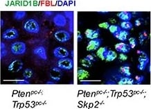
Immunofluorescence: Lysine (K)-specific Demethylase 5B/KDM5B/JARID1B Antibody - BSA Free [NB100-97821] -
SKP2 inactivation induces an accumulation of ubiquitinated JARID1B in nucleolus of cells in vitro and in vivo for cellular senescence(A) Immunofluorescence (IF) images show a co-localization of endogenous JARID1B and Fibrillarin (FBL) in nucleoli of PC3 cells upon SKP2 knockdown (Also see Supplementary Figure S6A). FBL indicates a nucleolar marker. Right panel: quantification of PC3 cells showing an increase of JARID1B localization in nucleolus. Error bars represent means +/- SD. (B) IF images show a co-localization of endogenous JARID1B and K63-Ub in nucleoli of PC3 cells upon SKP2 knockdown. (C) Western blotting assay shows an increase of beta -galactosidase ( beta -Gal) in PC3 cells upon SKP2 knockdown. (D) IF images show JARID1B in nucleolus as indicated by arrows and beta -Gal in cytoplasm in senescent cells upon SKP2 knockdown. (E) The co-localization of endogenous JARID1B and Fibrillarin (FBL) in nucleoli of prostate tissues in Ptenpc−/−;Trp53pc−/−;Skp2−/− mutant mice (Also see Supplementary Figure S6B). Scale bars represent 10 μm for panel A, B, D and E. (F) The positive staining of beta -galactosidase in prostate tissues of Ptenpc−/−;Trp53pc−/−;Skp2−/− mice but not in that of Ptenpc−/−;Trp53pc−/− mice. Scale bars represent 50 μm. Image collected and cropped by CiteAb from the following open publication (https://pubmed.ncbi.nlm.nih.gov/25596733), licensed under a CC-BY license. Not internally tested by Novus Biologicals.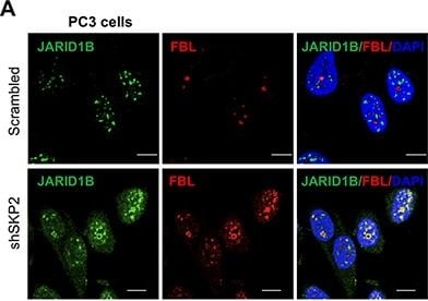
Immunofluorescence: Lysine (K)-specific Demethylase 5B/KDM5B/JARID1B Antibody - BSA Free [NB100-97821] -
SKP2 inactivation induces an accumulation of ubiquitinated JARID1B in nucleolus of cells in vitro and in vivo for cellular senescence(A) Immunofluorescence (IF) images show a co-localization of endogenous JARID1B and Fibrillarin (FBL) in nucleoli of PC3 cells upon SKP2 knockdown (Also see Supplementary Figure S6A). FBL indicates a nucleolar marker. Right panel: quantification of PC3 cells showing an increase of JARID1B localization in nucleolus. Error bars represent means +/- SD. (B) IF images show a co-localization of endogenous JARID1B and K63-Ub in nucleoli of PC3 cells upon SKP2 knockdown. (C) Western blotting assay shows an increase of beta -galactosidase ( beta -Gal) in PC3 cells upon SKP2 knockdown. (D) IF images show JARID1B in nucleolus as indicated by arrows and beta -Gal in cytoplasm in senescent cells upon SKP2 knockdown. (E) The co-localization of endogenous JARID1B and Fibrillarin (FBL) in nucleoli of prostate tissues in Ptenpc−/−;Trp53pc−/−;Skp2−/− mutant mice (Also see Supplementary Figure S6B). Scale bars represent 10 μm for panel A, B, D and E. (F) The positive staining of beta -galactosidase in prostate tissues of Ptenpc−/−;Trp53pc−/−;Skp2−/− mice but not in that of Ptenpc−/−;Trp53pc−/− mice. Scale bars represent 50 μm. Image collected and cropped by CiteAb from the following open publication (https://pubmed.ncbi.nlm.nih.gov/25596733), licensed under a CC-BY license. Not internally tested by Novus Biologicals.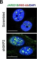
Immunofluorescence: Lysine (K)-specific Demethylase 5B/KDM5B/JARID1B Antibody - BSA Free [NB100-97821] -
SKP2 inactivation induces an accumulation of ubiquitinated JARID1B in nucleolus of cells in vitro and in vivo for cellular senescence(A) Immunofluorescence (IF) images show a co-localization of endogenous JARID1B and Fibrillarin (FBL) in nucleoli of PC3 cells upon SKP2 knockdown (Also see Supplementary Figure S6A). FBL indicates a nucleolar marker. Right panel: quantification of PC3 cells showing an increase of JARID1B localization in nucleolus. Error bars represent means +/- SD. (B) IF images show a co-localization of endogenous JARID1B and K63-Ub in nucleoli of PC3 cells upon SKP2 knockdown. (C) Western blotting assay shows an increase of beta -galactosidase ( beta -Gal) in PC3 cells upon SKP2 knockdown. (D) IF images show JARID1B in nucleolus as indicated by arrows and beta -Gal in cytoplasm in senescent cells upon SKP2 knockdown. (E) The co-localization of endogenous JARID1B and Fibrillarin (FBL) in nucleoli of prostate tissues in Ptenpc−/−;Trp53pc−/−;Skp2−/− mutant mice (Also see Supplementary Figure S6B). Scale bars represent 10 μm for panel A, B, D and E. (F) The positive staining of beta -galactosidase in prostate tissues of Ptenpc−/−;Trp53pc−/−;Skp2−/− mice but not in that of Ptenpc−/−;Trp53pc−/− mice. Scale bars represent 50 μm. Image collected and cropped by CiteAb from the following open publication (https://pubmed.ncbi.nlm.nih.gov/25596733), licensed under a CC-BY license. Not internally tested by Novus Biologicals.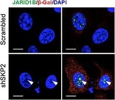
Immunofluorescence: Lysine (K)-specific Demethylase 5B/KDM5B/JARID1B Antibody - BSA Free [NB100-97821] -
SKP2 inactivation induces an accumulation of ubiquitinated JARID1B in nucleolus of cells in vitro and in vivo for cellular senescence(A) Immunofluorescence (IF) images show a co-localization of endogenous JARID1B and Fibrillarin (FBL) in nucleoli of PC3 cells upon SKP2 knockdown (Also see Supplementary Figure S6A). FBL indicates a nucleolar marker. Right panel: quantification of PC3 cells showing an increase of JARID1B localization in nucleolus. Error bars represent means +/- SD. (B) IF images show a co-localization of endogenous JARID1B and K63-Ub in nucleoli of PC3 cells upon SKP2 knockdown. (C) Western blotting assay shows an increase of beta -galactosidase ( beta -Gal) in PC3 cells upon SKP2 knockdown. (D) IF images show JARID1B in nucleolus as indicated by arrows and beta -Gal in cytoplasm in senescent cells upon SKP2 knockdown. (E) The co-localization of endogenous JARID1B and Fibrillarin (FBL) in nucleoli of prostate tissues in Ptenpc−/−;Trp53pc−/−;Skp2−/− mutant mice (Also see Supplementary Figure S6B). Scale bars represent 10 μm for panel A, B, D and E. (F) The positive staining of beta -galactosidase in prostate tissues of Ptenpc−/−;Trp53pc−/−;Skp2−/− mice but not in that of Ptenpc−/−;Trp53pc−/− mice. Scale bars represent 50 μm. Image collected and cropped by CiteAb from the following open publication (https://pubmed.ncbi.nlm.nih.gov/25596733), licensed under a CC-BY license. Not internally tested by Novus Biologicals.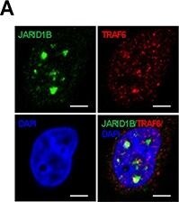
Immunofluorescence: Lysine (K)-specific Demethylase 5B/KDM5B/JARID1B Antibody - BSA Free [NB100-97821] -
SKP2 regulates the ubiquitination of JARID1B through TRAF6(A) Immunofluorescence images show a co-localization of endogenous JARID1B and TRAF6 in PC3 cells. Scale bars represent 10 μm. (B) and (C) Co-immunoprecipitation analysis shows that endogenous TRAF6 physically interacts with JARID1B in PC3 cells, as shown by reciprocal co-immunoprecipitation between the two proteins (Also see Supplementary Figure S5). (D)In vivo ubiquitination assay shows that TRAF6 increases K63-linked ubiquitination of JARID1B and SKP2 inhibits TRAF6-mediated JARID1B ubiquitination. Cells were transfected with Flag-JARID1B, HA-Ub-K63-only, Myc-TRAF6 and Myc-SKP2 constructs as indicated. WCL indicates the whole cell lysates. (E) TRAF6 mediates JARID1B ubiquitination through lysine residue 242. HEK293T cells were transfected with Flag-JARID1B WT or Flag-JARID1B-K242R, HA-Ub-K63-only, Myc-TRAF6 and Myc-SKP2 plasmids as indicated. In vivo ubiquitination assay was performed in a standard procedure. WCL indicates the whole cell lysates. Image collected and cropped by CiteAb from the following open publication (https://pubmed.ncbi.nlm.nih.gov/25596733), licensed under a CC-BY license. Not internally tested by Novus Biologicals.Applications for Lysine (K)-specific Demethylase 5B/KDM5B/JARID1B Antibody - BSA Free
Immunohistochemistry
Immunohistochemistry-Paraffin
Immunoprecipitation
Western Blot
Reviewed Applications
Read 1 review rated 5 using NB100-97821 in the following applications:
Flow Cytometry Panel Builder
Bio-Techne Knows Flow Cytometry
Save time and reduce costly mistakes by quickly finding compatible reagents using the Panel Builder Tool.
Advanced Features
- Spectra Viewer - Custom analysis of spectra from multiple fluorochromes
- Spillover Popups - Visualize the spectra of individual fluorochromes
- Antigen Density Selector - Match fluorochrome brightness with antigen density

Formulation, Preparation, and Storage
Purification
Formulation
Format
Preservative
Concentration
Shipping
Stability & Storage
Background: Lysine (K)-specific Demethylase 5B/KDM5B
Alternate Names
Gene Symbol
UniProt
Additional Lysine (K)-specific Demethylase 5B/KDM5B Products
Product Documents for Lysine (K)-specific Demethylase 5B/KDM5B/JARID1B Antibody - BSA Free
Certificate of Analysis
To download a Certificate of Analysis, please enter a lot or batch number in the search box below.
Product Specific Notices for Lysine (K)-specific Demethylase 5B/KDM5B/JARID1B Antibody - BSA Free
This product is for research use only and is not approved for use in humans or in clinical diagnosis. Primary Antibodies are guaranteed for 1 year from date of receipt.
Related Research Areas
Citations for Lysine (K)-specific Demethylase 5B/KDM5B/JARID1B Antibody - BSA Free
 Powered by Bioz
Customer Reviews for Lysine (K)-specific Demethylase 5B/KDM5B/JARID1B Antibody - BSA Free (1)
Have you used Lysine (K)-specific Demethylase 5B/KDM5B/JARID1B Antibody - BSA Free?
Submit a review and receive an Amazon gift card!
$25/€18/£15/$25CAN/¥2500 for a review with an image
$10/€7/£6/$10CAN/¥1110 for a review without an image
Submit a review
Customer Images
-
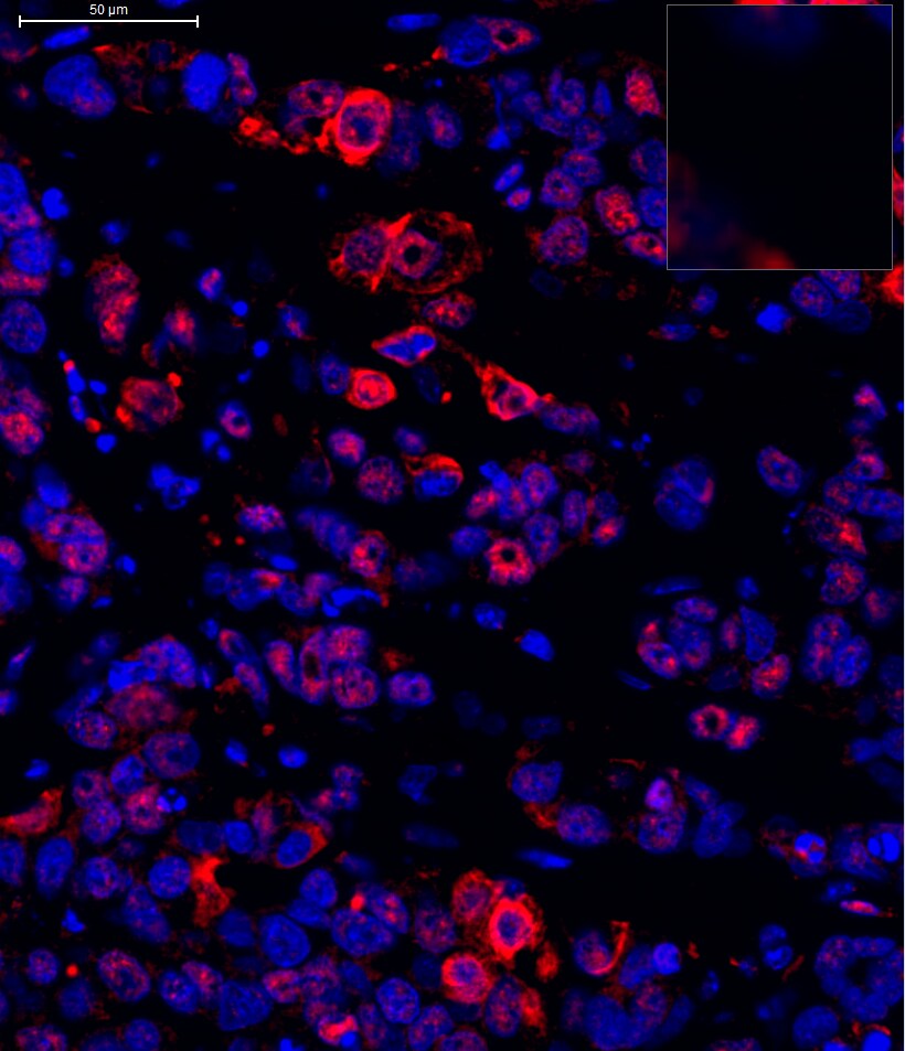
Application: Immunohistochemistry-ParaffinSample Tested: MCF7Species: HumanVerified Customer | Posted 10/06/2015MCF7 tumor with 1:1000 JARID1B antibody

There are no reviews that match your criteria.
Protocols
Find general support by application which include: protocols, troubleshooting, illustrated assays, videos and webinars.
- 7-Amino Actinomycin D (7-AAD) Cell Viability Flow Cytometry Protocol
- Antigen Retrieval Protocol (PIER)
- Antigen Retrieval for Frozen Sections Protocol
- Appropriate Fixation of IHC/ICC Samples
- Cellular Response to Hypoxia Protocols
- Chromogenic IHC Staining of Formalin-Fixed Paraffin-Embedded (FFPE) Tissue Protocol
- Chromogenic Immunohistochemistry Staining of Frozen Tissue
- ClariTSA™ Fluorophore Kits
- Detection & Visualization of Antibody Binding
- Extracellular Membrane Flow Cytometry Protocol
- Flow Cytometry Protocol for Cell Surface Markers
- Flow Cytometry Protocol for Staining Membrane Associated Proteins
- Flow Cytometry Staining Protocols
- Flow Cytometry Troubleshooting Guide
- Fluorescent IHC Staining of Frozen Tissue Protocol
- Graphic Protocol for Heat-induced Epitope Retrieval
- Graphic Protocol for the Preparation and Fluorescent IHC Staining of Frozen Tissue Sections
- Graphic Protocol for the Preparation and Fluorescent IHC Staining of Paraffin-embedded Tissue Sections
- Graphic Protocol for the Preparation of Gelatin-coated Slides for Histological Tissue Sections
- IHC Sample Preparation (Frozen sections vs Paraffin)
- Immunofluorescent IHC Staining of Formalin-Fixed Paraffin-Embedded (FFPE) Tissue Protocol
- Immunohistochemistry (IHC) and Immunocytochemistry (ICC) Protocols
- Immunohistochemistry Frozen Troubleshooting
- Immunohistochemistry Paraffin Troubleshooting
- Immunoprecipitation Protocol
- Intracellular Flow Cytometry Protocol Using Alcohol (Methanol)
- Intracellular Flow Cytometry Protocol Using Detergents
- Intracellular Nuclear Staining Flow Cytometry Protocol Using Detergents
- Intracellular Staining Flow Cytometry Protocol Using Alcohol Permeabilization
- Intracellular Staining Flow Cytometry Protocol Using Detergents to Permeabilize Cells
- Preparing Samples for IHC/ICC Experiments
- Preventing Non-Specific Staining (Non-Specific Binding)
- Primary Antibody Selection & Optimization
- Propidium Iodide Cell Viability Flow Cytometry Protocol
- Protocol for Heat-Induced Epitope Retrieval (HIER)
- Protocol for Liperfluo
- Protocol for Making a 4% Formaldehyde Solution in PBS
- Protocol for VisUCyte™ HRP Polymer Detection Reagent
- Protocol for the Characterization of Human Th22 Cells
- Protocol for the Characterization of Human Th9 Cells
- Protocol for the Preparation & Fixation of Cells on Coverslips
- Protocol for the Preparation and Chromogenic IHC Staining of Frozen Tissue Sections
- Protocol for the Preparation and Chromogenic IHC Staining of Frozen Tissue Sections - Graphic
- Protocol for the Preparation and Chromogenic IHC Staining of Paraffin-embedded Tissue Sections
- Protocol for the Preparation and Chromogenic IHC Staining of Paraffin-embedded Tissue Sections - Graphic
- Protocol for the Preparation and Fluorescent IHC Staining of Frozen Tissue Sections
- Protocol for the Preparation and Fluorescent IHC Staining of Paraffin-embedded Tissue Sections
- Protocol for the Preparation of Gelatin-coated Slides for Histological Tissue Sections
- Protocol: Annexin V and PI Staining by Flow Cytometry
- Protocol: Annexin V and PI Staining for Apoptosis by Flow Cytometry
- R&D Systems Quality Control Western Blot Protocol
- TUNEL and Active Caspase-3 Detection by IHC/ICC Protocol
- The Importance of IHC/ICC Controls
- Troubleshooting Guide: Fluorokine Flow Cytometry Kits
- Troubleshooting Guide: Immunohistochemistry
- Troubleshooting Guide: Western Blot Figures
- Western Blot Conditions
- Western Blot Protocol
- Western Blot Protocol for Cell Lysates
- Western Blot Troubleshooting
- Western Blot Troubleshooting Guide
- View all Protocols, Troubleshooting, Illustrated assays and Webinars