15-PGDH/HPGD Antibody - BSA Free
Novus Biologicals | Catalog # NBP1-87062
Loading...
Key Product Details
Validated by
Orthogonal Validation, Independent Antibodies
Species Reactivity
Validated:
Human
Predicted:
Mouse (97%), Rat (95%). Backed by our 100% Guarantee.
Applications
Immunohistochemistry, Immunohistochemistry-Paraffin, Western Blot
Label
Unconjugated
Antibody Source
Polyclonal Rabbit IgG
Format
BSA Free
Loading...
Product Specifications
Immunogen
This antibody was developed against Recombinant Protein corresponding to amino acids: VDWNLEAGVQCKAALDEQFEPQKTLFIQCDVADQQQLRDTFRKVVDHFGRLDILVNNAGVNNEKNWEKTLQINLVSVISGTYLGLDYMSKQNGGEGGIIINMSSLAGLMPVAQQPV
Clonality
Polyclonal
Host
Rabbit
Isotype
IgG
Scientific Data Images for 15-PGDH/HPGD Antibody - BSA Free
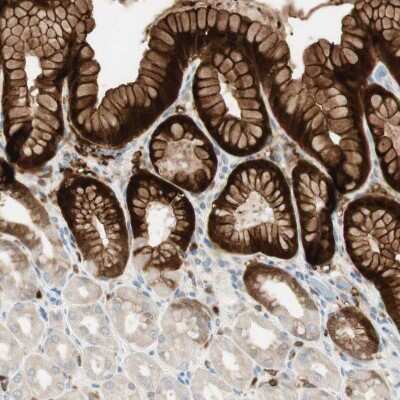
Immunohistochemistry-Paraffin: 15-PGDH/HPGD Antibody [NBP1-87062] - Staining of human stomach shows moderate to strong cytoplasmic and membranous positivity in glandular cells.
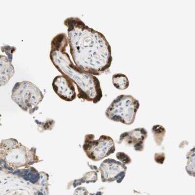
Immunohistochemistry-Paraffin: 15-PGDH/HPGD Antibody [NBP1-87062] - Staining of human placenta shows moderate to strong cytoplasmic positivity in trophoblastic cells.
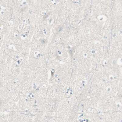
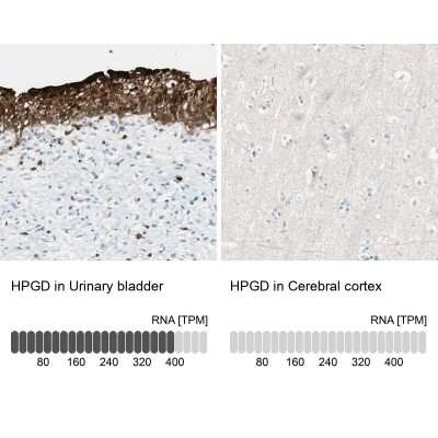
Immunohistochemistry-Paraffin: 15-PGDH/HPGD Antibody [NBP1-87062] - Staining of human cerebral cortex shows no positivity in neurons as expected.
![15-PGDH/HPGD Antibody - BSA Free Immunohistochemistry-Paraffin: 15-PGDH/HPGD Antibody - BSA Free [NBP1-87062]](https://resources.rndsystems.com/images/products/nbp1-87062_rabbit-polyclonal-15-pgdh-hpgd-antibody-304202513105884.jpg "Immunohistochemistry-Paraffin: 15-PGDH/HPGD Antibody - BSA Free [NBP1-87062]")
Immunohistochemistry-Paraffin: 15-PGDH/HPGD Antibody - BSA Free [NBP1-87062]
Staining of human urinary bladder shows strong cytoplasmic positivity in urothelial cells.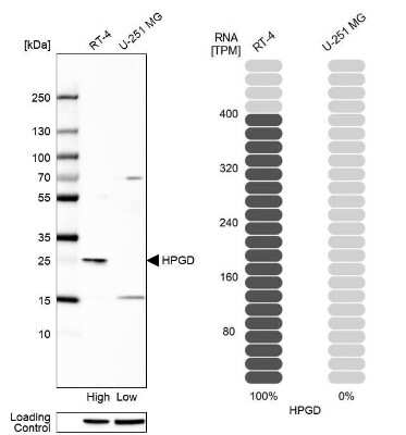
Western Blot: 15-PGDH/HPGD Antibody [NBP1-87062] - Analysis in human cell line RT-4 and human cell line U-251 MG.
![15-PGDH/HPGD Antibody - BSA Free Western Blot: 15-PGDH/HPGD Antibody - BSA Free [NBP1-87062]](https://resources.rndsystems.com/images/products/nbp1-87062_rabbit-polyclonal-15-pgdh-hpgd-antibody-30420251371456.jpg "Western Blot: 15-PGDH/HPGD Antibody - BSA Free [NBP1-87062]")
Applications for 15-PGDH/HPGD Antibody - BSA Free
Application
Recommended Usage
Immunohistochemistry
1:50 - 1:200
Immunohistochemistry-Paraffin
1:50 - 1:200
Western Blot
0.04-0.4 ug/ml
Application Notes
For IHC-Paraffin, HIER pH 6 retrieval is recommended.
Formulation, Preparation, and Storage
Purification
Affinity purified
Formulation
PBS (pH 7.2) and 40% Glycerol
Format
BSA Free
Preservative
0.02% Sodium Azide
Concentration
Concentrations vary lot to lot. See vial label for concentration. If unlisted please contact technical services.
Shipping
The product is shipped with polar packs. Upon receipt, store it immediately at the temperature recommended below.
Stability & Storage
Store at 4C short term. Aliquot and store at -20C long term. Avoid freeze-thaw cycles.
Background: 15-PGDH/HPGD
15-PGDH is a novel tumor suppressor in the COX-2 pathway. 15-PGDH is found in many normal tissues and appears to be down-regulated in colorectal and lung carcinoma tissue.
Long Name
15-Hydroxyprostaglandin Dehydrogenase
Alternate Names
15PGDH, HPGD, PGDH1, SDR36C1
Gene Symbol
HPGD
Additional 15-PGDH/HPGD Products
Product Documents for 15-PGDH/HPGD Antibody - BSA Free
Certificate of Analysis
To download a Certificate of Analysis, please enter a lot or batch number in the search box below.
Product Specific Notices for 15-PGDH/HPGD Antibody - BSA Free
This product is for research use only and is not approved for use in humans or in clinical diagnosis. Primary Antibodies are guaranteed for 1 year from date of receipt.
Related Research Areas
Citations for 15-PGDH/HPGD Antibody - BSA Free
 Powered by Bioz
Customer Reviews for 15-PGDH/HPGD Antibody - BSA Free
There are currently no reviews for this product. Be the first to review 15-PGDH/HPGD Antibody - BSA Free and earn rewards!
Have you used 15-PGDH/HPGD Antibody - BSA Free?
Submit a review and receive an Amazon gift card!
$25/€18/£15/$25CAN/¥2500 Yen for a review with an image
$10/€7/£6/$10CAN/¥1110 Yen for a review without an image
Submit a review
Protocols
Find general support by application which include: protocols, troubleshooting, illustrated assays, videos and webinars.
- Antigen Retrieval Protocol (PIER)
- Antigen Retrieval for Frozen Sections Protocol
- Appropriate Fixation of IHC/ICC Samples
- Cellular Response to Hypoxia Protocols
- Chromogenic IHC Staining of Formalin-Fixed Paraffin-Embedded (FFPE) Tissue Protocol
- Chromogenic Immunohistochemistry Staining of Frozen Tissue
- ClariTSA™ Fluorophore Kits
- Detection & Visualization of Antibody Binding
- Fluorescent IHC Staining of Frozen Tissue Protocol
- Graphic Protocol for Heat-induced Epitope Retrieval
- Graphic Protocol for the Preparation and Fluorescent IHC Staining of Frozen Tissue Sections
- Graphic Protocol for the Preparation and Fluorescent IHC Staining of Paraffin-embedded Tissue Sections
- Graphic Protocol for the Preparation of Gelatin-coated Slides for Histological Tissue Sections
- IHC Sample Preparation (Frozen sections vs Paraffin)
- Immunofluorescent IHC Staining of Formalin-Fixed Paraffin-Embedded (FFPE) Tissue Protocol
- Immunohistochemistry (IHC) and Immunocytochemistry (ICC) Protocols
- Immunohistochemistry Frozen Troubleshooting
- Immunohistochemistry Paraffin Troubleshooting
- Preparing Samples for IHC/ICC Experiments
- Preventing Non-Specific Staining (Non-Specific Binding)
- Primary Antibody Selection & Optimization
- Protocol for Heat-Induced Epitope Retrieval (HIER)
- Protocol for Making a 4% Formaldehyde Solution in PBS
- Protocol for VisUCyte™ HRP Polymer Detection Reagent
- Protocol for the Preparation & Fixation of Cells on Coverslips
- Protocol for the Preparation and Chromogenic IHC Staining of Frozen Tissue Sections
- Protocol for the Preparation and Chromogenic IHC Staining of Frozen Tissue Sections - Graphic
- Protocol for the Preparation and Chromogenic IHC Staining of Paraffin-embedded Tissue Sections
- Protocol for the Preparation and Chromogenic IHC Staining of Paraffin-embedded Tissue Sections - Graphic
- Protocol for the Preparation and Fluorescent IHC Staining of Frozen Tissue Sections
- Protocol for the Preparation and Fluorescent IHC Staining of Paraffin-embedded Tissue Sections
- Protocol for the Preparation of Gelatin-coated Slides for Histological Tissue Sections
- R&D Systems Quality Control Western Blot Protocol
- TUNEL and Active Caspase-3 Detection by IHC/ICC Protocol
- The Importance of IHC/ICC Controls
- Troubleshooting Guide: Immunohistochemistry
- Troubleshooting Guide: Western Blot Figures
- Western Blot Conditions
- Western Blot Protocol
- Western Blot Protocol for Cell Lysates
- Western Blot Troubleshooting
- Western Blot Troubleshooting Guide
- View all Protocols, Troubleshooting, Illustrated assays and Webinars
Loading...